. 2015 Apr 6;33(1):107-18.
doi: 10.1016/j.devcel.2015.01.025.

Transcriptome dynamics of the stomatal lineage: birth, amplification, and termination of a self-renewing population

Affiliations

Transcriptome dynamics of the stomatal lineage: birth, amplification, and termination of a self-renewing population

Jessika Adrian et al. Dev Cell. .

Abstract

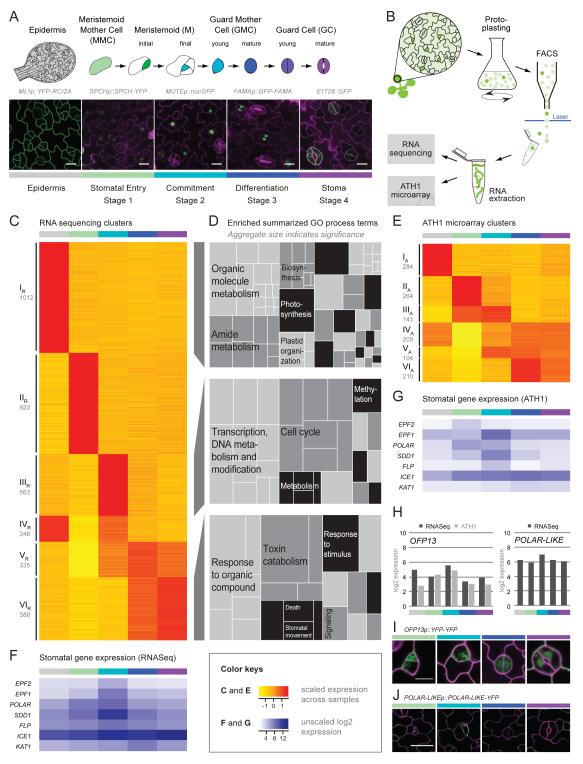
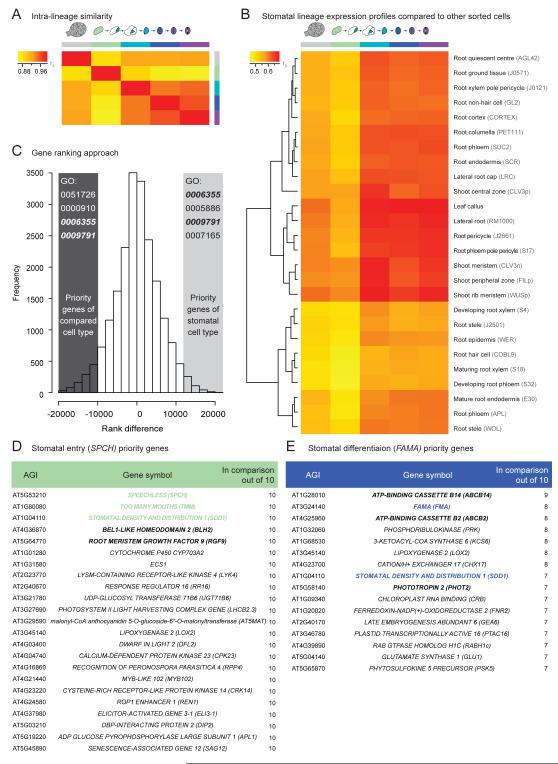
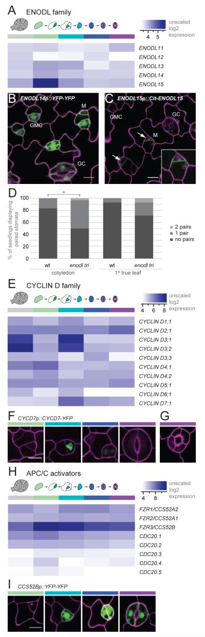
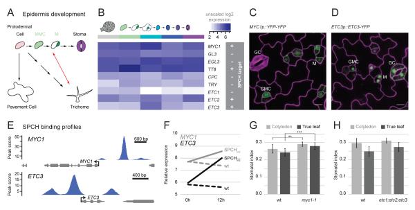
Developmental transitions can be described in terms of morphology and the roles of individual genes, but also in terms of global transcriptional and epigenetic changes. Temporal dissections of transcriptome changes, however, are rare for intact, developing tissues. We used RNA sequencing and microarray platforms to quantify gene expression from labeled cells isolated by fluorescence-activated cell sorting to generate cell-type-specific transcriptomes during development of an adult stem-cell lineage in the Arabidopsis leaf. We show that regulatory modules in this early lineage link cell types that had previously been considered to be under separate control and provide evidence for recruitment of individual members of gene families for different developmental decisions. Because stomata are physiologically important and because stomatal lineage cells exhibit exemplary division, cell fate, and cell signaling behaviors, this dataset serves as a valuable resource for further investigations of fundamental developmental processes.


Figures

Figure 1
Figure 1. Transcriptional profiling of stomatal lineage cells isolated by FACS
(A) Cartoon of stages in stomatal development with confocal images of markers used for FACS. Specific reporters used to mark cell stages are: ML1p::YFP-RCI2A, epidermal cells (including stomatal lineage cells), grey; SPCHp::SPCH-YFP, stomatal entry, green; MUTEp::nucGFP, commitment, light blue; FAMAp::GFP-FAMA, differentiation, violet; E1728::GFP, maturation, purple. Confocal images show cell-type specific expression of fluorescent markers (green) in 2nd true leaves of 14-day old seedlings. Cell outlines are in magenta; scale bars, 10μm (B) Scheme of cell isolation protocol. Aerial seedling tissues expressing markers were protoplasted and fluorescently labeled cells sorted for RNA extraction. Expression profiles of sorted cells were generated using RNA sequencing (RNA-Seq) and ATH1 microarrays (ATH1). (C, E) Clustering of differentially expressed genes identified six dominant expression patterns (clusters I-VI; indices R and A for RNA-Seq and ATH1, respectively). Heat maps showing expression of genes assigned to a cluster (clustering coefficient cut-off 0.6). Mean and median expression values are scaled per gene across samples; low expression is in yellow, high expression in red. The number of genes/cluster is indicated below the cluster name. (D) Enriched GO process terms for clusters IR, IIR, and VIR (from C) summarized using REVIGO (Supek et al., 2011). Related GO terms are displayed in similar colors; aggregate size indicates significance of overrepresentation of a group of GO terms. (F, G) Expression profiles of known stomatal genes generated from sorted cells profiles by RNASeq (F) and ATH1 microarray (G) are highly correlated with published in planta data. Heat maps show unscaled mean and median log2-transformed expression values; low expression is in white; high expression in blue. (H-J) Validation of transcriptional map by reporter analysis of two genes not previously assigned to the stomatal lineage: OVATE FAMILY PROTEIN 13 matches peak expression in stage 2 in both RNA-Seq and ATH1 profiles and POLAR-LIKE (not on ATH1) matches RNA-Seq expression. In (H) the y-axis represents log2-transformed expression values; in (I and J) YFP signal is depicted in green and cell walls in magenta; scale bars, 10μm.
Figure 2
Figure 2. Comparing expression profiles across different cell types in Arabidopsis
(A, B) Heat maps of Spearman’s rank correlation coefficients in pairwise comparisons; low correlation is in yellow; high correlation in red. Within the stomatal lineage, gene expression of stage 1 cells is least correlated to other stomatal lineage cell types (A). Lower correlations are seen comparing FACs-isolated root, shoot, and leaf callus cells to the stomatal lineage (particularly in stage 1) (B). (C-E) A ranking approach compares stomatal stages 1 and 3 with the 10 most highly correlated non-stomatal cell-type specific datasets from (B). Genes were ranked corresponding to their expression within a dataset and the difference in ranking calculated (C). High priority genes fall in the top and bottom 5% of the graph; from these, common enriched GO terms were used to enrich for genes that contribute to a similar process. Selection of genes prioritized in multiple comparisons in entry (D) and differentiation (E) samples (full gene lists in Table S8 and S9).
Figure 3
Figure 3. Genes differentially expressed in stages of the stomatal lineage map have roles in stomatal development
(A) Expression patterns of ENODLs suggest roles for ENODL15, ENODL14 and ENODL13 during stomatal lineage development. (B-C) Confocal images of ENODL14 transcriptional (B) and ENODL15 translational (C) reporters confirm cell-type specific expression in 4-day old cotyledons. White arrow points to ENODL15 accumulation at division planes. Inset shows meristemoid at higher magnification (D) enodl13-1;enodl14-1;enodl15-1 mutants exhibit a higher frequency of mispatterned stomata in cotyledons and true leaves. y-axis shows percentage of seedlings displaying stomatal pairs in a given leaf area. Mann Whitney test, *: p<0.05. (E) Stage-specific expression enrichment for some CYCD family members. (F) GMC-specific expression of CYCD7p::CYCD7;1-YFP; bars are color-coded as in Figure 1A. (G) Expression of CYCD7p::CYCD7;1-YFP promotes extra GC divisions. (H) Expression pattern of APC/C activator genes emphasizes uniquely high expression of CCS52B in the stomatal lineage. (I) Confocal images of CCS52B reporter expression in stomatal lineage cells. All heat maps show unscaled mean and median log2-transformed expression values; low expression is depicted in white; high expression in blue. In confocal images, reporter signal is in green; cell outlines in magenta; scale bars, 10μm.
Figure 4
Figure 4. Cross talk between developmental pathways in the leaf epidermis
(A) Classical (black arrows) and updated (red arrow) view of cell lineages in the leaf. Our data suggest a pluripotent stage from which both trichomes and stomata are derived. (B) Trichome specifying transcription factors MYC1, TT8, ETC2, and ETC3 show high transcript abundance in early stomatal cells. Heat map shows unscaled mean and median log2-transformed expression values; low expression is depicted in white; high expression in blue. (C, D) Expression of MYC1 and ETC3 reporters (green) at stage 1 and 2 of stomatal development in 4-day old cotyledons. scale bars, 10μm (E) MYC1 and ETC3 are direct targets of the stomatal lineage key regulator SPCH. The y-axis represents the computed enrichment score of SPCH binding and arrows indicate transcriptional start sites and orientation of genes; data derived from Lau et al., (2014). (F) Induction of SPCH leads to upregulation of MYC1 and ETC3 transcript. Y-axis represents relative expression; x-axis times after induction in hours; data derived from (Lau et al., 2014). (G) myc1 plants show a higher stomatal index than WT, suggesting interactions between regulation of trichomes and stomata. Stomatal index is shown as mean +/- standard deviation of 7-17 seedlings. Mann Whitney test, **: p<0.01, ***: p<0.001. (H) etc1;etc2;etc3 plants show a non-significant stomatal index increase relative to WT.

Similar articles

Cited by

References

    1. Anders S, Huber W. Differential expression analysis for sequence count data. Genome Biol. 2010;11:R106. - PMC - PubMed
    1. Bargmann BOR, Birnbaum KD. Fluorescence activated cell sorting of plant protoplasts. J Vis Exp. 2010;36 - PMC - PubMed
    1. Bergmann DC, Lukowitz W, Somerville CR. Stomatal development and pattern controlled by a MAPKK kinase. Science. 2004;304:1494–1497. - PubMed
    1. Birnbaum KD, Kussell E. Measuring cell identity in noisy biological systems. Nucleic Acids Research. 2011;39:9093–9107. - PMC - PubMed
    1. Birnbaum K, Shasha DE, Wang JYY, Jung JW, Lambert GM, Galbraith DW, Benfey PN. A gene expression map of the Arabidopsis root. Science. 2003;302:1956–1960. - PubMed

Publication types

MeSH terms

Associated data