Clinical Trial
. 2015 Jul 15;21(14):3149-59.
doi: 10.1158/1078-0432.CCR-14-1421. Epub 2015 Apr 7.

Phase I Hepatic Immunotherapy for Metastases Study of Intra-Arterial Chimeric Antigen Receptor-Modified T-cell Therapy for CEA+ Liver Metastases

Affiliations
Clinical Trial

Phase I Hepatic Immunotherapy for Metastases Study of Intra-Arterial Chimeric Antigen Receptor-Modified T-cell Therapy for CEA+ Liver Metastases

Steven C Katz et al. Clin Cancer Res. .

Abstract

Purpose: Chimeric antigen receptor-modified T cells (CAR-T) have demonstrated encouraging results in early-phase clinical trials. Successful adaptation of CAR-T technology for CEA-expressing adenocarcinoma liver metastases, a major cause of death in patients with gastrointestinal cancers, has yet to be achieved. We sought to test intrahepatic delivery of anti-CEA CAR-T through percutaneous hepatic artery infusions (HAIs).

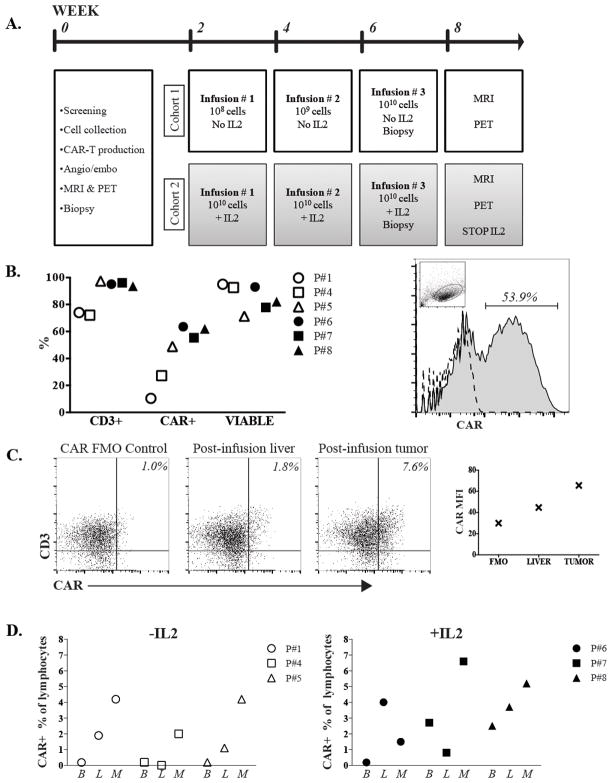
Experimental design: We conducted a phase I trial to test HAI of CAR-T in patients with CEA(+) liver metastases. Six patients completed the protocol, and 3 received anti-CEA CAR-T HAIs alone in dose-escalation fashion (10(8), 10(9), and 10(10) cells). We treated an additional 3 patients with the maximum planned CAR-T HAI dose (10(10) cells × 3) along with systemic IL2 support.

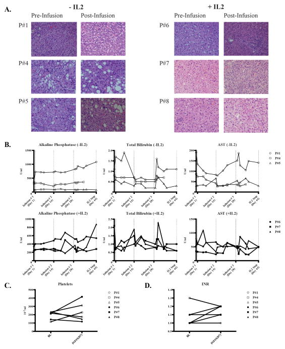
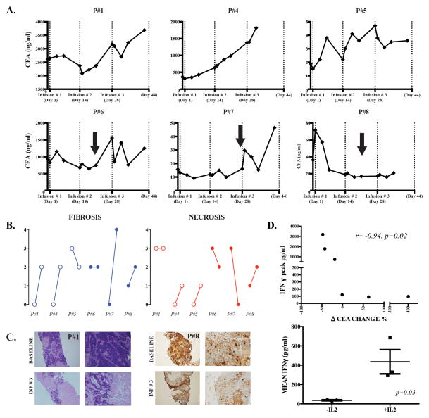
Results: Four patients had more than 10 liver metastases, and patients received a mean of 2.5 lines of conventional systemic therapy before enrollment. No patient suffered a grade 3 or 4 adverse event related to the CAR-T HAIs. One patient remains alive with stable disease at 23 months following CAR-T HAI, and 5 patients died of progressive disease. Among the patients in the cohort that received systemic IL2 support, CEA levels decreased 37% (range, 19%-48%) from baseline. Biopsies demonstrated an increase in liver metastasis necrosis or fibrosis in 4 of 6 patients. Elevated serum IFNγ levels correlated with IL2 administration and CEA decreases.

Conclusions: We have demonstrated the safety of anti-CEA CAR-T HAIs with encouraging signals of clinical activity in a heavily pretreated population with large tumor burdens. Further clinical testing of CAR-T HAIs for liver metastases is warranted.


Conflict of interest statement

Conflicts of Interest: The authors have no relevant disclosures.

Figures

Figure 1
Figure 1. HITM trial design and CAR- T cell trafficking data
(A) Patient treatment and evaluation schedule. (B) Quality control data for the CAR-T products is shown. The percentage of cells that expressed CD3 and the anti-CEA CAR, in addition to the viability fraction, are illustrated for each patient (left). Flow cytometry histogram of pre-infusion product from P#7 demonstrating CAR+ percentage, with the dashed line representing the FMO control and lymphocyte gating demonstrated in the inset dot plot (right). (C) Flow cytometry data from HITM P#7 to illustrate detection of CD3+CAR+ cells within normal liver and liver metastasis two weeks following the second CAR-T HAI. CAR gating was set based on the illustrated FMO control. Plot on right demonstrates CAR mean fluorescence intensity (MFI) values from each specimen for P#7. (D) Blood (B), normal liver (L), and liver metastasis (M) biopsies were harvested and analyzed by flow cytometry as illustrated for P#7. CAR-T percentages among lymphocytes are shown. Percentages were adjusted by subtracting background staining values obtained from FMO control samples. All values represent samples taken 2 weeks following the second infusion. The second dose for cohort 1, -IL2, was 109 cells and for cohort 2, +IL2, was 1010 cells.
Figure 2
Figure 2. Assessment of liver inflammation and injury following anti-CEA CAR-T HAIs
(A) Normal liver biopsies were obtained under CT guidance prior to the first infusion and just prior to the third infusion. Routine H&E staining is shown. (B) Alkaline phosphatase, total bilirubin, and aspartate aminotransferase levels are shown for the patients who did not or did receive systemic IL2. Dotted vertical lines indicate CAR-T infusion time points and the first data point represents the baseline value prior to CAR-T infusion. Platelet counts (C) and INR values (D) for each patient are shown.
Figure 3
Figure 3. Assessments of clinical activity of anti-CEA CAR-T HAIs
(A) Serum CEA levels are illustrated for patients who were treated with CAR-T HAIs alone (top row) or CAR-T with systemic IL2 support (bottom row). CAR-T infusion time points are indicated by dotted vertical lines and IL2 dose interruptions by black arrows. The first data point represents the baseline value prior to CAR-T infusion. (B) A blinded pathologist, comparing baseline to post-infusion, scored fibrosis and necrosis from normal and liver metastasis biopsies. For each patient, baseline and post-infusion scores are shown from left to right. (C) Routine H&E staining for patient # 1 (left panel) and CEA staining for patient # 8 (right panel) are shown, comparing baseline to post-infusion. (D) We measured serum IFNγ concentrations by ELISA before and after each hepatic artery CAR-T infusion. Peak IFNγ levels were correlated with the percentage change in CEA concentration from baseline to the time point before IL-2 dose interruptions or reductions (top). Mean IFNγ values were calculated for each patient and compared among those who did or did not receive systemic IL2 support in addition to hepatic artery CAR-T infusions (bottom).

References

    1. Katz SC, Bamboat ZM, Maker AV, Shia J, Pillarisetty VG, Yopp AC, et al. Regulatory T cell infiltration predicts outcome following resection of colorectal cancer liver metastases. Annals of surgical oncology. 2013;20:946–55. - PMC - PubMed
    1. Katz SC, Donkor C, Glasgow K, Pillarisetty VG, Gonen M, Espat NJ, et al. T cell infiltrate and outcome following resection of intermediate-grade primary neuroendocrine tumours and liver metastases. HPB (Oxford) 2010;12:674–83. - PMC - PubMed
    1. Katz SC, Pillarisetty V, Bamboat ZM, Shia J, Hedvat C, Gonen M, et al. T cell infiltrate predicts long-term survival following resection of colorectal cancer liver metastases. Annals of surgical oncology. 2009;16:2524–30. - PubMed
    1. Wagner P, Koch M, Nummer D, Palm S, Galindo L, Autenrieth D, et al. Detection and functional analysis of tumor infiltrating T-lymphocytes (TIL) in liver metastases from colorectal cancer. Annals of surgical oncology. 2008;15:2310–7. - PubMed
    1. Turcotte S, Katz SC, Shia J, Jarnagin WR, Kingham TP, Allen PJ, et al. Tumor MHC class I expression improves the prognostic value of T-cell density in resected colorectal liver metastases. Cancer immunology research. 2014;2:530–7. - PMC - PubMed

Publication types

Substances