Review
. 2015 Sep;20(8):932-42.
doi: 10.1177/1087057115579638. Epub 2015 Apr 7.

Electroporation Knows No Boundaries: The Use of Electrostimulation for siRNA Delivery in Cells and Tissues

Affiliations
Review

Electroporation Knows No Boundaries: The Use of Electrostimulation for siRNA Delivery in Cells and Tissues

Christin Luft et al. J Biomol Screen. 2015 Sep.

Abstract

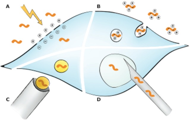
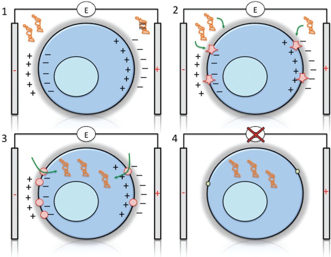
The discovery of RNA interference (RNAi) has enabled several breakthrough discoveries in the area of functional genomics. The RNAi technology has emerged as one of the major tools for drug target identification and has been steadily improved to allow gene manipulation in cell lines, tissues, and whole organisms. One of the major hurdles for the use of RNAi in high-throughput screening has been delivery to cells and tissues. Some cell types are refractory to high-efficiency transfection with standard methods such as lipofection or calcium phosphate precipitation and require different means. Electroporation is a powerful and versatile method for delivery of RNA, DNA, peptides, and small molecules into cell lines and primary cells, as well as whole tissues and organisms. Of particular interest is the use of electroporation for delivery of small interfering RNA oligonucleotides and clustered regularly interspaced short palindromic repeats/Cas9 plasmid vectors in high-throughput screening and for therapeutic applications. Here, we will review the use of electroporation in high-throughput screening in cell lines and tissues.

Keywords: RNA interference; cell transfection; electroporation; high-throughput screening.


Conflict of interest statement

Declaration of Conflicting Interests: The authors declared no potential conflicts of interest with respect to the research, authorship, and/or publication of this article.

Figures

Figure 1.
Figure 1.
General gene delivery mechanisms. (A) Electroporation. During electroporation, cell membranes are destabilized allowing nucleic acid entry into the cell. (B) Reagent-based techniques. The reagents used form complexes with the negatively charged nucleic acids, which are then taken up by the cell via endocytosis. Reagents include cationic lipids, cationic polymers, and calcium phosphate. Cationic lipids form liposomes, which will fuse with the cell membrane and endosomes causing the release of the nucleic acids into the cytoplasm. Cationic polymers such as polyethylenimine condense nucleic acids. They act as a proton sponge, thus buffering acidic endolysosomes and possibly causing their rupture. How calcium phosphate/DNA precipitates are taken up and released into the cytoplasm is not well understood so far. (C) Biolistic particle delivery. Nucleic acid–coated gold particles are shot at target cells. (D) Microinjection. Via an injection needle, nucleic acids can be directly delivered into the nucleus or cytoplasm. Less frequently used methods to deliver genetic material into cells like magnetofection or laserfection as well as viral transduction methods are not displayed.
Figure 2.
Figure 2.
Electroporation of cells. Electroporation occurs through four main steps: (1) polarization of the cell, (2) rapture of the membrane creating nanopores, (3) entry of the macromolecules, and (4) resealing of the membrane. (1) Application of short electrical pulses will result in membrane charging, creating an electrical field and resulting in polarization of the cell. The strong electrical field will result in structural rearrangements of the membrane, creation of water-filled membrane structures (“aqueous pores”) and “nanopores” with a size of more than 1 nm that allow ionic transport. (2) Larger pores are formed in the membrane that allows influx of macromolecules such as DNA or RNA. Generally, more pores are formed at the site facing the negative electrode. (3) Large macromolecules can enter the cell. The negative charge of DNA/RNA can act as a drag to enhance uptake, although, on the other hand, positive ions such as calcium can enhance proximity to the negatively charged membrane prior to uptake. (4) Electroporation is reversible, and once the electric field is switched off, the membrane has the capacity to reseal and keep the macromolecules inside the cell. Resealing occurs on a much longer time frame (minutes to hours), whereas pore formation can occur within milliseconds. Low temperature can enhance resealing, although this may not be practical for eukaryotic cells in some applications.

Similar articles

Cited by

References

    1. Ketteler R., Glaser S., Sandra O., et al. , Enhanced Transgene Expression in Primitive Hematopoietic Progenitor Cells and Embryonic Stem Cells Efficiently Transduced by Optimized Retroviral Hybrid Vectors. Gene Ther. 2002, 9, 477–487. - PubMed
    1. Schiefsky M. J. Hippocrates on Ancient Medicine. In Studies in Ancient Medicine; Brill, Leiden: Boston, 2005. - PubMed
    1. Kilcher S., Schmidt F. I., Schneider C., et al. siRNA Screen of Early Poxvirus Genes Identifies the AAA+ ATPase D5 as the Virus Genome-Uncoating Factor. Cell Host Microbe 2014, 15, 103–112. - PubMed
    1. Collinet C., Stoter M., Bradshaw C. R., et al. Systems Survey of Endocytosis by Multiparametric Image Analysis. Nature 2010, 464, 243–249. - PubMed
    1. Neumann B., Walter T., Heriche J. K., et al. Phenotypic Profiling of the Human Genome by Time-Lapse Microscopy Reveals Cell Division Genes. Nature 2010, 464, 721–727. - PMC - PubMed

Publication types

Substances