Review
. 2015 May;38(5):319-29.
doi: 10.1016/j.tins.2015.03.002. Epub 2015 Apr 4.

Extending the amygdala in theories of threat processing

Affiliations
Review

Extending the amygdala in theories of threat processing

Andrew S Fox et al. Trends Neurosci. 2015 May.

Abstract

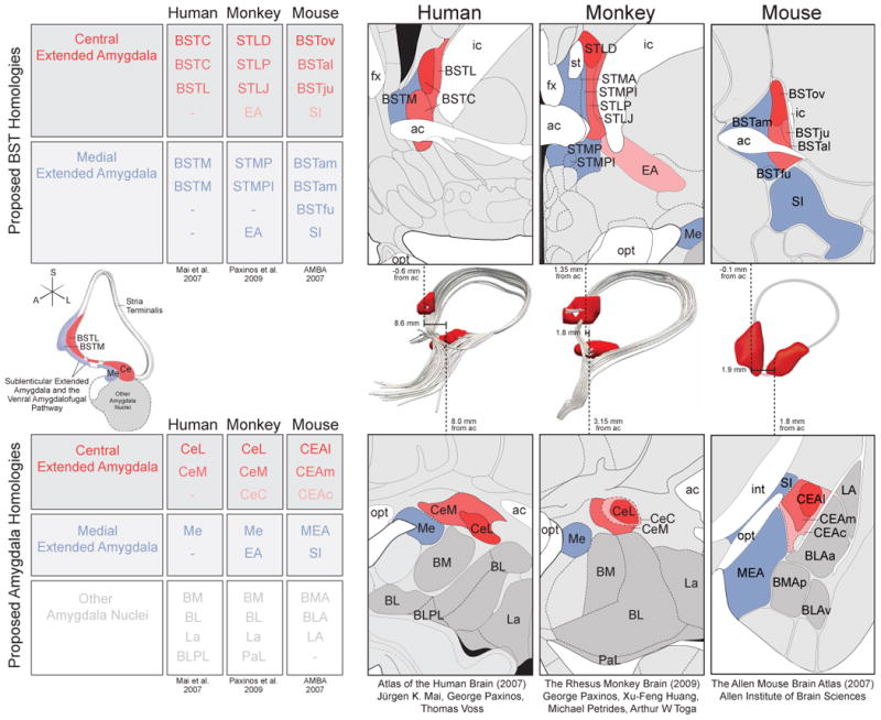
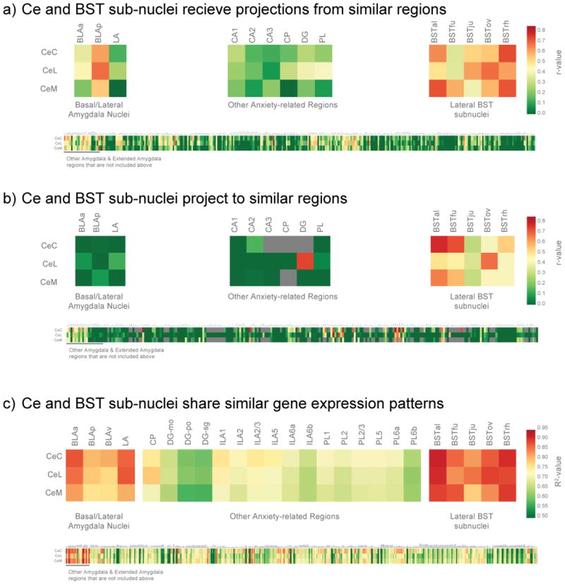
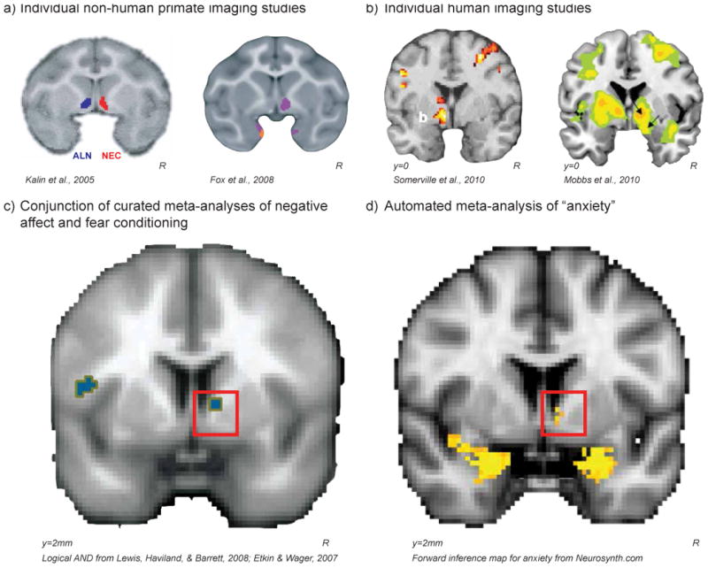
The central extended amygdala is an evolutionarily conserved set of interconnected brain regions that play an important role in threat processing to promote survival. Two core components of the central extended amygdala, the central nucleus of the amygdala (Ce) and the lateral bed nucleus of the stria terminalis (BST) are highly similar regions that serve complimentary roles by integrating fear- and anxiety-relevant information. Survival depends on the ability of the central extended amygdala to rapidly integrate and respond to threats that vary in their immediacy, proximity, and characteristics. Future studies will benefit from understanding alterations in central extended amygdala function in relation to stress-related psychopathology.

Keywords: anxiety; cross-species; extended amygdala; gene expression connectivity; neuroimaging.


Figures

Figure 1
Figure 1. Central extended amygdala (red/pink) and medial extended amygdala (blue) both encompass portions of the BST (top), Ce-BST pathways (middle), and amygdala (bottom)
Although nomenclature varies by species, and by researcher, many regions are thought to have cross-species homology, which is denoted by color throughout and depicted in tables for BST (top left) and amygdala (bottom left). Atlas slices were adapted to highlight the proposed extended amygdala homologies across human ([115]; left), rhesus monkey ([116]; middle) and mouse ([35]; right). Ce-BST connections are show in 3D across the middle row, depicted with: a schematic from Heimer et al. ([117]; left), human DTI, rhesus monkey DTI, and a corresponding mouse drawing (right). Importantly, dashes in the tables do not always denote a lack of homology, but rather a lack of correspondence across atlases. For example, although the CeC can be seen in the human brain [117], this sub-region of the Ce is not included in the Mai et al. 2007 human brain atlas. All figures reprinted with permission.
Figure 2
Figure 2. The Ce and BST show similar patterns of connectivity in rats (a & b), and similar gene expression in mice (c)
We identified those brain structures that most often projected to, or received projections from, the same regions as the individual Ce sub-nuclei (CeL, CeM, and CeC). To accomplish this, we extracted information about efferent and afferent connectivity strength (ranging from does not exist to very strong) for all available brain regions. The efferent and afferent connectivity patterns for each region were correlated with the respective patterns of connectivity for the Ce subnuclei. Because the database is incomplete and we used correlational statistics, we restricted correlations to those based on at least 20 data-points. Results demonstrated that BST subnuclei shared similar inputs (a) and outputs (b) with Ce subnuclei, as compared to other amygdala nuclei (top left), other anxiety-related regions (top middle), or the rest of the brain (bottom). To understand how gene expression within the Ce relates to that in the BST, and the exclusivity of this relationship, we performed a large-scale analysis with the Allen Institute for Brain Science (AIBS) atlas of mouse gene expression [35]. Specifically, we examined the correlation between gene expression profiles in Ce subnuclei with gene expression profiles in other brain regions. Results demonstrated BST subnuclei to have similar gene expression profiles to Ce subnuclei, as compared to other amygdala nuclei (top left), other anxiety-related regions (top middle), or the rest of the brain (bottom). For details of the relationships between Ce subnuclei and other brain regions, zoom in on the online version to read text in the bottom panels.
Figure 3
Figure 3. Individual neuroimaging studies in non-human primates (a) and humans (b) as well as meta-analyses of human neuroimaging data (c-d) demonstrate BST involvement anxiety-related behavior and prolonged threat preparedness
In non-human primates individual differences in brain metabolism in the BST-region were correlated with variation in freezing (a, left) while animals were alone experiencing separation stress (ALN; blue) as well as during exposure to a potentially threatening human intruder making no-eye-contact (NEC; red); and variation in anxious temperament (a, right) during both ALN & NEC (purple). In humans, fMRI activation in the BST-region while potential threats were getting closer, as participants were earning shocks (b, left) or as a spider was approaching their foot (b, right). A Logical AND conjunction (p<.005) of curated meta-analyses of human neuroimaging studies examining 1) the experience of negative affect, and 2) threat conditioning in anxiety-disorder patients compared to controls reveal activation in the BST region (c). In addition, an automated meta-analysis using Neurosynth [92] found that the 222 brain imaging papers using the word “anxiety” were more likely to report activations in coordinates near the BST in comparison to all of the papers in the database (d). Although we do not interpret these studies as conclusive evidence for involvement of the central extended amygdala in anxiety, together these studies provide the necessary initial data to motivate further study of this region in the primate brain. All figures were reprinted with permission.
Figure 4
Figure 4. The number of fMRI articles calling attention to the “Amygdala” as compared to “BST” or extended amygdala
More specifically the total number of fMRI articles (solid) and percentage of total articles using fMRI (dashed), where pubmed searches matched: Amygdala (blue), and (“Extended amygdale” OR BNST OR BST OR “Bed Nucleus of Stria Terminalis” OR “Bed Nucleus of the Stria Terminalis”) (red).

References

    1. Adolphs R. The biology of fear. Curr Biol. 2013;23:R79–93. - PMC - PubMed
    1. Ledoux J. The Emotional Brain: The Mysterious Underpinnings of Emotional Life. Simon and Schuster; 1998.
    1. Lindquist KA, et al. The brain basis of emotion: a meta-analytic review. Behav Brain Sci. 2012;35:121–143. - PMC - PubMed
    1. Paré D, et al. New vistas on amygdala networks in conditioned fear. J Neurophysiol. 2004;92:1–9. - PubMed
    1. Phelps EA. Emotion and cognition: insights from studies of the human amygdala. Annu Rev Psychol. 2006;57:27–53. - PubMed

Publication types