. 2015 Jun;83(6):1165-79.
doi: 10.1002/prot.24810. Epub 2015 Apr 28.

Deformability in the cleavage site of primary microRNA is not sensed by the double-stranded RNA binding domains in the microprocessor component DGCR8

Affiliations

Deformability in the cleavage site of primary microRNA is not sensed by the double-stranded RNA binding domains in the microprocessor component DGCR8

Kaycee A Quarles et al. Proteins. 2015 Jun.

Abstract

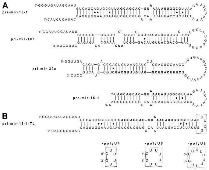
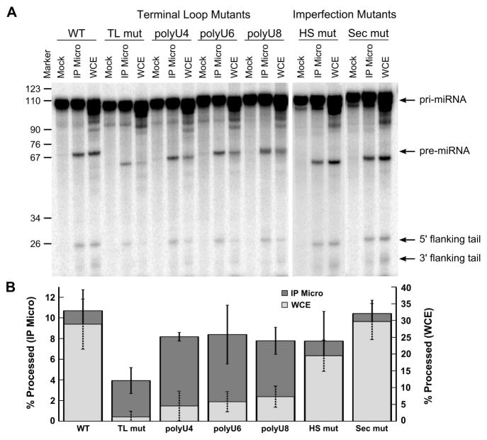
The prevalence of double-stranded RNA (dsRNA) in eukaryotic cells has only recently been appreciated. Of interest here, RNA silencing begins with dsRNA substrates that are bound by the dsRNA-binding domains (dsRBDs) of their processing proteins. Specifically, processing of microRNA (miRNA) in the nucleus minimally requires the enzyme Drosha and its dsRBD-containing cofactor protein, DGCR8. The smallest recombinant construct of DGCR8 that is sufficient for in vitro dsRNA binding, referred to as DGCR8-Core, consists of its two dsRBDs and a C-terminal tail. As dsRBDs rarely recognize the nucleotide sequence of dsRNA, it is reasonable to hypothesize that DGCR8 function is dependent on the recognition of specific structural features in the miRNA precursor. Previously, we demonstrated that noncanonical structural elements that promote RNA flexibility within the stem of miRNA precursors are necessary for efficient in vitro cleavage by reconstituted Microprocessor complexes. Here, we combine gel shift assays with in vitro processing assays to demonstrate that neither the N-terminal dsRBD of DGCR8 in isolation nor the DGCR8-Core construct is sensitive to the presence of noncanonical structural elements within the stem of miRNA precursors, or to single-stranded segments flanking the stem. Extending DGCR8-Core to include an N-terminal heme-binding region does not change our conclusions. Thus, our data suggest that although the DGCR8-Core region is necessary for dsRNA binding and recruitment to the Microprocessor, it is not sufficient to establish the previously observed connection between RNA flexibility and processing efficiency.

Keywords: DGCR8; Drosha; RNA interference; binding affinity; dsRBD; dsRNA; heme binding domain; in vitro processing; microRNA; protein interactions.


Figures

Figure 1
Figure 1
Bending model for pri-miRNA recognition by DGCR8-Core currently supported in the literature. The DGCR8-Core crystal structure (PDB 2YT4, with loops built-in as previously described) is shown with dsRBD1 in red and dsRBD2 in blue. Approximately one turn of idealized A-form dsRNA (tan) has been modeled in contact with each dsRNA binding face of DGCR8, with an additional turn of dsRNA shown in between to bridge the space separating the dsRBDs. The flanking tails, flexible regions in the stem, and terminal loop are implied by dotted lines to suggest the full make-up of a pri-miRNA bound to DGCR8-Core. This model suggests that the pri-miRNA must undergo extreme bending in order to accommodate binding by DGCR8, which may occur at the hot spot and secondary imperfection sites (labeled). Approximate Drosha and Dicer cut sites are also labeled.
Figure 2
Figure 2
Secondary structures of in vitro transcribed RNA models for (A) native pri-miRNAs and pre-mir-16-1; and (B) non-native pri-mir-16-1 stem-loop constructs (region of mutation boxed). Secondary structures shown are as predicted experimentally by combined SHAPE/MC-Pipeline analysis. For all constructs, the mature miRNA is indicated in bold.
Figure 3
Figure 3
Secondary structures of perfect Watson-Crick duplexes derived from pri-mir-16-1 (which is displayed at the top for reference). The mature miRNA strand is shown in bold and the dotted line indicates where each construct’s sequence aligns relative to pri-mir-16-1; these are consistent for all RNA model constructs throughout.
Figure 4
Figure 4
Electrophoretic mobility shift assays used to examine binding by DGCR8 to varying lengths of perfect Watson-Crick RNA duplexes. Representative gels are shown for both (A) DGCR8-Core and (C) DGCR8-dsRBD1 binding to ds44. The leftmost lane in the gels contains RNA, but no protein (labeled “RNA” above the gel). In all other lanes, the concentration of protein increases from left to right (triangle above gel). Bands corresponding to free and bound RNA are indicated to the left of the gels. (B,D) The corresponding fits to the EMSA data for all lengths of duplex (for best-fit parameters, see Table 2; fitting procedure is described in the Materials and Methods). Representative gels for all dsRNA lengths contributing to panels B and D are provided in Supporting Figures S5 and S6.
Figure 5
Figure 5
Competition processing assays were used to corroborate the EMSA results (Fig. 4) in a more biological context. (A) A representative gel using ds22 as the competitor is shown with the concentration of competitor increasing from left to right (triangle above gel). The leftmost lanes report a ladder, pri-mir-16-1 processing in the absence of transfected Microprocessor (Mock), and pri-mir-16-1 processed in the presence of transfected Microprocessor but the absence of any competitor duplex (-Comp). The positions of the pri-miRNA substrate and cleaved pre-miRNA and flanking tails are indicated to the right of the gels. (B) The corresponding fits to a single exponential decay model for all competitors are shown with solid lines (IC50 values reported in Table 3); ds12 and ds16 estimated fits are displayed as dashed lines because these assays only yielded lower limits for IC50. Representative competition processing gels for all RNA constructs are provided in Supporting Figures S7 and S8.
Figure 6
Figure 6
Secondary structures of (A) flanking and (B) terminal loop duplexes derived from pri-mir-16-1 (top). The various terminal loops are boxed to highlight the extent of the mutations.
Figure 7
Figure 7
Drosha processing assays show that the secondary structure of pri-miRNAs is an important determinant of Microprocessor cleavage efficiency in vitro. (A) Denaturing gels for the processing of native pri-mir-16-1 and its mutants: pri-mir-16-1-WT (WT), thermally stable tetraloop mutant (TL mut), polyU4 mutant (polyU4), polyU6 mutant (polyU6), polyU8 mutant (polyU8), “hot spot” mutant (HS mut), and secondary mutant (Sec mut). In each assay, lanes represent RNA exposed to FLAG beads with addition of cell lysate that did not express FLAG-tagged proteins (Mock), exposed to the FLAG-tagged immunopurified Microprocessor (IP Micro), and exposed to whole cell extract containing overexpressed Microprocessor (WCE). The positions of the pri-miRNA substrate, cleaved pre-miRNA, and flanking tails are indicated to the right of the gel. (B) Percentages of pri-miRNAs cleaved by the Microprocessor in vitro averaged over three independent experiments. Processing efficiencies are graphed for the immunopurified Microprocessor (left axis) and the whole cell extract (right axis), with error bars to one standard deviation (see Materials and Methods for details).
Figure 8
Figure 8
Secondary structures of constructs mimicking the imperfections found in pri-mir-16-1: (A) in the context of full-length pri-mir-16-1 and (B) in the context of short duplexes. The regions of mutation are boxed.
Figure 9
Figure 9
Drosha processing assays show that the cysteine residue C352 is an important determinant of Microprocessor cleavage efficiency in vitro. (A) Denaturing gels for the processing of native pri-mir-16-1 and its mutants: pri-mir-16-1-WT (WT), thermostable tetraloop mutant (TL mut), and “hot spot” mutant (HS mut). In each assay, lanes represent RNA exposed to FLAG beads with addition of cell lysate that did not express FLAG-tagged proteins (Mock), exposed to either the FLAG-tagged immunopurified Microprocessor or to whole cell extract containing overexpressed Microprocessor for both the wild-type Microprocessor (IP Micro and WCE Micro, respectively) and for the Microprocessor containing the C352A mutation in DGCR8 (IP C352A and WCE C352A, respectively). The positions of the pri-miRNA substrate, cleaved pre-miRNA, and flanking tails are indicated to the right of the gel. (B) Percentages of pri-miRNAs cleaved by the Microprocessor in vitro averaged over three independent experiments. Processing efficiencies are graphed for the immunopurified Microprocessor (left axis) and the whole cell extract (right axis), with error bars to one standard deviation (see Materials and Methods for details).

References

    1. Zangi L, Lui KO, von Gise A, Ma Q, Ebina W, Ptaszek LM, Spater D, Xu H, Tabebordbar M, Gorbatov R, Sena B, Nahrendorf M, Briscoe DM, Li RA, Wagers AJ, Rossi DJ, Pu WT, Chien KR. Modified mRNA directs the fate of heart progenitor cells and induces vascular regeneration after myocardial infarction. Nat Biotechnol. 2013;31:898–907. - PMC - PubMed
    1. Li Z, Rana TM. Therapeutic targeting of microRNAs: current status and future challenges. Nature reviews Drug discovery. 2014;13:622–638. - PubMed
    1. Bora RS, Gupta D, Mukkur TK, Saini KS. RNA interference therapeutics for cancer: challenges and opportunities (review) Molecular medicine reports. 2012;6:9–15. - PubMed
    1. Castel SE, Martienssen RA. RNA interference in the nucleus: roles for small RNAs in transcription, epigenetics and beyond. Nat Rev Genet. 2013;14:100–112. - PMC - PubMed
    1. Ha M, Kim VN. Regulation of microRNA biogenesis. Nat Rev Mol Cell Biol. 2014;15:509–524. - PubMed

Publication types

LinkOut - more resources