Review
. 2015 Jul-Aug;17(4):646-52.
doi: 10.4103/1008-682X.153299.

Mechanisms of fertilization elucidated by gene-manipulated animals

Affiliations
Review

Mechanisms of fertilization elucidated by gene-manipulated animals

Masaru Okabe. Asian J Androl. 2015 Jul-Aug.

Abstract

Capacitation and the acrosome reaction are key phenomena in mammalian fertilization. These phenomena were found more than 60 years ago. However, fundamental questions regarding the nature of capacitation and the timing of the acrosome reaction remain unsolved. Factors were postulated over time, but as their roles were not verified by gene-disruption experiments, widely accepted notions concerning the mechanism of fertilization are facing modifications. Today, although in vitro fertilization systems remain our central research tool, the importance of in vivo observations must be revisited. Here, primarily focusing on our own research, I summarize how in vivo observations using gene-manipulated animals have elucidated new concepts in the mechanisms of fertilization.


Figures

Figure 1
Figure 1
The mechanisms of fertilization, elucidated by gene-manipulated animals. (a) Spermatozoa that present Adam3 (or some unknown factor(s)) can migrate into the oviduct and reach the vicinity of the eggs. Acrosome reaction is induced before spermatozoa reach the zona pellucida and the fusion-related sperm protein Izumo1 on the outer acrosomal membrane migrates out to sperm surface (indicated by red color). (b) Spermatozoa bind to zona pellucida when mixed with cumulus-free oocytes. However, this binding (mostly observed between the acrosome-intact spermatozoa and zona pellucida) was dispensable. The spermatozoa that lost the so-called “zona-binding” ability remained able to fertilize eggs in vivo once the oviduct migration step was bypassed. Moreover, the timing of the acrosome reaction is flexible, as acrosome-reacted spermatozoa recovered from the perivitelline space could penetrate the zona pellucida a second time and fertilize eggs. The mechanism of sperm penetration of zona pellucida is largely unknown. (c) Only acrosome-reacted spermatozoa can fuse with eggs. Spermatozoa without Izumo1 never fused with eggs. Cd9 on the egg played an important role in fertilization, but Cd9-disrupted females were not completely infertile. In addition, no direct interaction between Cd9 and Izumo1 was observed. This led us to predict a real counterpart for Izumo1. Using the newly established AVEXIS assay, JUNO was recently found to be a counterpart for Izumo1 on the egg. Modified from review.
Figure 2
Figure 2
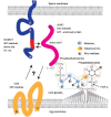
Factors involved in sperm-egg fusion. Izumo1, migrated outward from the outer acrosomal membrane to the sperm surface, tending to localize in the equatorial segment of spermatozoa. Various segments of Izumo1 were examined for their binding ability to eggs and residue 57–113 was indicated to contain an active binding site. Using the AVEXIS assay, JUNO was identified as an Izumo1 binding protein and its role in fusion was verified by gene-disruption experiments. JUNO is a 244-residue protein but is cleaved at 222 to form a GPI (glycosylphosphatidylinositol)-anchored protein. GPI-anchored proteins are initially formed on the cytosolic side and flipped over to the outer membrane side in the final maturation stage. The next helpful piece of information will be the elucidation of the active site of JUNO. Since Izumo1 (57–113) bound to Cd9-disrupted eggs normally, the elucidation of Cd9's role(s) will offer further clarification.
Figure 3
Figure 3
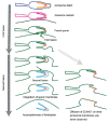
Fertilization requires two independent fusions. Intact spermatozoa have a plasma membrane (blue) and an acrosomal membrane (orange). After the acrosome reaction, these two membranes fuse and form a new sperm membrane (pink). The first fusion takes place between the pink membrane and egg plasma membrane (black). After the first fusion, egg and sperm membrane form a new consecutive membrane (green). If fusion is accomplished in this step, Izumo1 on the acrosomal cap of the inner acrosomal membrane (indicated by red) should spread on the newly-formed egg surface (green). However, the second fusion (invagination) follows the first fusion that separates the acrosomal cap and acrosomal sheath areas (light blue) from the fused membrane (green). Thus, Izumo1 on the inner acrosomal membrane is invaginated into the cytoplasm of the eggs. From live imaging, Izumo1 seems to be required for the first fusion. The nature of the second fusion remains totally unknown.

References

    1. Clift D, Schuh M. Restarting life: fertilization and the transition from meiosis to mitosis. Nat Rev Mol Cell Biol. 2013;14:549–62. - PMC - PubMed
    1. Austin CR. Observations on the penetration of the sperm in the mammalian egg. Aust J Sci Res B. 1951;4:581–96. - PubMed
    1. Chang MC. Fertilizing capacity of spermatozoa deposited into the fallopian tubes. Nature. 1951;168:697–8. - PubMed
    1. Dan J. Studies on the acrosome reaction. I. Reaction to egg water and other stimuli. Biol Bull. 1952;103:54–66.
    1. Yanagimachi R, Chang MC. Fertilization of hamster eggs in vitro. Nature. 1963;200:281–2. - PubMed

Publication types