Review
. 2015 Sep;84(9):1685-93.
doi: 10.1016/j.ejrad.2015.03.016. Epub 2015 Mar 21.

Ultrasound molecular imaging: Moving toward clinical translation

Affiliations
Review

Ultrasound molecular imaging: Moving toward clinical translation

Lotfi Abou-Elkacem et al. Eur J Radiol. 2015 Sep.

Abstract

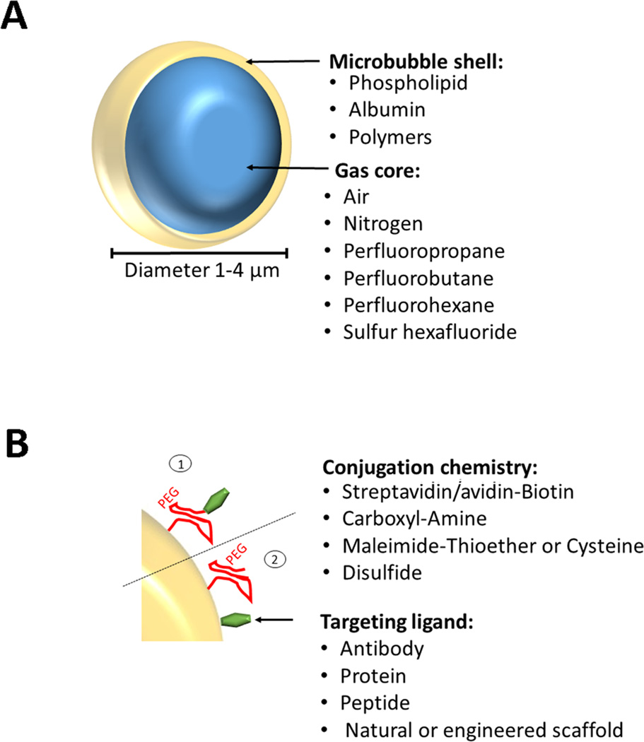
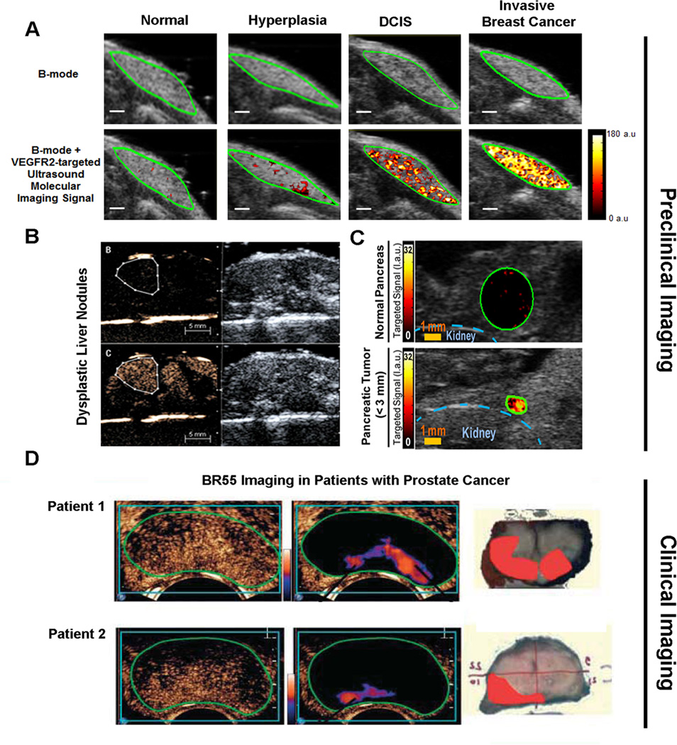
Ultrasound is a widely available, cost-effective, real-time, non-invasive and safe imaging modality widely used in the clinic for anatomical and functional imaging. With the introduction of novel molecularly-targeted ultrasound contrast agents, another dimension of ultrasound has become a reality: diagnosing and monitoring pathological processes at the molecular level. Most commonly used ultrasound molecular imaging contrast agents are micron sized, gas-containing microbubbles functionalized to recognize and attach to molecules expressed on inflamed or angiogenic vascular endothelial cells. There are several potential clinical applications currently being explored including earlier detection, molecular profiling, and monitoring of cancer, as well as visualization of ischemic memory in transient myocardial ischemia, monitoring of disease activity in inflammatory bowel disease, and assessment of arteriosclerosis. Recently, a first clinical grade ultrasound contrast agent (BR55), targeted at a molecule expressed in neoangiogenesis (vascular endothelial growth factor receptor type 2; VEGFR2) has been introduced and safety and feasibility of VEGFR2-targeted ultrasound imaging is being explored in first inhuman clinical trials in various cancer types. This review describes the design of ultrasound molecular imaging contrast agents, imaging techniques, and potential future clinical applications of ultrasound molecular imaging.

Keywords: Cancer; Clinical translation; Inflammation; Microbubbles; Molecular imaging; Ultrasound.


Figures

Figure 1
Figure 1. Design of molecularly-targeted microbubbles
(A) Microbbubles are 1–4 µm microspheres with a shell composed of various materials and a core that can contain different types of gases. (B) Incorporated PEG chains stabilize the microbubble shell, form a steric barrier to prevent coalescence, minimize adsorption of macromolecules to the microbubble surface, and provide spacing between the shell and binding ligands. Various types of ligands (e.g., antibodies, proteins, peptides) can be attached non-covalently or covalently by using biotin/streptavidin, biotin/avidin, amine (NH2)/amide, maleimide/thioether, 2-(Pridylthio)propionyl (PDP)/disulfide, either via the PEG arm (1) or directly onto the shell surface (2).
Figure 2
Figure 2. Examples of quantification techniques of ultrasound molecular imaging signal
(A) Destruction-replenishment technique shown graphically in a vessel expressing molecular imaging targets (red) and after injection of molecularly-targeted microbubbles (green). (B) Imaging signal in field of view increases after intravenous injection of targeted microbubbles and is composed of signal from attached and freely circulating microbubbles as well as tissue background signal. After a few minutes, a high pressure destructive pulse destroys all microbubbles within the beam elevation and after an additional few seconds freely circulating microbubbles have replenished into the field of view. The difference in imaging signal pre and post destruction corresponds to the signal from attached microbubbles (targeted MB signal). Modified from [13]. (C) Assessment of the attenuation independent residual-to-saturation ratio using acoustic radiation force. After injection of molecularly-targeted microbubbles (green) an acoustic radiation force (ARF) pulse gently pushes targeted microbubbles to the vascular endothelial cell wall thereby enhancing molecular target attachment of microbubbles. After terminating the push pulse, unbound microbubbles are released due to flow shear forces and only firmly attaching microbubbles stay attached. (D) The initial signal Minitial represents the background signal of the tissue in the absence of adherent microbubbles. After ARF pulses, the imaging signal from locally accumulating microbubbles enhances up to full saturation (Msaturation). After termination of the ARF pulse, non-attached microbubbles float away and the imaging signal from attached microbubbles (Mresidual) can be measured. Modified from [18].
Figure 3
Figure 3. Molecular imaging of inflammation in inflammatory bowel disease
(A) Schematic representation shows molecularly-targeted microbubbles (blue) binding to molecular markers expressed on the vascular endothelial cells of inflamed capillaries in a bowel segment. Modified from [22]. (B) Ultrasound molecular imaging of Mucosal Addressin Cellular Adhesion Molecule (MAdCAM-1) in an IBD model of spontaneous ileitis in mice shows weak background signal in non-inflamed ileum (left) and strong signal in acute ileitis (middle); histology confirms active inflammation (right). Reproduced with permission from [20]. (C) Transverse dual P- and E-selectin targeted ultrasound images obtained in mice with chemically induced acute colitis scanned at day 1 (severe colitis), day 5 (mild colitis), and in mouse with normal colon, compared to imaging with control, non-targeted microbubbles. Note strong signal in acute inflammation at day 1 which decreases when inflammation is reduced at day 5 (scale bar = 1 mm). Representative confocal micrographs overlaid on differential interference contrast images show accumulation of fluorescently labeled selectin-targeted microbubbles (red) in mucosal capillaries (green) in acute inflammation but not in non-inflamed colon tissue (scale bar = 100 µm). Reprinted with permission from [22]. (D) Cross-modality intra-animal comparison of dual-selectin-targeted ultrasound and FDG PET-CT imaging shows good quantitative correlation of both modalities (scale bar, 1 mm) with histology confirming imaging results. (E) Translational study in acute terminal ileitis model in a pig shows feasibility of dual-selectin targeted ultrasound imaging signal (middle) with substantially increased imaging signal compared to control non-targeted contrast agent (left) in inflamed ileum. Histology confirms inflammation (right).
Figure 4
Figure 4. Preclinical and clinical examples of ultrasound molecular imaging of cancer and cancer development using clinical grade vascular endothelial cell receptor type 2 (VEGFR2) targeted microbubbles (BR55)
(A) Ultrasound molecular images using BR55 in transgenic mouse model of breast cancer development shows substantially increasing imaging signal in the mammary gland with breast tissue progressing from normal to hyperplasia, ductal carcinoma in situ, and invasive breast cancer, suggesting that the magnitude of imaging signal at the cancer stage may help earlier detection of breast cancer using ultrasound molecular imaging. Reprinted with permission from [32]. (B) In a transgenic mouse model of hepatocellular carcinoma development, VEGFR2-targeted ultrasound imaging allowed diagnosing of dysplastic nodule based on magnitude of imaging signal (lower row) while non-targeted contrast-enhanced ultrasound imaging could not differentiate between healthy and dysplastic liver tissue. Reprinted with permission from [34]. (C) Ultrasound imaging also allows visualization of early pancreatic adenocarcinoma in a transgenic mouse model of spontaneous pancreatic cancer development with small foci of cancer showing substantially higher signal (lower row) than normal pancreatic tissue (upper row) due to strong expression of VEGFR2, suggesting that this technology could be used for screening purposes in high risk populations. Reprinted with permisison from [33]. (D) Examples of transrectal transverse VEGFR2-targeted ultrasound molecular images in two patients with biospy-proven prostate cancer, imaged 11 min following intravenous contrast agent injection. Raw data images (left, showing mixed signal from freely circulating and attached microbubbles) and post-processed images (middle; highligthening signal from stationary (attached) microbubbles) show foci of enhanced signal in the peripheral zone suggesting presence of cancer. Right images show corresponding macroscopy slices following radical prostatectomy with the extent of prostate cancer assess by pathological analysis overlaid in red. Reprinted with permission from [47].

Similar articles

Cited by

References

    1. Wilson SR, Burns PN. Microbubble-enhanced US in body imaging: what role? Radiology. 2010;257(1):24–39. - PubMed
    1. Pysz MA, Willmann JK. Targeted contrast-enhanced ultrasound: an emerging technology in abdominal and pelvic imaging. Gastroenterology. 2011;140(3):785–790. - PMC - PubMed
    1. Kiessling F, Fokong S, Bzyl J, Lederle W, Palmowski M, Lammers T. Recent advances in molecular, multimodal and theranostic ultrasound imaging. Adv Drug Deliv Rev. 2014;72:15–27. - PMC - PubMed
    1. Deshpande N, Needles A, Willmann JK. Molecular ultrasound imaging: current status and future directions. Clin Radiol. 2010;65(7):567–581. - PMC - PubMed
    1. Klibanov AL. Ligand-carrying gas-filled microbubbles: ultrasound contrast agents for targeted molecular imaging. Bioconjug Chem. 2005;16(1):9–17. - PubMed

Publication types

MeSH terms

LinkOut - more resources