Clinical Trial
. 2015 Jun 4;125(23):3618-26.
doi: 10.1182/blood-2015-01-620781. Epub 2015 Apr 7.

Characteristic repartition of monocyte subsets as a diagnostic signature of chronic myelomonocytic leukemia

Affiliations
Clinical Trial

Characteristic repartition of monocyte subsets as a diagnostic signature of chronic myelomonocytic leukemia

Dorothée Selimoglu-Buet et al. Blood. .

Abstract

Chronic myelomonocytic leukemia (CMML) is a myelodysplastic syndrome/ myeloproliferative neoplasm whose diagnosis is currently based on the elevation of peripheral blood monocytes to >1 × 10(9)/L, measured for ≥3 months. Diagnosis can be ambiguous; for example, with prefibrotic myelofibrosis or reactive monocytosis. We set up a multiparameter flow cytometry assay to distinguish CD14(+)/CD16(-) classical from CD14(+)/CD16(+) intermediate and CD14(low)/CD16(+) nonclassical monocyte subsets in peripheral blood mononucleated cells and in total blood samples. Compared with healthy donors and patients with reactive monocytosis or another hematologic malignancy, CMML patients demonstrate a characteristic increase in the fraction of CD14(+)/CD16(-) cells (cutoff value, 94.0%). The associated specificity and sensitivity values were 95.1% and 90.6% in the learning cohort (175 samples) and 94.1% and 91.9% in the validation cohort (307 samples), respectively. The accumulation of classical monocytes, which demonstrate a distinct gene expression pattern, is independent of the mutational background. Importantly, this increase disappears in patients who respond to hypomethylating agents. We conclude that an increase in the fraction of classical monocytes to >94.0% of total monocytes is a highly sensitive and specific diagnostic marker that rapidly and accurately distinguishes CMML from confounding diagnoses.


Figures

Figure 1
Figure 1
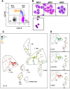
Monocyte subsets in PBMCs were explored. (A) PBMCs from a representative healthy blood donor were labeled with anti-CD45, -CD24, -CD14, -CD16, -CD56, -CD115, -CD62L, -CD64, -CCR2, and -CX3CR1 antibodies. Monocytes were identified using an exclusion gating strategy (described in supplemental Figure 1), and subsets were separated on CD14 and CD16 expression. The percentage of each subset is indicated. (B) May-Grünwald-Giemsa staining of sorted MO1s, MO2s, MO3s, and DN (remaining double-negative CD14/CD16) cells. (C) Multiparametric analysis of single cells monitored with 10 surface markers (supplemental Table 1) using the SPADE algorithm, which organizes cells in a hierarchy of related phenotypes. Flow cytometry data from 19 healthy donor PBMCs were gated on morphology, then on CD45+/SSC intermediate, and used to construct the SPADE tree that automatically separates, on the basis of the hierarchy of related phenotypes, MO1s (CD14+/CD16), MO2s (CD14+/CD16+), MO3s (CD14low/CD16+), natural killer cells (NK; CD56+), B lymphocytes (CD24+), and residual granulocytes (Gran; CD24+/CD16+). The percentage of each subset in the monocyte population is indicated. Circles indicate the size of cell populations, and colors are based on CD14 expression. (D) Color representation of CD16, CX3CR1, and CCR2 expression in the monocyte subsets delineated in the SPADE tree.
Figure 2
Figure 2
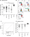
Abnormal repartition of monocyte subsets in CMML. (A) Percentage of MO1s in a learning cohort of CMML patients as compared to healthy blood donors (Co), age-matched healthy donors (Aged-Co), patients with diverse hematologic malignancies (Non-CMML), and those with reactive monocytosis (Reactive). Data are presented as the mean ± SEM; ***P < .0001; Kruskal-Wallis test. (B) Multicolor representation of monocyte subset repartition in PBMCs collected from the distinct groups of the learning cohort. The percentage of each monocyte subset is indicated. (C) ROC curve analysis of diagnostic sensitivity and specificity of MO1 percentage in peripheral blood monocytes established on the learning cohort (young and age-matched healthy donors, other hematologic malignancies, reactive monocytosis, and CMML) defined in panel A. (D) Percentage of MO1s in a validation cohort of CMML as compared to age-matched healthy donors (Aged-Co), patients with MDS or MPN (Non-CMML), and those with reactive monocytosis (Reactive). Data are presented as the mean ± SEM. SEM, standard error of the mean.
Figure 3
Figure 3
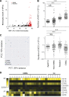
CMML patients accumulate abnormal MO1s at the expense of MO3s. (A) Lack of significant correlation between the percentage of MO1s and peripheral blood monocyte count in reactive monocytosis (black circles) and in CMML (red circles). Reactive monocytoses and CMML samples from the learning and validation cohorts were pooled. (B) Absolute number of MO1s and MO3s in the peripheral blood of CMML patients as compared to age-matched healthy donors (Aged-Co), non-CMML patients, and patients with reactive monocytosis. The learning and validation cohorts were pooled (***P < .0001; Student t test). (C) Component principal analysis of gene expression after RNA sequencing (DESSeq2 analysis) in MO1s sorted from the blood of healthy donors (green) and patients with reactive monocytosis (blue) or CMML (red). (D) Heatmap established by using Cytobank software to summarize the flow cytometry analysis of 8 markers at the surface of MO1s from 6 age-matched healthy donors and 17 CMML patients.
Figure 4
Figure 4
Monocyte subset profile as a biomarker of disease evolution. (A) Repeated evaluation of MO1 fraction in 21 untreated CMML patients followed from 6 to 26 months. (B) Evaluation of MO1 fraction in 7 CMML patients before and after treatment with azacytidine (AZA); all 7 patients responded to treatment. (C-E) Evolution of monocyte subset repartition and monocyte count in CMML patients during treatment with a demethylating agent. (C) Patient who responded to azacytidine. (D) Patient who only transiently responded to azacytidine. (E) Patient who relapsed after initial response.
Figure 5
Figure 5
Comparison between monocyte subset profile and monocyte count diagnosis. (A) ROC curves of the “MO1 percentage >94.0%” and “monocyte count >1 × 10⁹/L” criteria to identify CMML at initial workup in the validation cohort. The difference between the 2 curves was highly significant (P < .0001, χ2 test). (B) Monocyte subset repartition analyzed in 2 independent samples obtained from 3 patients classified as having MDS and showing a fluctuating monocyte count of ∼1 × 10⁹/L that precludes their classification as CMML according to the WHO criteria. (C) Monocyte subset repartition analyzed in 2 independent samples obtained from a patient who could be classified as having CMML according to the WHO criteria but who, based on cytologic and molecular analyses, demonstrates sideroblastic anemia with monocytosis.

References

    1. Vardiman JW, Thiele J, Arber DA, et al. The 2008 revision of the World Health Organization (WHO) classification of myeloid neoplasms and acute leukemia: rationale and important changes. Blood. 2009;114(5):937–951. - PubMed
    1. Itzykson R, Solary E. An evolutionary perspective on chronic myelomonocytic leukemia. Leukemia. 2013;27(7):1441–1450. - PubMed
    1. Ziegler-Heitbrock L, Ancuta P, Crowe S, et al. Nomenclature of monocytes and dendritic cells in blood. Blood. 2010;116(16):e74–e80. - PubMed
    1. Cros J, Cagnard N, Woollard K, et al. Human CD14dim monocytes patrol and sense nucleic acids and viruses via TLR7 and TLR8 receptors. Immunity. 2010;33(3):375–386. - PMC - PubMed
    1. Wong KL, Tai JJ-Y, Wong W-C, et al. Gene expression profiling reveals the defining features of the classical, intermediate, and nonclassical human monocyte subsets. Blood. 2011;118(5):e16–e31. - PubMed

Publication types

Substances