

High-spin Mn-oxo complexes and their relevance to the oxygen-evolving complex within photosystem II
- PMID: 25852147
- PMCID: PMC4418911
- DOI: 10.1073/pnas.1422800112
High-spin Mn-oxo complexes and their relevance to the oxygen-evolving complex within photosystem II
Abstract
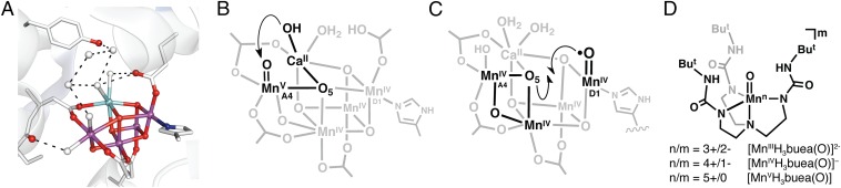
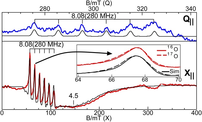
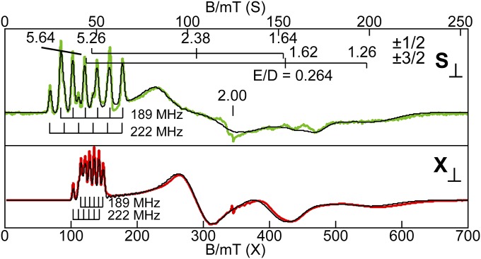
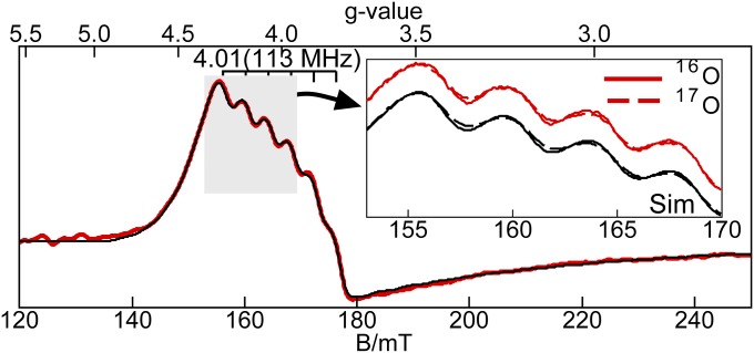
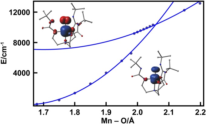
The structural and electronic properties of a series of manganese complexes with terminal oxido ligands are described. The complexes span three different oxidation states at the manganese center (III-V), have similar molecular structures, and contain intramolecular hydrogen-bonding networks surrounding the Mn-oxo unit. Structural studies using X-ray absorption methods indicated that each complex is mononuclear and that oxidation occurs at the manganese centers, which is also supported by electron paramagnetic resonance (EPR) studies. This gives a high-spin Mn(V)-oxo complex and not a Mn(IV)-oxy radical as the most oxidized species. In addition, the EPR findings demonstrated that the Fermi contact term could experimentally substantiate the oxidation states at the manganese centers and the covalency in the metal-ligand bonding. Oxygen-17-labeled samples were used to determine spin density within the Mn-oxo unit, with the greatest delocalization occurring within the Mn(V)-oxo species (0.45 spins on the oxido ligand). The experimental results coupled with density functional theory studies show a large amount of covalency within the Mn-oxo bonds. Finally, these results are examined within the context of possible mechanisms associated with photosynthetic water oxidation; specifically, the possible identity of the proposed high valent Mn-oxo species that is postulated to form during turnover is discussed.
Keywords: inorganic chemistry; metal–oxo complexes; oxygen-evolving complex; photosynthesis; water oxidation.
Conflict of interest statement
The authors declare no conflict of interest.
Figures
Comment in
-
An Mn(V)-oxo role in splitting water?Proc Natl Acad Sci U S A. 2015 Apr 28;112(17):5265-6. doi: 10.1073/pnas.1505223112. Epub 2015 Apr 16. Proc Natl Acad Sci U S A. 2015. PMID: 25883270 Free PMC article. No abstract available.
References
-
- Britt RD, et al. Recent pulsed EPR studies of the Photosystem II oxygen-evolving complex: Implications as to water oxidation mechanisms. Biochim Biophys Acta Bioenerg. 2004;1655(1–3):158–171. - PubMed
-
- Umena Y, Kawakami K, Shen J-R, Kamiya N. Crystal structure of oxygen-evolving photosystem II at a resolution of 1.9 Å. Nature. 2011;473(7345):55–60. - PubMed
-
- Suga M, et al. Native structure of photosystem II at 1.95 Å resolution viewed by femtosecond X-ray pulses. Nature. 2015;517(7532):99–103. - PubMed
-
- Rapatskiy L, et al. Detection of the water-binding sites of the oxygen-evolving complex of Photosystem II using W-band 17O electron-electron double resonance-detected NMR spectroscopy. J Am Chem Soc. 2012;134(40):16619–16634. - PubMed
Publication types
MeSH terms
Substances
Grants and funding
LinkOut - more resources
Full Text Sources
Other Literature Sources
