. 2015 Apr 7;21(13):3876-87.
doi: 10.3748/wjg.v21.i13.3876.

Fucosylation is a common glycosylation type in pancreatic cancer stem cell-like phenotypes

Affiliations

Fucosylation is a common glycosylation type in pancreatic cancer stem cell-like phenotypes

Naoko Terao et al. World J Gastroenterol. .

Abstract

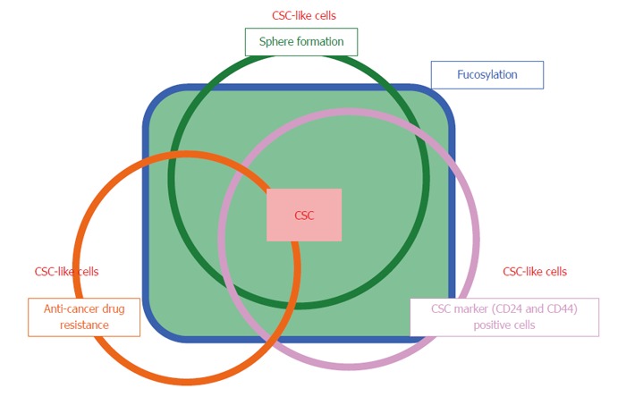
Aim: To evaluate/isolate cancer stem cells (CSCs) from tissue or cell lines according to various definitions and cell surface markers.

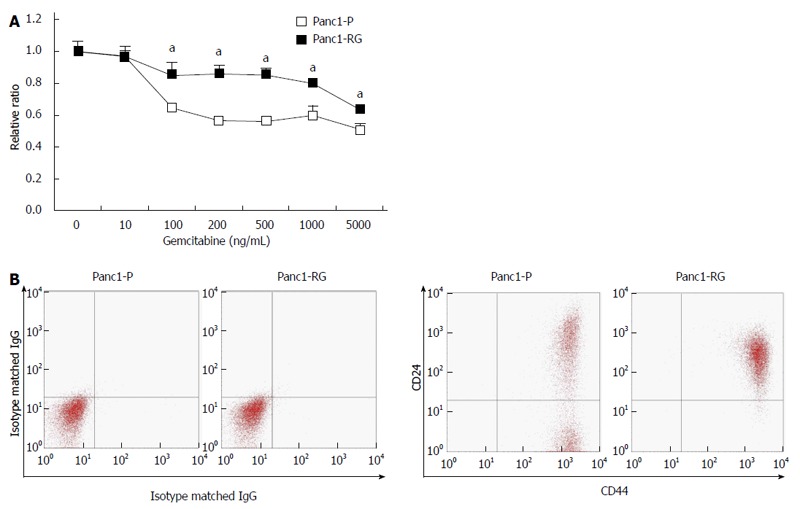
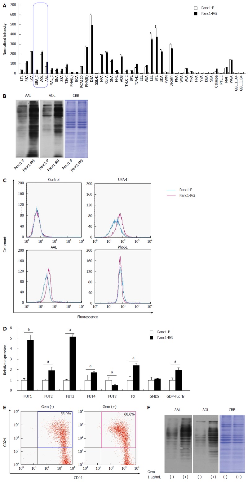
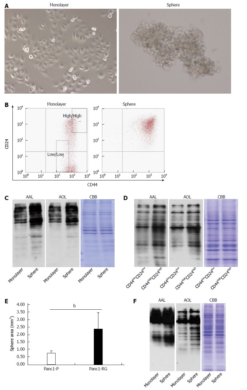
Methods: Lectin microarray analysis was conducted on CSC-like fractions of the human pancreatic cancer cell line Panc1 by establishing anti-cancer drug-resistant cells. Changes in glycan structure of CSC-like cells were also investigated in sphere-forming cells as well as in CSC fractions obtained from overexpression of CD24 and CD44.

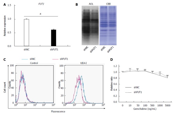
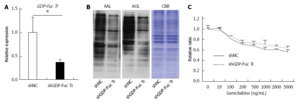
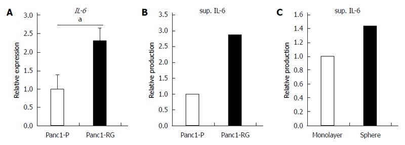
Results: Several types of fucosylation were increased under these conditions, and the expression of fucosylation regulatory genes such as fucosyltransferases, GDP-fucose synthetic enzymes, and GDP-fucose transporters were dramatically enhanced in CSC-like cells. These changes were significant in gemcitabine-resistant cells and sphere cells of a human pancreatic cancer cell line, Panc1. However, downregulation of cellular fucosylation by knockdown of the GDP-fucose transporter did not alter gemcitabine resistance, indicating that increased cellular fucosylation is a result of CSC-like transformation.

Conclusion: Fucosylation might be a biomarker of CSC-like cells in pancreatic cancer.

Keywords: Anti-cancer drug resistance; Cancer stem cells; Fucosylation; Glycosylation; Pancreatic cancer; Sphere formation.


Figures

Figure 1
Figure 1
Cancer stem cells are enriched in established gemcitabine-resistant cells. A: Panc1-RG cells were established by long-term step-wise gemcitabine treatment. WST assay showed that Panc1-RG cells exhibited gemcitabine resistance compared with Panc1-P cells; B: Flow cytometric analysis showing a higher ratio of CD24+/CD44+ cells in Panc1-RG cells compared with Panc1-P cells. All results are expressed as mean ± SD; aP < 0.05 vs Panc1-P cells.
Figure 2
Figure 2
Fucosylation is enhanced in gemcitabine-treated pancreatic cancer cells. A: Differential glycan profiles of Panc1-P and Panc1-RG cells were analyzed by lectin microarray. The fluorescence intensity of each lectin was normalized to the intensity of wheat germ agglutinin; B: Aleuria aurantia lectin (AAL) or Aspergillus oryzae lectin (AOL) lectin blot of Panc1-P and Panc1-RG. Coomassie brilliant blue (CBB) staining confirms equal protein loading among lanes; C: Cell-surface fucosylated glycoproteins in Panc1-P and Panc1-RG cells were assessed by flow cytometry using Ulex europaeus agglutinin (UEA)-I, AAL, and Pholiota squarrosa lectin (PhoSL) lectins; D: Fucosylation-related gene expression, determined by real-time reverse transcription PCR in Panc1-P and Panc1-RG cells, and expressed relative to that of Panc1-P cells; E: Flow cytometric analysis represented gemcitabine treatment increased the ratio of CD24+/CD44+ cells in PK59 cells; F: AAL or AOL lectin blot of PK59 cells with (+) or without (-) gemcitabine treatment. All results are expressed as mean ± SD; aP < 0.05 vs Panc1-P cells.
Figure 3
Figure 3
Fucosylation is enhanced in cancer stem cell-like cells fractions of pancreatic cancer cells. A: Phase-contrast images of monolayer (left panel) and sphere cells (right panel) derived from Panc1 cells; B: Results of flow cytometric analysis in monolayer and sphere Panc1 cells using CD24 and CD44 antibodies. CD24+/CD44+ cells were increased in sphere cells compared with monolayer cells; C: Aleuria aurantia lectin (AAL) or Aspergillus oryzae lectin (AOL) lectin blot of monolayer and sphere Panc1 cells; D: AAL or AOL lectin blot of CD24high/CD44high or CD24low/CD44low Panc1 cells obtained by cell sorting. Each region is indicated as a square field in (B); E: Comparison of sphere cell-forming ability between Panc1-P and Panc1-RG; F: AAL or AOL lectin blot of monolayer and PSN-1 cells. Results are expressed as mean ± SD; bP < 0.01 vs Panc1-P cells. CBB: Coomassie brilliant blue.
Figure 4
Figure 4
FUT1 knockdown in Panc1-RG cells does not alter gemcitabine resistance. A: Expression of fucosyltransferase (FUT)1 mRNA in Panc1-RG cells stably expressing small hairpin (sh)RNA; B: Aspergillus oryzae lectin (AOL) blot of shNC- or shFUT1-expressing Panc1-RG cells; C: Flow cytometric analysis using Ulex europaeus agglutinin (UEA)-I; D: WST assay results of transfectant cells treated with various gemcitabine concentrations. Results are expressed as mean ± SD; aP < 0.05 vs shNC. CBB: Coomassie brilliant blue; NC: Negative control.
Figure 5
Figure 5
GDP-fucose transporter gene knockdown does not affect gemcitabine resistance in Panc1-RG cells. A: Expression of GDP-fucose transporter (GDP-Fuc Tr) mRNA in small hairpin (sh)RNA-expressing Panc1-RG cells; B: Aleuria aurantia lectin (AAL) or Aspergillus oryzae lectin (AOL) blots for shRNA transfectant cells; C: WST assay results for cells treated with various gemcitabine concentrations. Results are expressed as mean ± SD; aP < 0.05 vs shNC. CBB: Coomassie brilliant blue; NC: Negative control.
Figure 6
Figure 6
Increased interleukin-6 production in anti-cancer drug-resistance and sphere-forming cells. A: Expression of interleukin (IL)-6 mRNA was measured by real-time reverse transcription PCR in Panc1-P and Panc1-RG; B: The concentration of IL-6 in conditioned media from Panc1-P or Panc1-RG cultures was assessed by enzyme-linked immunosorbent assay (ELISA); C: The concentration of IL-6 in conditioned media from monolayer or sphere-forming Panc1-P cells was measured by ELISA. All results are expressed as mean ± SD; aP < 0.05 vs Panc1-P cells.
Figure 7
Figure 7
Overview of the relationship between fucosylation and three types of cancer stem cell-like cells. Cancer stem cells (CSCs) are a limited fraction of cancer cells, which show asymmetric and slow growth, anti-cancer drug resistance, and sphere formation. CSC-like cells are differentiated from and have similar biologic characteristics of CSCs. Fucosylation is common type of glycosylation in the CSC-like phenotype of pancreatic cancer under various conditions.

Similar articles

Cited by

References

    1. Reya T, Morrison SJ, Clarke MF, Weissman IL. Stem cells, cancer, and cancer stem cells. Nature. 2001;414:105–111. - PubMed
    1. Ailles LE, Weissman IL. Cancer stem cells in solid tumors. Curr Opin Biotechnol. 2007;18:460–466. - PubMed
    1. Dean M, Fojo T, Bates S. Tumour stem cells and drug resistance. Nat Rev Cancer. 2005;5:275–284. - PubMed
    1. Singh A, Settleman J. EMT, cancer stem cells and drug resistance: an emerging axis of evil in the war on cancer. Oncogene. 2010;29:4741–4751. - PMC - PubMed
    1. Jemal A, Siegel R, Ward E, Murray T, Xu J, Smigal C, Thun MJ. Cancer statistics, 2006. CA Cancer J Clin. 2006;56:106–130. - PubMed

Publication types

MeSH terms