. 2015 Feb 28:10:97.
doi: 10.1186/s11671-015-0781-y. eCollection 2015.

Angular shaping of fluorescence from synthetic opal-based photonic crystal

Affiliations

Angular shaping of fluorescence from synthetic opal-based photonic crystal

Vitalii Boiko et al. Nanoscale Res Lett. .

Abstract

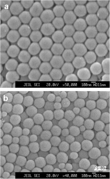
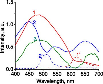
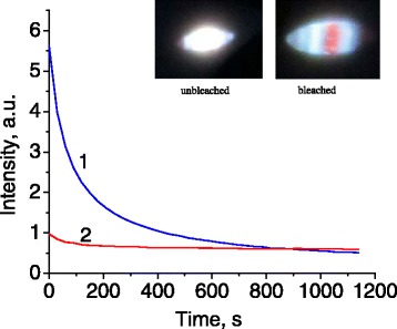
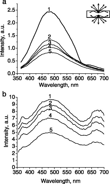
Spectral, angular, and temporal distributions of fluorescence as well as specular reflection were investigated for silica-based artificial opals. Periodic arrangement of nanosized silica globules in the opal causes a specific dip in the defect-related fluorescence spectra and a peak in the reflectance spectrum. The spectral position of the dip coincides with the photonic stop band. The latter is dependent on the size of silica globules and the angle of observation. The spectral shape and intensity of defect-related fluorescence can be controlled by variation of detection angle. Fluorescence intensity increases up to two times at the edges of the spectral dip. Partial photobleaching of fluorescence was observed. Photonic origin of the observed effects is discussed.

Keywords: Angular dependence; Fluorescence; Photobleaching; Photonic crystal; Refractive index; Stop band; Synthetic opal.


Figures

Figure 1
Figure 1
SEM image of silica globules. They are ordered in sample 1 (a) and disordered in photonic glass (b).
Figure 2
Figure 2
Spectra of light reflectance for photonic samples having different sizes of silica globules. Numbering of spectra corresponds to the numbering of samples.
Figure 3
Figure 3
Angular dependence of light reflectance for sample 1. Wavelengths of incident light were 532 nm (a) and 593 nm (b). Both s-polarized (1) and p-polarized (2) light were tested.
Figure 4
Figure 4
Angular dependence of light reflectance for sample 2. Wavelength of incident light were 402 nm (a) and 593 nm (b). Both s-polarized (1) and p-polarized (2) light were tested.
Figure 5
Figure 5
Angular dependence of light reflectance for sample 1. Wavelength of incident light was 402 nm. Both s-polarized (1) and p-polarized (2) light were tested.
Figure 6
Figure 6
Light reflectance spectra for sample 2 measured at the angles 10° and 20°.
Figure 7
Figure 7
Fluorescence spectra of photonic crystal samples with different spectral positions of stop zones. Numbering of spectra 1 and 2 corresponds to the numbering of samples. Spectrum 3 is fluorescence of a soda lime glass slab. Spectra 1′ and 2′ are reflectance spectra of samples 1 and 2, respectively (plotted not to scale with fluorescence).
Figure 8
Figure 8
Photobleaching of photonic crystal fluorescence in time. Unequal kinetics of blue and red fluorescence is illustrated on the example of 450 nm (1) and 650 nm (2) spectral bands excited by steady laser irradiation at 266 nm. Images of the fluorescent spot on the surface of sample 2 in the initial and bleached states are shown in the inset.
Figure 9
Figure 9
Fluorescence spectra of sample 2 measured at different detection angles. (1) 0°; (2) 10°; (3) 20°; (4) 30°; (5) 40°; (6) 70° (λ exc = 266 nm).
Figure 10
Figure 10
Fluorescence spectra of photonic glass (a) and soda lime glass slab (b), measured at different angles. (1) 0°; (2) 40°; (3) 50°; (4) 60°; (5) 70°. The inset depicts total internal reflection of light at high angles.

References

    1. Chaudhery V, George S, Lu M, Pokhriya A, Cunningham B. Nanostructured surfaces and detection instrumentation for photonic crystal enhanced fluorescence. Sensors. 2013;13:5561–84. doi: 10.3390/s130505561. - DOI - PMC - PubMed
    1. Ganesh N, Zhang W, Mathias P, Chow E, Soares J, Malyarchuk V, et al. Enhanced fluorescence emission from quantum dots on a photonic crystal surface. Nat Nanotech. 2007;2:515–20. doi: 10.1038/nnano.2007.216. - DOI - PubMed
    1. Knabe S, Soleimani N, Markvart T, Bauer G. Efficient light trapping in a fluorescence solar collector by 3D photonic crystal. Phys Status Solidi RRL. 2010;4(5–6):118–20. doi: 10.1002/pssr.201004047. - DOI
    1. Barnes W. Fluorescence near interfaces: the role of photonic mode density. J Mod Optic. 1998;45(4):661–99. doi: 10.1080/09500349808230614. - DOI
    1. Badugu R, Nowaczyk K, Descrovi E, Lakowicz J. Radiative decay engineering 6: fluorescence on one-dimensional photonic crystals. Anal Biochem. 2013;442:83–96. doi: 10.1016/j.ab.2013.07.021. - DOI - PMC - PubMed

LinkOut - more resources