Review
. 2015 Mar 23:9:98.
doi: 10.3389/fnins.2015.00098. eCollection 2015.

Proteoglycans and neuronal migration in the cerebral cortex during development and disease

Affiliations
Review

Proteoglycans and neuronal migration in the cerebral cortex during development and disease

Nobuaki Maeda. Front Neurosci. .

Abstract

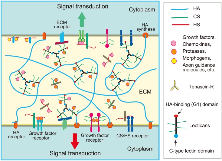
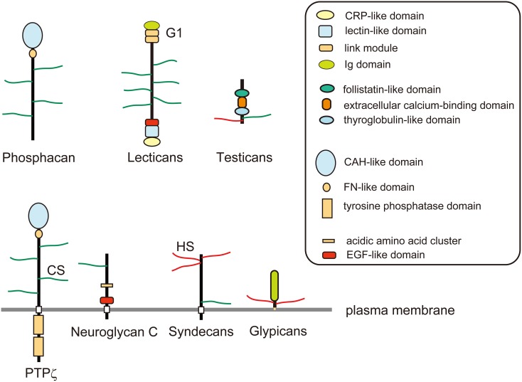
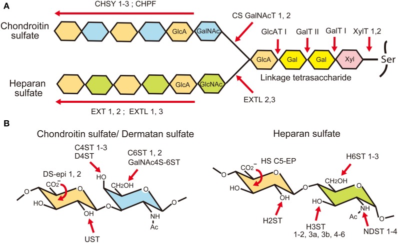
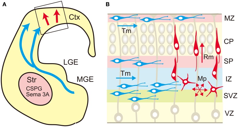
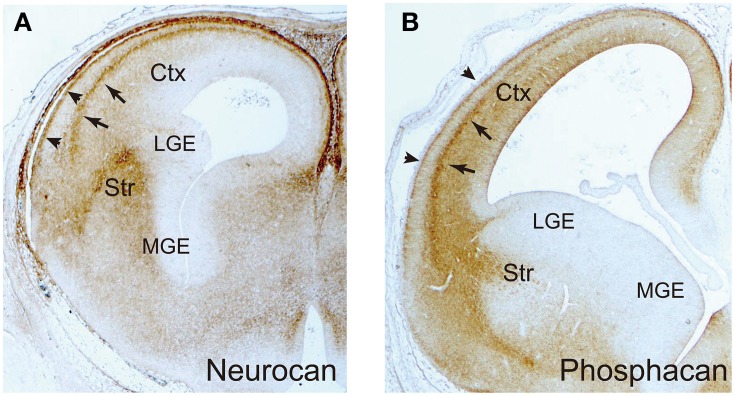
Chondroitin sulfate proteoglycans and heparan sulfate proteoglycans are major constituents of the extracellular matrix and the cell surface in the brain. Proteoglycans bind with many proteins including growth factors, chemokines, axon guidance molecules, and cell adhesion molecules through both the glycosaminoglycan and the core protein portions. The functions of proteoglycans are flexibly regulated due to the structural variability of glycosaminoglycans, which are generated by multiple glycosaminoglycan synthesis and modifying enzymes. Neuronal cell surface proteoglycans such as PTPζ, neuroglycan C and syndecan-3 function as direct receptors for heparin-binding growth factors that induce neuronal migration. The lectican family, secreted chondroitin sulfate proteoglycans, forms large aggregates with hyaluronic acid and tenascins, in which many signaling molecules and enzymes including matrix proteases are preserved. In the developing cerebrum, secreted chondroitin sulfate proteoglycans such as neurocan, versican and phosphacan are richly expressed in the areas that are strategically important for neuronal migration such as the striatum, marginal zone, subplate and subventricular zone in the neocortex. These proteoglycans may anchor various attractive and/or repulsive cues, regulating the migration routes of inhibitory neurons. Recent studies demonstrated that the genes encoding proteoglycan core proteins and glycosaminoglycan synthesis and modifying enzymes are associated with various psychiatric and intellectual disorders, which may be related to the defects of neuronal migration.

Keywords: chondroitin sulfate; extracellular matrix; heparan sulfate; neuronal migration; proteoglycan.


Figures

Figure 1
Figure 1
Schematic structure of extracellular matrix (ECM) in the brain. The ECM of the brain is mainly composed of chondroitin sulfate (CS) and heparan sulfate (HS) proteoglycans, hyaluronic acid (HA), and glycoproteins such as tenascins. Lectican family CS proteoglycans form large aggregates with HA and tenascins, which store various proteins such as chemokines, growth factors and axon guidance molecules. The ECM proteoglycans may bind with a CS/HS receptor on the cell surface such as RPTPσ. Cell surface proteoglycans may function as receptors or co-receptors for growth factors.
Figure 2
Figure 2
Schematic structures of proteoglycans. Phosphacan and lectican family chondroitin sulfate (CS) proteoglycans are major constituents of brain extracellular matrix. PTPζ is a CS proteoglycan-type protein tyrosine phosphatase, which is a splice variant of phosphacan. Neuroglycan C is a transmembrane CS proteoglycan that is classified as Neuregulin-6. Glypicans are a family of glycosyl-phosphatidylinositol (GPI)-anchored heparan sulfate (HS) proteoglycans. Syndecans are a family of transmembrane HS proteoglycans, some members of which may also be modified by CS chains. Testicans are a family of secreted CS/HS proteoglycans.
Figure 3
Figure 3
Biosynthesis of glycosaminoglycans. (A) Chondroitin sulfate (CS) and heparan sulfate (HS) chains are covalently attached to the proteoglycan core proteins through a common linkage tetrasaccharide. It is considered that the biosynthesis of a chondroitin chain starts with the addition of an N-acetylgalactosamine (GalNAc) residue to the linkage tetrasaccharide by CS N-acetylgalactosaminyltransferases (CSGalNAcT1, 2). After that, glucuronic acid (GlcA) and GalNAc residues are co-polymerized by chondroitin sulfate synthases (CHSY-1, -2, -3) and chondroitin polymerization factor (CHPF). When an N-acetylglucosamine (GlcNAc) residue is added to the linkage tetrasaccharide instead of GalNAc by EXTL 2 or 3, a heparan chain is polymerized by EXT family members. (B) After polymerization, they are modified by sulfation and epimerization reactions by many glycosaminoglycan modifying enzymes.
Figure 4
Figure 4
Migration routes of excitatory and inhibitory neurons in the cerebrum. (A) The excitatory neurons are generated in the ventricular zone of the neocortex (Ctx) and migrate radially toward the brain surface (red arrow). The cortical inhibitory neurons are generated mainly in the medial ganglionic eminence (MGE), and migrate tangentially toward the neocortex (blue arrows). The migrating interneurons avoid the striatum (Str) that expresses chondroitin sulfate proteoglycans (CSPG) and semaphorin 3A (Sema 3A). (B) In the neocortex, the excitatory neurons (red cells) born in the ventricular zone (VZ) show multipolar morphology and migrate in random directions (Mp) in the subventricular (SVZ) and intermediate (IZ) zones. When the multipolar neurons reach the subplate (SP), they transform into a bipolar shape and migrate radially (Rm) in the cortical plate (CP) toward the marginal zone (MZ). On the other hand, the tangential migration (Tm) of interneurons (blue cells) occurs in a layer-specific manner, in which interneurons prefer MZ, SP, lower IZ, and SVZ.
Figure 5
Figure 5
Immunohistochemical localization of neurocan and phosphacan. The frontal sections of embryonic day 16 rat brains were immunohistochemically stained with anti-neurocan (A) and anti-phosphacan (B) monoclonal antibodies. They are selectively expressed in the marginal zone (arrowheads) and subplate (arrows) in the neocortex, and the striatum (Str). The medial (MGE) and lateral (LGE) ganglionic eminences are negative.

References

    1. Ai X., Do A. T., Lozynska O., Kusche-Gullberg M., Lindahl U., Emerson C. P. (2003). QSulf1 remodels the 6-O sulfation states of cell surface heparan sulfate proteoglycans to promote Wnt signaling. J. Cell Biol. 162, 341–351. 10.1083/jcb.200212083 - DOI - PMC - PubMed
    1. Ai X., Kitazawa T., Do A. T., Kusche-Gullberg M., Labosky P. A., Emerson C. P. (2007). SULF1 and SULF2 regulate heparan sulfate-mediated GDNF signaling for esophageal innervation. Development 134, 3327–3338. 10.1242/dev.007674 - DOI - PubMed
    1. Aono S., Tokita Y., Shuo T., Yamauchi S., Matsui F., Nakanishi K., et al. (2004). Glycosylation site for chondroitin sulfate on the neural part-time proteoglycan, neuroglycan C. J. Biol. Chem. 279, 46536–46541. 10.1074/jbc.M403263200 - DOI - PubMed
    1. Aspberg A., Miura R., Bourdoulous S., Shimonaka M., Heinegard D., Schachner M., et al. (1997). The C-type lectin domains of lecticans, a family of aggregating chondroitin sulfate proteoglycans, bind tenascin-R by protein-protein interactions independent of carbohydrate moiety. Proc. Natl. Acad. Sci. U.S.A. 94, 10116–10121. 10.1073/pnas.94.19.10116 - DOI - PMC - PubMed
    1. Berretta S. (2012). Extracellular matrix abnormalities in schizophrenia. Neuropharmacology 62, 1584–1597. 10.1016/j.neuropharm.2011.08.010 - DOI - PMC - PubMed