Review
. 2015 Mar 17:9:91.
doi: 10.3389/fncel.2015.00091. eCollection 2015.

Researching glutamate - induced cytotoxicity in different cell lines: a comparative/collective analysis/study

Affiliations
Review

Researching glutamate - induced cytotoxicity in different cell lines: a comparative/collective analysis/study

Aristeidis A Kritis et al. Front Cell Neurosci. .

Abstract

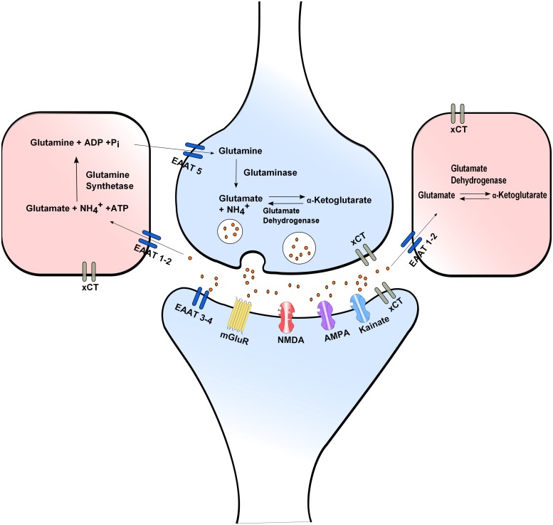
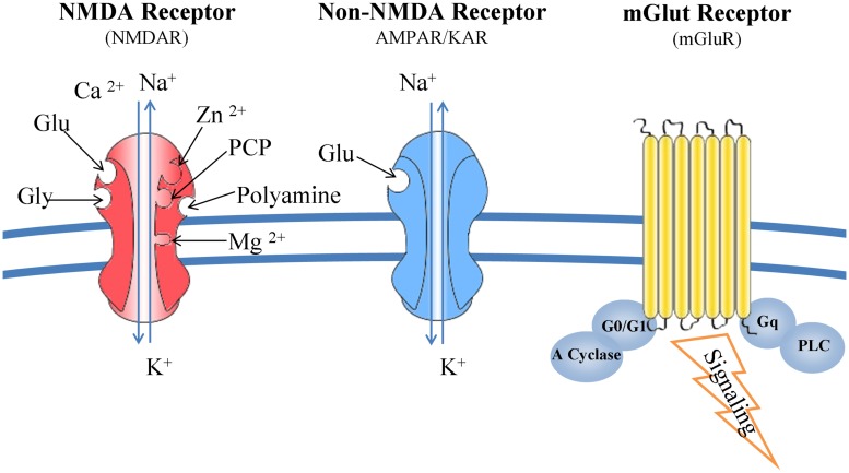
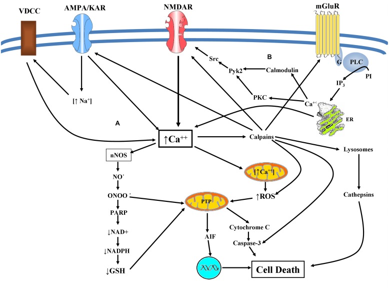
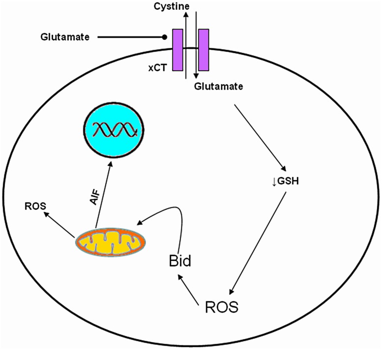
Although glutamate is one of the most important excitatory neurotransmitters of the central nervous system, its excessive extracellular concentration leads to uncontrolled continuous depolarization of neurons, a toxic process called, excitotoxicity. In excitotoxicity glutamate triggers the rise of intracellular Ca(2+) levels, followed by up regulation of nNOS, dysfunction of mitochondria, ROS production, ER stress, and release of lysosomal enzymes. Excessive calcium concentration is the key mediator of glutamate toxicity through over activation of ionotropic and metabotropic receptors. In addition, glutamate accumulation can also inhibit cystine (CySS) uptake by reversing the action of the CySS/glutamate antiporter. Reversal of the antiporter action reinforces the aforementioned events by depleting neurons of cysteine and eventually glutathione's reducing potential. Various cell lines have been employed in the pursuit to understand the mechanism(s) by which excitotoxicity affects the cells leading them ultimately to their demise. In some cell lines glutamate toxicity is exerted mainly through over activation of NMDA, AMPA, or kainate receptors whereas in other cell lines lacking such receptors, the toxicity is due to glutamate induced oxidative stress. However, in the greatest majority of the cell lines ionotropic glutamate receptors are present, co-existing to CySS/glutamate antiporters and metabotropic glutamate receptors, supporting the assumption that excitotoxicity effect in these cells is accumulative. Different cell lines differ in their responses when exposed to glutamate. In this review article the responses of PC12, SH-SY5Y, HT-22, NT-2, OLCs, C6, primary rat cortical neurons, RGC-5, and SCN2.2 cell systems are systematically collected and analyzed.

Keywords: HT-22; NT-2; PC12; RGC-5; SCN2.2; SH-SY5Y; excitotoxicity; glutamate oxidative toxicity.


Figures

FIGURE 1
FIGURE 1
Glutamate release and uptake, the Xc- antiporter and the glutamate/glutamine cycle. In glial cells reuptaken glutamate is converted to glutamine by glutamine synthetase. Glial glutamine is taken up into the presynaptic neuron via Na+-dependent glutamine uptake systems, where it is converted to glutamate by glutaminase. Extra- and intra-cellular glutamate concentrations are modulated through the XC- antiporter. Neurotransmission is ended by efficient glutamate reuptake via Na+-dependent high affinity glutamate membrane EAATs.
FIGURE 2
FIGURE 2
Glutamate receptors: structure and function. NMDARs bind glutamate, glycine, Mg2+, Zn2+, and polyamines. Composed from seven subunits (one NR1, four NR2, and two NR3), their function is determined by the combination of NR1 and NR2 subunits. NMDARs form channels that are more permeable to Ca2+ than Na+ and K+. Kainate and AMPA receptors interact only with glutamate and their specific agonists, and their associated channels are more permeable to Na+ and K+ than Ca2+. mGluRs are G-protein coupled receptors and trigger a second messenger cascade. They are found both at the pre- and post-synaptic neurons, subunits of metabotropic receptors are also expressed in microglia.
FIGURE 3
FIGURE 3
(A) Excitotoxicity mediated cell death: glutamate excitotoxicity causes Ca2+ mediated NO production leading to mitochondrial dysfunction resulting in superoxide production. Peroxynitrite is produced causing lipid peroxidation, direct DNA damage, and protein dysfunction. Peroxynitrite inhibits the mitochondrial electron transport chain, cytochrome c normal activity as well as of superoxide dismutase via protein nitration. Activation of ryanodine receptors in conjunction with accumulation of misfolded proteins and depletion of endoplasmic Ca2+ storage, results in ER dysfunction (ER-stress). These facts can provoke caspase mediated cell death and an eventual apoptotic cell death. Alternatively augmented intracellular Ca2+ concentration can lead to calpain activation engaging both calpain dependent and cathepsin dependent cell death. (B) mGluRs and NMDAR crosstalk: both mGluR1 and mGluR5 lead to the potentiation of NMDA receptor currents via Src-dependent mechanism in a PKC or calmodulin depended manner.
FIGURE 4
FIGURE 4
Molecular mechanism of oxidative glutamate toxicity. Increased extracellular glutamate concentration leads to reverse action of Cys glutamate antiporter. GSH depletion, follows due to decrease of intracellular Cys influencing the capacity of cells to scavenge free radicals, rendering them vulnerable mitochondrial dysfunction and to secondary events such as accumulation of ROS, production of Bid and AIF, resulting in cell death.

References

    1. Ahern G. P., Klyachko V. A., Jackson M. B. (2002). cGMP and S-nitrosylation: two routes for modulation of neuronal excitability by NO. Trends Neurosci. 25 510–517 10.1016/S0166-2236(02)02254-3 - DOI - PubMed
    1. Akundi R. S., Hüll M., Clement H. W., Fiebich B. L. (2003). 1-trichloromethyl-1,2,3,4-tetrahydro-beta-carboline (TaClo) induces apoptosis in human neuroblastoma cell lines. Ann. N. Y. Acad. Sci. 1010 304–306 10.1196/annals.1299.053 - DOI - PubMed
    1. Albasanz J. L., Ros M., Martin M. (1997). Characterization of metabotropic glutamate receptors in rat C6 glioma cells. Eur. J. Pharmacol. 326 85–91 10.1016/S0014-2999(97)00154-4 - DOI - PubMed
    1. Alberdi E., Sánchez-Gómez M. V., Marino A., Matute C. (2002). Ca(2+) influx through AMPA or kainate receptors alone is sufficient to initiate excitotoxicity in cultured oligodendrocytes. Neurobiol. Dis. 9 234–243 10.1006/nbdi.2001.0457 - DOI - PubMed
    1. Ali D. W., Salter M. W. (2001). NMDA receptor regulation by Src kinase signalling in excitatory synaptic transmission and plasticity. Curr. Opin. Neurobiol. 11 336–342 10.1016/S0959-4388(00)00216-6 - DOI - PubMed