. 2015 Mar 19:9:93.
doi: 10.3389/fncel.2015.00093. eCollection 2015.

Dendritic patch-clamp recordings from cerebellar granule cells demonstrate electrotonic compactness

Affiliations

Dendritic patch-clamp recordings from cerebellar granule cells demonstrate electrotonic compactness

Igor Delvendahl et al. Front Cell Neurosci. .

Abstract

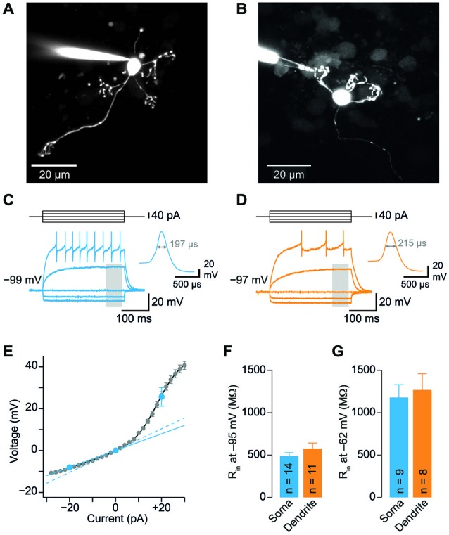
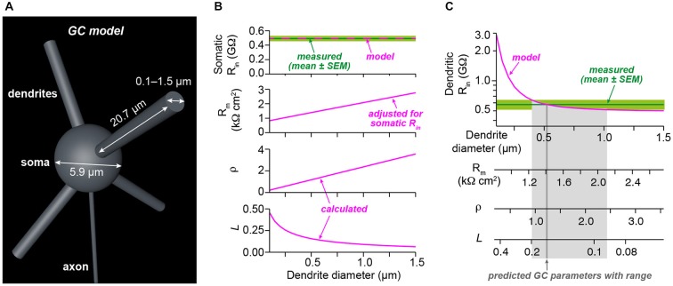
Cerebellar granule cells (GCs), the smallest neurons in the brain, have on average four short dendrites that receive high-frequency mossy fiber inputs conveying sensory information. The short length of the dendrites suggests that GCs are electrotonically compact allowing unfiltered integration of dendritic inputs. The small average diameter of the dendrites (~0.7 µm), however, argues for dendritic filtering. Previous studies based on somatic recordings and modeling indicated that GCs are electrotonically extremely compact. Here, we performed patch-clamp recordings from GC dendrites in acute brain slices of mice to directly analyze the electrotonic properties of GCs. Strikingly, the input resistance did not differ significantly between dendrites and somata of GCs. Furthermore, spontaneous excitatory postsynaptic potentials (EPSP) were similar in amplitude at dendritic and somatic recording sites. From the dendritic and somatic input resistances we determined parameters characterizing the electrotonic compactness of GCs. These data directly demonstrate that cerebellar GCs are electrotonically compact and thus ideally suited for efficient high-frequency information transfer.

Keywords: cerebellum; dendrites; electrophysiology; granule cell; patch-clamp techniques.


Figures

Figure 1
Figure 1
Somatic and dendritic input resistances are similar. (A) Cerebellar granule cell (GC) filled with a fluorescent dye via the patch-pipette during a somatic recording. Maximum intensity projection of a two-photon stack of 11 images, z-step 2 µm. (B) GC filled with a fluorescent dye via the patch-pipette during a dendritic recording. Maximum intensity projection of a two-photon stack of 8 images, z-step 3 µm. (C) Voltage transients obtained in response to tonic current injections (steps of ± 20 pA) in a somatic GC recording. Action potentials are truncated for clarity. Inset shows action potential on enlarged time scale, the duration at the half-maximal amplitude is indicated. Same cell as in (A). The gray area indicates time window used for analysis of input resistance. (D) Corresponding data from a dendritic recording. Same cell as in (B). (E) Voltage-current relation determined with small current steps in n = 35 GC somata in a different set of experiments (gray markers). Blue markers represent data obtained with ± 20 pA steps. Input resistance determined using small (2 pA, dashed line) and larger steps (−20 pA, continuous line) differed by a factor of 1.3. Input resistance measured using −20 pA steps was corrected by this factor (see Material and Methods). (F) Average input resistance (Rin) at GC somata and dendrites was not significantly different. Rin was determined at a membrane potential of −95.4 ± 1.1 mV (corresponding to 0 pA in panel E). Bars represent means ± SEM (number of somatic and dendritic GC recordings is indicated). (G) Rin recorded from somata and dendrites at a membrane potential of −62.4 ± 3.2 mV (corresponding to +20 pA in panel E).
Figure 2
Figure 2
GC dendrites do not significantly filter spontaneous EPSPs. (A) Representative spontaneous excitatory postsynaptic potentials (EPSP) recorded from the soma (blue) or dendrite (orange) of GCs. Traces were digitally filtered to 8 kHz (−3 dB cut-off) for display. The recording configurations are illustrated on the left. (B) Dendritic EPSP amplitude as a function of distance from soma. For comparison, the somatically recorded EPSP amplitude is plotted in blue (n = 12 and n = 11 somatic and dendritic recordings, respectively). (C) EPSP amplitude was comparable at somatic and dendritic recording sites. (D) The 20–80% EPSP rise time did not differ significantly between soma and dendrites. (E) EPSP decay time constants were similar for somatic and dendritic recording sites. All bargraphs show means ± SEM (number of somatic and dendritic GC recordings is indicated).
Figure 3
Figure 3
Analysis of electrotonic properties demonstrates electrical compactness. (A) Illustration of our GC model. The model consisted of a spherical soma and four dendrites. The indicated diameter of the soma and the length of dendrites were measured from stacks of two-photon microscopic images obtained during dendritic recordings. Axon diameter was taken from the literature. The diameter of the dendrites was systematically varied between 0.1 and 1.5 µm. (B) Superposition of the measured somatic input resistance (Rin) with the prediction of the model. For each diameter of the dendrites, the specific membrane resistance (Rm) was adjusted to ensure the correct somatic Rin. The dendrite-to-soma conductance ratio (ρ) and the electrotonic length of the dendrites (L) were calculated as a function of dendrite diameter. (C) Superposition of the measured dendritic Rin with the prediction of the model as a function of dendrite diameter. Comparison of the model prediction with the mean and the SEM of the dendritic Rin revealed estimates with confidence ranges for the dendrite diameter, Rm, ρ, and L (see Table 1).

References

    1. Abbott L. F., Regehr W. G. (2004). Synaptic computation. Nature 431, 796–803. 10.1038/nature03010 - DOI - PubMed
    1. Abrahamsson T., Cathala L., Matsui K., Shigemoto R., DiGregorio D. A. (2012). Thin dendrites of cerebellar interneurons confer sublinear synaptic integration and a gradient of short-term plasticity. Neuron 73, 1159–1172. 10.1016/j.neuron.2012.01.027 - DOI - PubMed
    1. Billings G., Piasini E., Lorincz A., Nusser Z., Silver R. A. (2014). Network structure within the cerebellar input layer enables lossless sparse encoding. Neuron 83, 960–974. 10.1016/j.neuron.2014.07.020 - DOI - PMC - PubMed
    1. Blot A., Barbour B. (2014). Ultra-rapid axon-axon ephaptic inhibition of cerebellar Purkinje cells by the pinceau. Nat. Neurosci. 17, 289–295. 10.1038/nn.3624 - DOI - PubMed
    1. Brickley S. G., Revilla V., Cull-Candy S. G., Wisden W., Farrant M. (2001). Adaptive regulation of neuronal excitability by a voltage-independent potassium conductance. Nature 409, 88–92. 10.1038/35051086 - DOI - PubMed

LinkOut - more resources