. 2015 Mar 19:6:49.
doi: 10.3389/fphar.2015.00049. eCollection 2015.

IL-1 interacts with ethanol effects on GABAergic transmission in the mouse central amygdala

Affiliations

IL-1 interacts with ethanol effects on GABAergic transmission in the mouse central amygdala

Michal Bajo et al. Front Pharmacol. .

Abstract

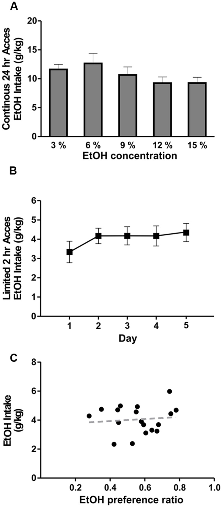
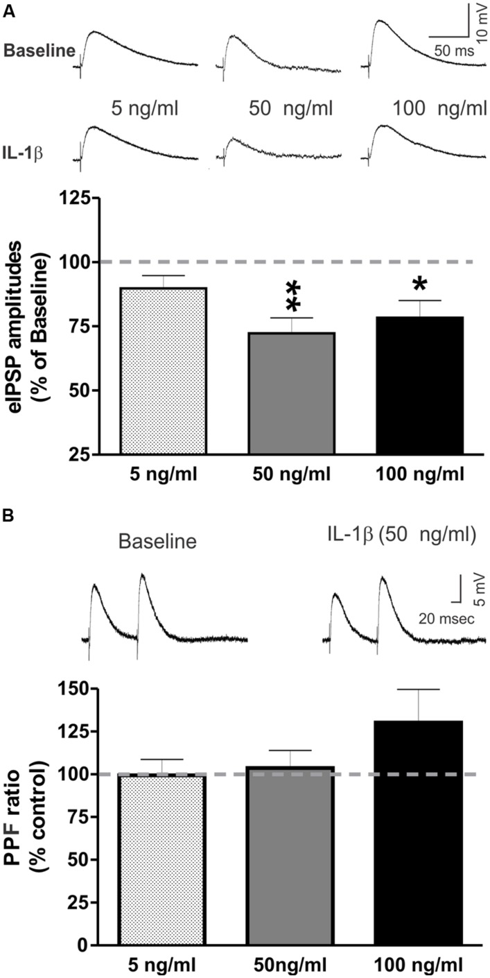
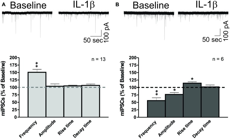
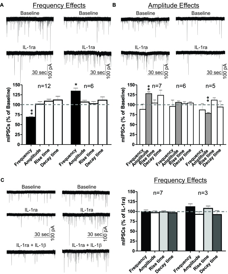
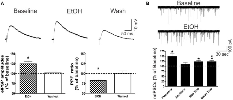
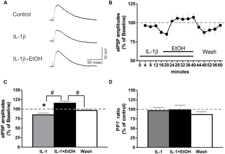
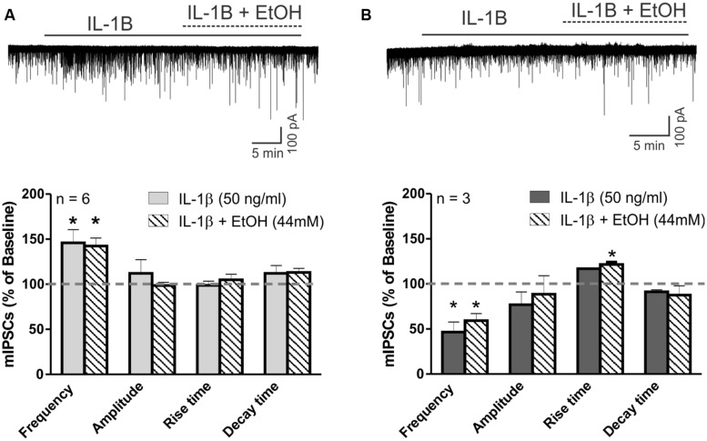
Neuroinflammation is hypothesized to enhance alcohol consumption and contribute to the development of alcoholism. GABAergic transmission in the central amygdala (CeA) plays an important role in the transition to alcohol dependence. Therefore, we studied the effects of interleukin-1β (IL-1β), a proinflammatory cytokine mediating ethanol-induced neuroinflammation, and its interaction with ethanol on CeA GABAegic transmission in B6129SF2/J mice. We also assessed ethanol intake in B6129SF2/J mice. Intake with unlimited (24 h) ethanol access was 9.2-12.7 g/kg (3-15% ethanol), while limited (2 h) access produced an intake of 4.1 ± 0.5 g/kg (15% ethanol). In our electrophysiology experiments, we found that recombinant IL-1β (50 and 100 ng/ml) significantly decreased the amplitude of evoked inhibitory postsynaptic potentials (eIPSPs), with no significant effects on paired-pulse facilitation (PPF). IL-1β (50 ng/ml) had dual effects on spontaneous miniature inhibitory postsynaptic currents (mIPSCs): increasing mIPSC frequencies in most CeA neurons, but decreasing both mIPSC frequencies and amplitudes in a few cells. The IL-1β receptor antagonist (IL-1ra; 100 ng/ml) also had dual effects on mIPSCs and prevented the actions of IL-1β on mIPSC frequencies. These results suggest that IL-1β can alter CeA GABAergic transmission at pre- and postsynaptic sites. Ethanol (44 mM) significantly increased eIPSP amplitudes, decreased PPFs, and increased mIPSC frequencies. IL-1β did not alter ethanol's enhancement of the eIPSP amplitude, but, in IL-1β-responsive neurons, the ethanol effects on mIPSC frequencies were lost. Overall, our data suggest that the IL-1 system is involved in basal GABAergic transmission and that IL-1β interacts with the ethanol-induced facilitation of CeA GABAergic transmission.

Keywords: GABAA; IL-1ra; IL-1β; IPSCs; central amygdala; cytokine; eIPSPs; interleukin.


Figures

FIGURE 1
FIGURE 1
Ethanol drinking behavior of B6129SF2/J mice. The ethanol intake of B6129SF2/J mice was tested using unlimited (24 h) and limited (2 h) 2-bottle choice (2BC) paradigms. (A) The intake of 3, 6, 9, 12, and 15% ethanol was measured by 2BC with unlimited access to ethanol. On average, the mice consumed 11.6 ± 0.9 g/kg/day of 3% ethanol solution, 12.7 ± 1.8 g/kg/day of 6% ethanol solution, 10.7 ± 1.1 g/kg/day of 9% ethanol solution, 9.2 ± 1.1 g/kg/day at 12% ethanol solution, and 9.3 ± 1.0 g/kg/day of 15% ethanol solution. There was significant main difference in ethanol intake between the ethanol concentrations [F(4,19) = 2.5, n = 20], but Tukey post hoc analysis did not reveal significant differences between specific ethanol concentrations. (B) For limited access measurements of ethanol consumption, we used 15% ethanol solution and intake was measured for 2 h daily (starting 3 h after lights off) for a period of 5 days. Consumption was 3.3 ± 0.6 g/kg of ethanol on day 1; 4.2 ± 0.4 g/kg of ethanol on day 2; 0.4.2 ± 0.5 g/kg on days 3 and 4; 4.4 ± 0.5 on day 5. Repeated measure one-way ANOVA showed no significant difference in ethanol intake between testing days [F(4,17) = 0.7, n = 18]. (C) Ethanol intake of individual mice is plotted as a function of preference. The average ethanol preference ratio (volume of ethanol consumed/total volume of fluid consumed) was 0.56 ± 0.05. There was no significant correlation (R2 = 0.01) between ethanol preference and intake in B6129SF2/J mice.
FIGURE 2
FIGURE 2
IL-1β decreases evoked GABAA-receptor mediated IPSPs in the CeA. (A) Recombinant mouse IL-1β decreased the mean amplitude of evoked IPSPs (eIPSPs) by 27.8 ± 6.0% (n = 11; t-test: p < 0.01) at 50 ng/ml and by 21.6 ± 6.7% (n = 5; t-test: p < 0.05) at 100 ng/ml. A lower concentration of IL-1β (5 ng/ml) had no significant effects on the eIPSPs (89.8 ± 5.0% of baseline, n = 7). (Top) Representative eIPSPs taken during baseline and IL-1β superfusion. (Bottom) Summary of the maximal effects of IL-1β elicited by the tested doses of IL-1β as compared to baseline. The statistical significance (p < 0.05 and ∗∗p < 0.01) was calculated by t-test. (B) IL-1β did not alter significantly paired-pulse facilitation (PPF; using a 100 ms interstimulus interval) at any of the tested concentrations (5 ng/ml: 99.4 ± 9.3% of baseline, n = 6; 50 ng/ml: 103.7 ± 7.2% of baseline, n = 8; 100 ng/ml: 130.1 ± 19.4% of baseline, n = 4). (Top) Representative recordings of PPF from a CeA neuron superfused with 50 ng/ml IL-1β. (Bottom) The PPF results are summarized on the bar graph with the PPF ratio during IL-1β superfusion compared to the baseline levels.
FIGURE 3
FIGURE 3
IL-1β has dual effects on miniature IPSCs in CeA neurons. (A) IL-1β increases mIPSC frequencies in the CeA. (Top) A representative whole- cell voltage clamp recording showing an increase in mIPSC frequency induced by 50 ng/ml IL-1β superfusion. (Bottom) The bar graphs present normalized mIPSC parameters from 62% of CeA neurons (13 of 21 cells) responding to IL-1β with an increase in mIPSC frequency (150.7 ± 10.0% of baseline). mIPSC amplitude (104.0 ± 7.8% of baseline) and kinetics were not significantly changed in these CeA neurons, although some cells showed individual changes in mIPSC amplitude. We calculated statistical significance (∗∗p < 0.01) by t-test. (B) IL-1β decreases mIPSC frequencies and amplitudes in CeA neurons. (Top) Representative recording of a CeA neuron responding to IL-1β with a decrease in mIPSC frequency. (Bottom) Acute application of IL-1β significantly decreased the mIPSC frequency (55.9 ± 9.5% of baseline) and amplitude (76.7 ± 6.2% of baseline) in 29% of CeA neurons (6 of 21 cells). In addition, IL-1β increased the mIPSC rise time (114.3 ± 4.7% of baseline) in these neurons. Statistical significance (p < 0.05) and (∗∗p < 0.01) were calculated by t-test.
FIGURE 4
FIGURE 4
IL-1ra has dual effects on basal mIPSCs and prevents IL-1β-induced modulation of mIPSCs in the CeA. IL-1ra-induced changes in mIPSC frequencies and/or amplitudes vary among CeA neurons, indicating cell-specific differences in IL-1ra modulation of GABAergic transmission. (A) Dual IL-1ra-induced changes in mIPSC frequencies. (Top) Representative recordings of two CeA neurons showing a decrease (left column) or increase (right column) in mIPSC frequencies following IL-1ra application (100 ng/ml). (Bottom) Summary bar graph showing IL-1ra decreased significantly (t-test, p < 0.01) the mean mIPSC frequency by 31.3 ± 2.1% in 12 of 18 (67%) neurons. In the remaining CeA cells (6 of 18), IL-1ra increased the mIPSC frequency by 34.1 ± 7.7% (t-test, p < 0.05). The changes in mIPSC frequencies were not associated with significant changes in mIPSC amplitudes or kinetics. (B) The IL-1ra induced changes in the mIPSC amplitudes were also variable. (Top) Representative recordings of two cells responding to IL-1ra with increased (left column) or decreased (right column) mIPSC amplitudes. (Bottom) Summary bar graph showing IL-1ra increased (t-test, p < 0.01) mIPSC amplitudes by 27.9 ± 4.5% in 7 of 18 cells (39%) and decreased (t-test, p < 0.01) by 21.7 ± 5.9% in 5 of 18 cells (28%). There were no significant changes in the mean mIPSC frequencies and kinetics across all cell groups. The statistical significance (p < 0.05) and (∗∗p < 0.01) was calculated by t-test. (C) To examine the effects of IL-1ra on the IL-1β-induced modulation of mIPSCs, we compared the mIPSC parameters recorded within 9–15 min of 100 ng/ml IL-1ra and 50 ng/ml IL-1β co-application to the last 6 min (9–15 min) of IL-1ra application alone. We divided the CeA neurons into two groups according to their cellular responses (mIPSCs frequency) to IL-1ra alone: the cells that responded to IL-1ra with decreased mIPSC frequency [by 31.8 ± 3%; F(2,23) = 7.5, p < 0.05; n = 7] and the cells that responded to IL-1ra with increased mIPSC frequency [by 27.9 ± 7%; F(2,8) = 1.7, p < 0.05; n = 3]. (Left) Representative recordings from two CeA neurons responding to IL-1ra with decreased (left column) or increased (right column) mIPSCs frequencies. (Right) IL-1ra prevented the IL-1β-induced modulation of mIPSCs, as there were no significance differences in mIPSCs after co-application of IL-1ra and IL-1β compared to IL-1ra alone. The statistical significance was set at (p < 0.05) and was calculated by repeated measurement one-way ANOVA followed by a Tukey post hoc test.
FIGURE 5
FIGURE 5
Ethanol potentiates CeA GABAergic transmission. (A) Ethanol potentiated eIPSPs via a presynaptic mechanism in CeA neurons. (Top) Representative recordings of eIPSPs from a CeA neuron showing an ethanol-induced increase in eIPSP amplitude that is reversed upon drug washout. (Bottom) On average, 44 mM ethanol significantly increased the mean eIPSP amplitude by 22.5 ± 5.9% (left column: n = 8; t-test: p < 0.05) and decreased the PPF ratio to 82.2 ± 4.0% of baseline in six of eight neurons (right column: n = 6; t-test: p < 0.05), indicating that ethanol-induced eIPSP potentiation is mediated by increased GABA release. (B) Ethanol increases spontaneous miniature GABA transmission in the CeA by both pre- and postsynaptic mechanisms. (Top) Representative mIPSC recordings from a CeA neuron showing an ethanol-induced increase in frequency. (Bottom) Superfusion of 44 mM ethanol induced a significant increase in the mean mIPSC frequency (140.7 ± 17.5% of baseline), but had no effect on the mean amplitude (102.6 ± 9.5% of baseline; n = 4, t-test: p < 0.05), supporting the finding that ethanol’s mechanism of action is predominantly presynaptic. However, ethanol significantly altered mIPSC kinetics, with a 14.7 ± 4.2% increase in the rise time and a 25.4 ± 7.0% increase in the decay time, indicating additional postsynaptic changes (t-test: p < 0.05 and ∗∗p < 0.01).
FIGURE 6
FIGURE 6
IL-1β and ethanol have opposing effects on eIPSP amplitudes. (A) Representative eIPSPs from a CeA neuron showing a 50 ng/ml IL-1β-induced decrease in eIPSP amplitude, and its subsequent reversal to baseline levels by the addition of ethanol (44 mM). (B) Time course presenting the averaged eIPSP amplitudes over 3 min bin periods. (C) Co-application of ethanol reversed the IL-1β-induced decrease in mean eIPSP amplitude [84.8 ± 4.7% of baseline, n = 9; F(2,26) = 12.1, p < 0.01] to slightly above baseline levels (115.4 ± 5.3% of baseline). Statistical significance [p < 0.05; (comparisons to baseline) and #(comparison of the effects of ethanol plus IL-1β co-application to IL-1β alone or washout)] was calculated by repeated measurement one-way ANOVA followed by a Tukey post hoc test. (D) There were no significant effects on the PPF ratio (100 ms interstimulus interval) of IL-1β alone, or when it was co-applied with ethanol [F(2,23) = 0.24].
FIGURE 7
FIGURE 7
IL-1β occludes ethanol’s facilitation of mIPSCs. (A) The effects of ethanol on mIPSCs are blocked by IL-1β in cells that previously showed increased mIPSC frequency in the presence of IL-1β alone. (Top) Voltage clamp recordings of mIPSCs from a CeA neuron showing an IL-1β-induced increase in mIPSC frequency that is unaltered by the addition of ethanol. (Bottom) Summary of the normalized mIPSC maximal effects of IL-1β (50 ng/ml) alone, and IL-1β and ethanol (44 mM) co-application. IL-1β alone significantly increases mIPSC frequency by 45.9 ± 14.6% in 6 of 10 CeA neurons. The co-application of ethanol did not further change the mIPSC frequency (142.4 ± 8.9% of baseline; [F(2,5) = 6.9, p < 0.05; Tukey post hoc test]. There were no differences in mIPSC amplitudes and kinetics across all treatments. Statistical significance (p < 0.05) was calculated by one-way ANOVA followed by a Tukey post hoc test. (B) Ethanol’s effects on mIPSC frequency are blocked by IL-1β in cells that previously showed decreased mIPSC frequency in the presence of IL-1β alone. (Top) Representative recordings from a CeA neuron showing a reduction in mIPSC frequency elicited by IL-1β and the co-application of IL-1β and ethanol. (Bottom) In 3 of 10 CeA neurons, mIPSC frequency was significantly decreased by IL-1β (46.5 ± 11.3% of baseline) alone, as well as with the co-application of IL-1β and ethanol [59.2 ± 7.9% of baseline; F(2,2) = 35.5, p < 0.05; Tukey post hoc test]. There was no significant difference between the effects of IL-1β alone and co-application of IL-1β and ethanol (Tukey post hoc test, p < 0.05), but co-application of IL-1β and ethanol significantly increased the mean rise time of mIPSCs [116.4 ± 5.4% of baseline; F(2,2) = 7.9, p < 0.05; Tukey post hoc test]. The statistical significance (p < 0.05) was calculated by one-way ANOVA followed by a Tukey post hoc test.

References

    1. Allan S. M., Tyrrell P. J., Rothwell N. J. (2005). Interleukin-1 and neuronal injury. Nat. Rev. Immunol. 5 629–640 10.1038/nri1664 - DOI - PubMed
    1. Bajo M., Cruz M. T., Siggins G. R., Messing R., Roberto M. (2008). Protein kinase C epsilon mediation of CRF- and ethanol-induced GABA release in central amygdala. Proc. Natl. Acad. Sci. U.S.A. 105 8410–8415 10.1073/pnas.0802302105 - DOI - PMC - PubMed
    1. Bajo M., Herman M. A., Varodayan F. P., Oleata C. S., Madamba S. G., Harris R. A., et al. (2014a). Role of the IL-1 receptor antagonist in ethanol-induced regulation of GABAergic transmission in the central amygdala. Brain Behav. Immun. 45 189–197 10.1016/j.bbi.2014.11.011 - DOI - PMC - PubMed
    1. Bajo M., Madamba S. G., Roberto M., Blednov Y. A., Sagi V. N., Roberts E., et al. (2014b). Innate immune factors modulate ethanol interaction with GABAergic transmission in mouse central amygdala. Brain Behav. Immun. 40 191–202 10.1016/j.bbi.2014.03.007 - DOI - PMC - PubMed
    1. Bajo M., Roberto M., Madamba S. G., Siggins G. R. (2011). Neuroadaptation of GABAergic transmission in the central amygdala during chronic morphine treatment. Addict. Biol. 16 551–564 10.1111/j.1369-1600.2010.00269.x - DOI - PMC - PubMed

LinkOut - more resources