Case Reports
. 2015 Mar 23:6:102.
doi: 10.3389/fgene.2015.00102. eCollection 2015.

Long-term survival in a child with severe encephalopathy, multiple respiratory chain deficiency and GFM1 mutations

Affiliations
Case Reports

Long-term survival in a child with severe encephalopathy, multiple respiratory chain deficiency and GFM1 mutations

Sara Brito et al. Front Genet. .

Erratum in

Abstract

Background: Mitochondrial diseases due to deficiencies in the mitochondrial oxidative phosphorylation system (OXPHOS) can be associated with nuclear genes involved in mitochondrial translation, causing heterogeneous early onset and often fatal phenotypes.

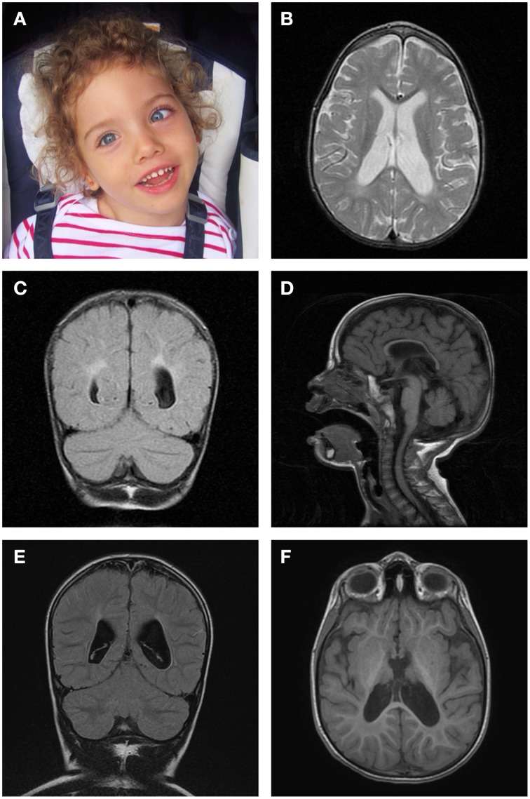
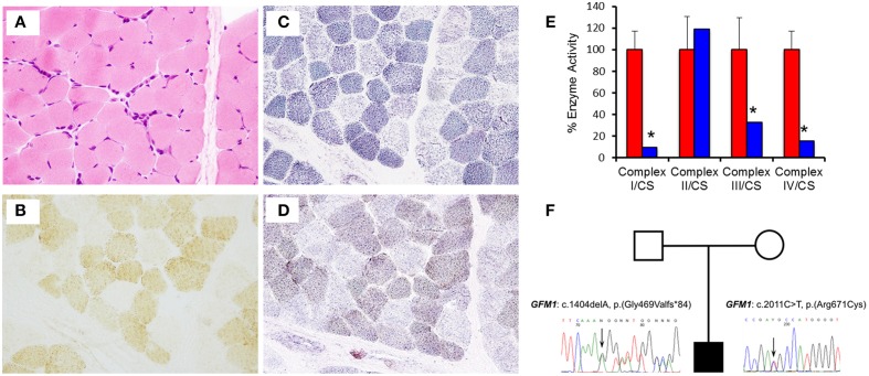
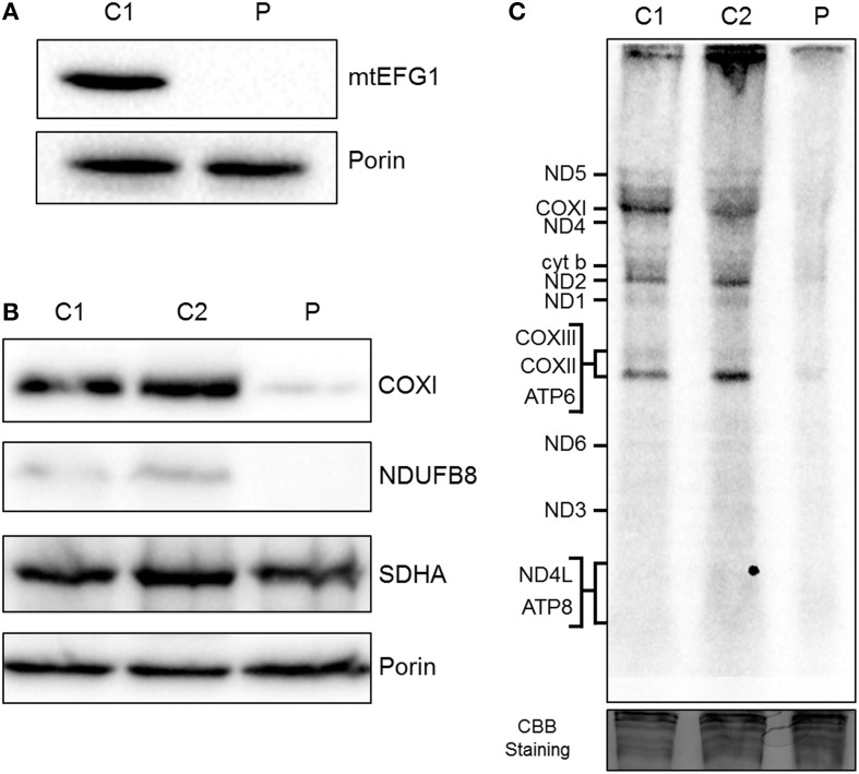
Case report: The authors describe the clinical features and diagnostic workup of an infant who presented with an early onset severe encephalopathy, spastic-dystonic tetraparesis, failure to thrive, seizures and persistent lactic acidemia. Brain imaging revealed thinning of the corpus callosum and diffuse alteration of white matter signal. Genetic investigation confirmed two novel mutations in the GFM1 gene, encoding the mitochondrial translation elongation factor G1 (mtEFG1), resulting in combined deficiencies of OXPHOS.

Discussion: The patient shares multiple clinical, laboratory and radiological similarities with the 11 reported patients with mutations involving this gene, but presents with a stable clinical course without metabolic decompensations, rather than a rapidly progressive fatal course. Defects in GFM1 gene confer high susceptibility to neurologic or hepatic dysfunction and this is, to the best of our knowledge, the first described patient who has survived beyond early childhood. Reporting of such cases is essential so as to delineate the key clinical and neuroradiological features of this disease and provide a more comprehensive view of its prognosis.

Keywords: GFM1; brain MRI; encephalopathy; mitochondrial disorders; mtEFG1.


Figures

Figure 1
Figure 1
Brain magnetic resonance imaging. (A) Photograph of the patient aged 5 years demonstrating left convergent squint and normal facial appearance. (B) Axial T2-weighted image at 29 months, revealing enlarged lateral ventricles. (C) Coronal T2-FLAIR image at 29 months, displaying abnormal white matter signal in a periventricular distribution. (D) Midsagittal T1-FLAIR image at 29 months, showing thinning of the corpus callosum with enlarged subarachnoid spaces. (E) Coronal T2-FLAIR image at 5 years 6 months, showing abnormal periatrial signal and enlarged lateral ventricles. (F) Axial 3D FSPGR image at 5 years 6 months, displaying abnormal signal in the posterolateral region of the putamen nucleus.
Figure 2
Figure 2
Muscle histology and histochemistry reveals mitochondrial abnormalities. (A) Haematoxylin and Eosin (H&E) histology shows mild variation in fiber size. (B) Cytochrome c oxidase (COX) histocytochemical activity revealed a mosaic pattern of COX deficiency without subsarcolemmal mitochondrial aggregates. (C) Succinate dehydrogenase (SDH) reaction indicates a population of pale fibers within the section. (D) Sequential COX-SDH histochemistry shows a population of weak “blue” fibers corresponding to the COX-deficient, SDH-positive fibers detected following the individual enzyme reactions. (E) The assessment of individual respiratory chain enzyme activities in fibroblasts identified a severe OXPHOS deficiency affecting complex I, III, and IV in the patient (blue) compared with controls (red). Mean enzyme activities of control fibroblasts (n = 8) are set at 100% and error bars represent the standard deviation. *denotes values outside of the normal range. (F) Pedigree and sequence analysis of the two mutations identified in the patient.
Figure 3
Figure 3
Biochemical assessment of patient fibroblasts. (A) Mitochondria isolated from control (C1) and patient (P) fibroblasts were subjected to SDS-PAGE and western blot analysis using an anti-mtEFG1 antibody. Anti-porin (VDAC1) antibody was used as a loading control. (B) Whole cell lysate from control (C1 and C2) and patient (P) primary fibroblasts were subjected to western blot analysis. Antibodies against COXI (mitochondrial encoded subunit of Complex IV), NDUFB8 (nuclear encoded subunit of Complex I), SDHA (nuclear encoded subunit of Complex II) and porin/VDAC1 (mitochondrial loading control). (C) Control (C1 and C2) and patient (P) fibroblasts were treated with emetine dihydrochloride to inhibit cytosolic translation and mitochondrial protein synthesis analyzed by [35S] met/cys incorporation (1 h followed by a 10 min chase). Cell lysate (50 μg) was separated through a 15% polyacrylamide gel. The gel was stained with Coomassie blue (CBB) to confirm equal loading. Post fixation and drying the signal was visualized by Typhoon FLA9500 PhosphorImaging. Signals were ascribed following established migration patterns (Chomyn, 1996).

References

    1. Anderson S., Bankier A. T., Barrell B. G., De Bruijn M. H., Coulson A. R., Drouin J., et al. (1981). Sequence and organization of the human mitochondrial genome. Nature 290, 457–465 10.1038/290457a0 - DOI - PubMed
    1. Antonicka H., Sasarman F., Kennaway N. G., Shoubridge E. A. (2006). The molecular basis for tissue specificity of the oxidative phosphorylation deficiencies in patients with mutations in the mitochondrial translation factor EFG1. Hum. Mol. Genet. 15, 1835–1846. 10.1093/hmg/ddl106 - DOI - PubMed
    1. Balasubramaniam S., Choy Y. S., Talib A., Norsiah M. D., Van Den Heuvel L. P., Rodenburg R. J. (2012). Infantile progressive Hepatoencephalomyopathy with combined OXPHOS deficiency due to mutations in the mitochondrial translation elongation factor gene GFM1. JIMD Rep. 5, 113–122 10.1007/8904_2011_107 - DOI - PMC - PubMed
    1. Calvo S. E., Compton A. G., Hershman S. G., Lim S. C., Lieber D. S., Tucker E. J., et al. . (2012). Molecular diagnosis of infantile mitochondrial disease with targeted next-generation sequencing. Sci. Trans. Med. 4, 118ra110. 10.1126/scitranslmed.3003310 - DOI - PMC - PubMed
    1. Chomyn A. (1996). In vivo labeling and analysis of human mitochondrial translation products Methods Enzymol. 264, 197–211. - PubMed

Publication types