

Rodent models of osteoporosis
- PMID: 25852854
- PMCID: PMC4388108
- DOI: 10.1038/bonekey.2014.109
Rodent models of osteoporosis
Abstract
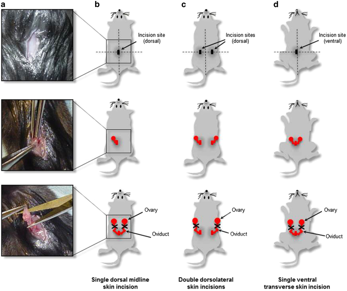
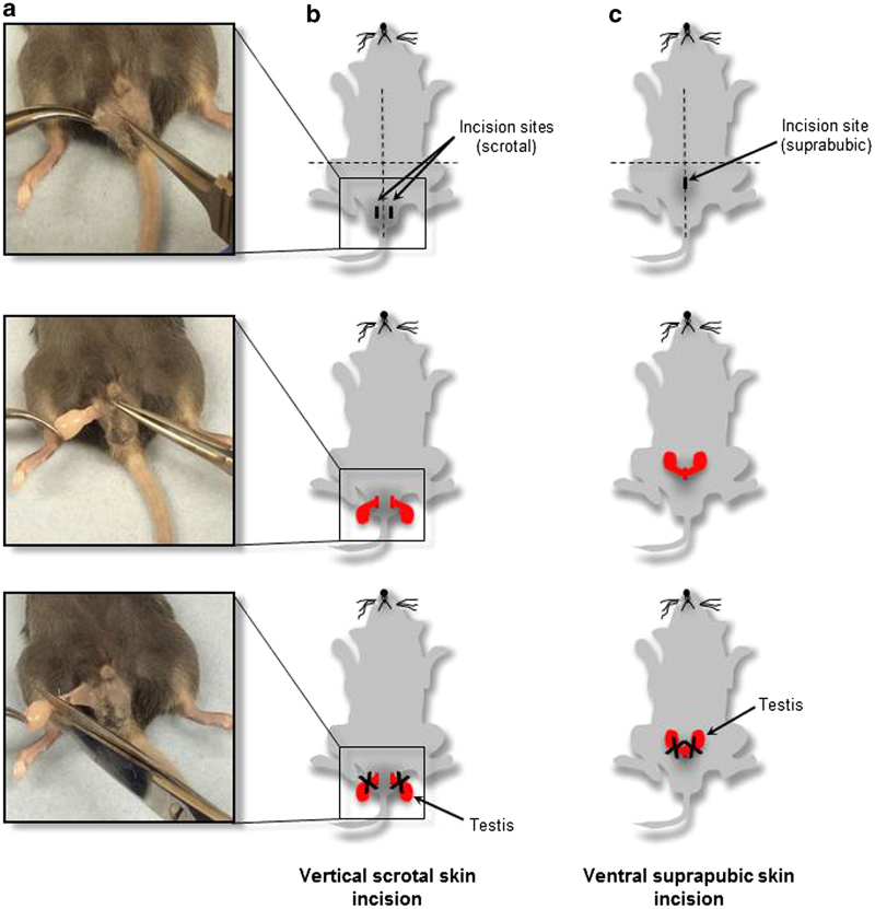
The aim of this protocol is to provide a detailed description of male and female rodent models of osteoporosis. In addition to indications on the methods of performing the surgical procedures, the choice of reliable and safe anaesthetics is also described. Post-operative care, including analgesia administration for pain management, is also discussed. Ovariectomy in rodents is a procedure where ovaries are surgically excised. Hormonal changes resulting from ovary removal lead to an oestrogen-deprived state, which enhances bone remodelling, causes bone loss and increases bone fracture risk. Therefore, ovariectomy has been considered as the most common preclinical model for understanding the pathophysiology of menopause-associated events and for developing new treatment strategies for tackling post-menopausal osteoporosis. This protocol also provides a detailed description of orchidectomy, a model for androgen-deficient osteoporosis in rodents. Endocrine changes following testes removal lead to hypogonadism, which results in accelerated bone loss, increasing osteoporosis risk. Orchidectomised rodent models have been proposed to mimic male osteoporosis and therefore remain a valuable tool for understanding androgen deficiency in aged men. Although it would have been particularly difficult to assemble an internationally acceptable description of surgical procedures, here we have attempted to provide a comprehensive guide for best practice in performing ovariectomy and orchidectomy in laboratory rodents. Research scientists are reminded that they should follow their own institution's interpretation of such guidelines. Ultimately, however, all animal procedures must be overseen by the local Animal Welfare and Ethical Review Body and conducted under licences approved by a regulatory ethics committee.
Conflict of interest statement
The authors declare no conflict of interest.
Figures
Similar articles
-
Description of Ovariectomy Protocol in Mice.Methods Mol Biol. 2019;1916:303-309. doi: 10.1007/978-1-4939-8994-2_29. Methods Mol Biol. 2019. PMID: 30535707
-
Poor histological healing of a femoral fracture following 12 months of oestrogen deficiency in rats.Osteoporos Int. 2013 Oct;24(10):2581-9. doi: 10.1007/s00198-013-2345-2. Epub 2013 Apr 6. Osteoporos Int. 2013. PMID: 23563933
-
The laboratory rat as an animal model for osteoporosis research.Comp Med. 2008 Oct;58(5):424-30. Comp Med. 2008. PMID: 19004367 Free PMC article. Review.
-
Orchidectomy models of osteoporosis.Methods Mol Biol. 2008;455:125-34. doi: 10.1007/978-1-59745-104-8_9. Methods Mol Biol. 2008. PMID: 18463815
-
Pharmaceutical treatment of bone loss: From animal models and drug development to future treatment strategies.Pharmacol Ther. 2023 Apr;244:108383. doi: 10.1016/j.pharmthera.2023.108383. Epub 2023 Mar 16. Pharmacol Ther. 2023. PMID: 36933702 Review.
Cited by
-
A novel modified RANKL variant can prevent osteoporosis by acting as a vaccine and an inhibitor.Clin Transl Med. 2021 Mar;11(3):e368. doi: 10.1002/ctm2.368. Clin Transl Med. 2021. PMID: 33784004 Free PMC article.
-
A Pre-clinical Standard Operating Procedure for Evaluating Orthobiologics in an In Vivo Rat Spinal Fusion Model.J Orthop Sports Med. 2022;4(3):224-240. doi: 10.26502/josm.511500060. Epub 2022 Sep 5. J Orthop Sports Med. 2022. PMID: 36203492 Free PMC article.
-
Effects of Early Life Stress on Bone Homeostasis in Mice and Humans.Int J Mol Sci. 2020 Sep 10;21(18):6634. doi: 10.3390/ijms21186634. Int J Mol Sci. 2020. PMID: 32927845 Free PMC article.
-
Influence of the Peripheral Nervous System on Murine Osteoporotic Fracture Healing and Fracture-Induced Hyperalgesia.Int J Mol Sci. 2022 Dec 28;24(1):510. doi: 10.3390/ijms24010510. Int J Mol Sci. 2022. PMID: 36613952 Free PMC article.
-
Anti-Osteoporotic Effects of Polysaccharides Isolated from Persimmon Leaves via Osteoclastogenesis Inhibition.Nutrients. 2018 Jul 13;10(7):901. doi: 10.3390/nu10070901. Nutrients. 2018. PMID: 30011853 Free PMC article.
References
-
- Ruggiero RJ, Likis FE. Estrogen: physiology, pharmacology, and formulations for replacement therapy. J Midwifery Womens Health 2002; 47:130–138. - PubMed
-
- Riggs BL, Khosla S, Melton LJ III. Sex steroids and the construction and conservation of the adult skeleton. Endocr Rev 2002; 23:279–302. - PubMed
-
- Felicio LS, Nelson JF, Finch CE. Longitudinal studies of estrous cyclicity in aging C57BL/6 J mice: II. Cessation of cyclicity and the duration of persistent vaginal cornification. Biol Reprod 1984; 31:446–453. - PubMed
-
- vom Saal FS, Finch CE, Nelson JF. Natural history and mechanism of reproductive aging in humans, laboratory rodents, and other selected vertebrates. In: Knobil E, Neill JD (eds). The Physiology of Reproduction, 2nd edn, Vol. 2. Raven Press: New York, 1994, pp 1213–1314.
LinkOut - more resources
Full Text Sources
Other Literature Sources