Review
. 2015 Mar;20(1):25-40.
doi: 10.15430/JCP.2015.20.1.25.

Review of atrophic gastritis and intestinal metaplasia as a premalignant lesion of gastric cancer

Affiliations
Review

Review of atrophic gastritis and intestinal metaplasia as a premalignant lesion of gastric cancer

Yo Han Park et al. J Cancer Prev. 2015 Mar.

Abstract

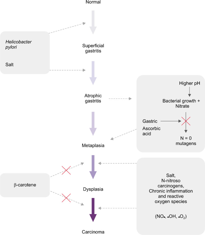
Atrophic gastritis (AG) and intestinal metaplasia (IM) are the main precursor lesions of gastric cancer as the incidence of gastric cancer increases in the gastric mucosa involved with AG and IM. The prevalence of AG and IM vary depending on countries, even it represents diverse results in the same nation. Usually AG is antecedent of IM but the etiologies of AG and IM are not always the same. The sensitivity and specificity of diagnostic methods to detect AG and IM are different. Furthermore, the management strategy of AG and IM has not been established, yet. Helicobacter pylori infection has been proved as the most important cause of AG and IM. Thus the eradication of H. pylori is very important to prevent the progression to gastric cancer which is still placed in the high rank in morbidity and mortality among cancers. However, the reversibility of AG and IM by eradication of H. pylori which was assumed to be certain by meta-analysis is; however, controversial now. Therefore, the understanding and early diagnosis of AG and IM are very important, especially, in high incidence area of gastric cancer such as Republic of Korea.

Keywords: Atrophic gastritis; Gastric cancer; Helicobacter pylori; Intestinal metaplasia.


Figures

Figure 1.
Figure 1.
Correa’s theory regarding human gastric carcinogenesis: a multistep and multifactorial process. Gastritis begins from superficial gastritis and progresses into atrophic gastritis, metaplasia, dysplasia and gastric cancer. Adapted from Correa et al.
Figure 2.
Figure 2.
Classificataion of gastritis (H&E, ×400). (A) Normal, (B) superficial gastritis, (C) atrophic gastritis, (D) intestinal metaplasia. Adapted from Schindler.
Figure 3.
Figure 3.
Grading of gastritis by Sydney System: acute inflammation, chronic inflammation, atrophic gastritis, intestinal metaplasia, and Helicobacter pylori densitiy. Adapted from Dixon et al.
Figure 4.
Figure 4.
The optimal gastric biopsy sites recommend by updated Sydney system. Biopsy specimens are taken at five different sites. A, lesser curvature of the antrum; B, greater curvature of the antrum; C, lesser curvature of the corpus; D, greater curvature of the corpus; and E, incisura angularis. Adapted from Dixon et al.
Figure 5.
Figure 5.
Gastritis staging: the OLGA system. Atrophy is defined as loss of appropriate glands (with or without metaplasia). In each compartment, atrophy is scored in a four-tiered scale (0–3) according to the visual analogue scale of the updated Sydney system. Adapted from Rugge et al.
Figure 6.
Figure 6.
Kimura-Takemoto classification of chronic atrophic gastirits: (A) Atrophic gastritis (AG) is classified into six types. The closed-type AG indicates that the atrophic border remains on the lesser curvature of the stomach, while the open-type AG means that the atrophic border no longer exits on the lesser curvature but extends along the anterior and posterior walls of the stomach, (B) the boundaries of the mucosal atrophy is called the F line, (C) progression of atrophic mucosal change from antrum to the corpus along with lesser curvature. Adapted from Kimura and Takemoto.
Figure 7.
Figure 7.
Phenotype of intestinal metaplasia (IM) classifying by mucin: type I IM expresses only sialomucins (bright blue) and type II, III expresses sialomucins (bright blue) and sulfomucins (black) (High iron diamine and alcian blue [pH 2.5] [HID-AB2.5] staining, ×400). Adapted from Kang et al.

References

    1. Ferlay J, Shin HR, Bray F, Forman D, Mathers C, Parkin DM. Estimates of worldwide burden of cancer in 2008: GLOBOCAN 2008. Int J Cancer. 2010;127:2893–917. doi: 10.1002/ijc.25516. - DOI - PubMed
    1. Statistics Korea. Daejeon, Statistics Korea. 2013 Available from: http://kostat.go.kr. Accessed December 3, 2014.
    1. Yuasa N, Nimura Y. Survival after surgical treatment of early gastric cancer, surgical techniques, and long-term survival. Langenbecks Arch Surg. 2005;390:286–93. doi: 10.1007/s00423-004-0482-y. - DOI - PubMed
    1. Park JM, Ryu WS, Kim JH, Park SS, Kim SJ, Kim CS, et al. Prognostic factors for advanced gastric cancer: stage-stratified analysis of patients who underwent curative resection. Cancer Res Treat. 2006;38:13–8. doi: 10.4143/crt.2006.38.1.13. - DOI - PMC - PubMed
    1. Hundahl SA, Phillips JL, Menck HR. The National Cancer Data Base Report on poor survival of U.S. gastric carcinoma patients treated with gastrectomy: fifth edition American Joint Committee on Cancer staging, proximal disease, and the “different disease” hypothesis. Cancer. 2000;88:921–32. doi: 10.1002/(SICI)1097-0142(20000215)88:4<921::AID-CNCR24>3.0.CO;2-S. - DOI - PubMed

LinkOut - more resources