. 2015 Apr 8:5:9713.
doi: 10.1038/srep09713.

Potent effects of dioscin against liver fibrosis

Affiliations

Potent effects of dioscin against liver fibrosis

Xiaoling Zhang et al. Sci Rep. .

Abstract

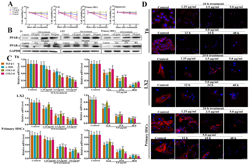
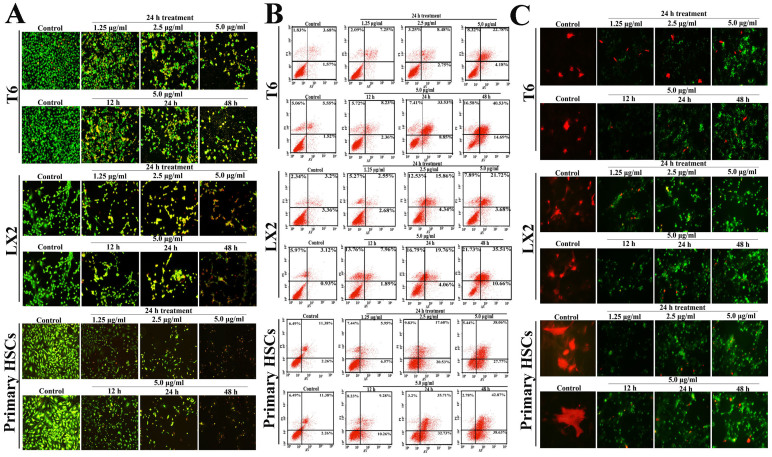
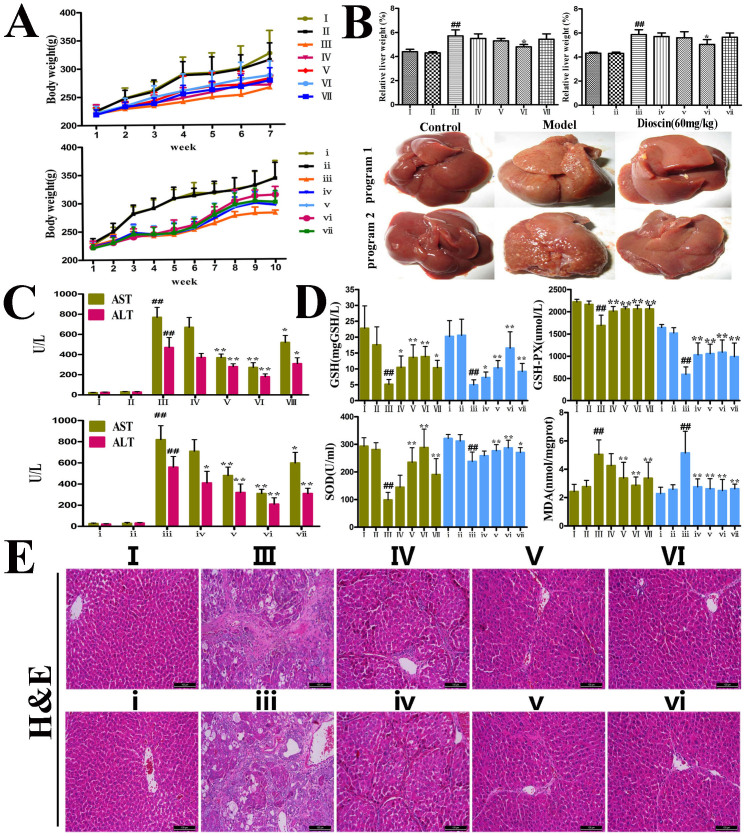
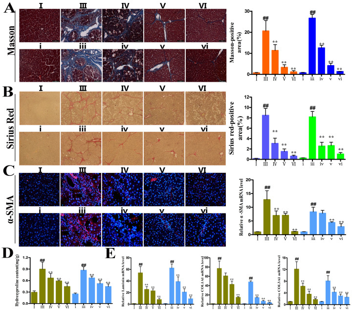
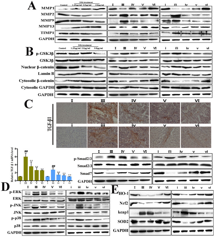
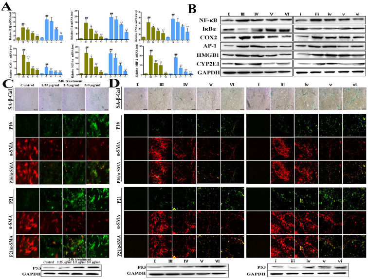
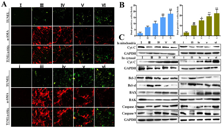
We previously reported the promising effects of dioscin against liver injury, but its effect on liver fibrosis remains unknown. The present work investigated the activities of dioscin against liver fibrosis and the underlying molecular mechanisms. Dioscin effectively inhibited the cell viabilities of HSC-T6, LX-2 and primary rat hepatic stellate cells (HSCs), but not hepatocytes. Furthermore, dioscin markedly increased peroxisome proliferator activated receptor-γ (PPAR-γ) expression and significantly reduced a-smooth muscle actin (α-SMA), transforming growth factor-β1 (TGF-β1), collagen α1 (I) (COL1A1) and collagen α1 (III) (COL3A1) levels in vitro. Notably, dioscin inhibited HSCs activation and induced apoptosis in activated HSCs. In vivo, dioscin significantly improved body weight and hydroxylproline, laminin, α-SMA, TGF-β1, COL1A1 and COL3A1 levels, which were confirmed by histopathological assays. Dioscin facilitated matrix degradation, and exhibited hepatoprotective effects through the attenuation of oxidative stress and inflammation, in addition to exerting anti-fibrotic effects through the modulation of the TGF-β1/Smad, Wnt/β-catenin, mitogen-activated protein kinase (MAPK) and mitochondrial signaling pathways, which triggered the senescence of activated HSCs. In conclusion, dioscin exhibited potent effects against liver fibrosis through the modulation of multiple targets and signaling pathways and should be developed as a novel candidate for the treatment of liver fibrosis in the future.


Conflict of interest statement

The authors declare no competing financial interests.

Figures

Figure 1
Figure 1. Effects of dioscin on HSCs activation of HSC-T6, LX2 and primary HSCs.
(A) Impacts of dioscin on the cell viabilities of HSC-T6, LX2, primary rat HSCs. After incubating for 24 14h, the cells were treated with various concentrations of dioscin (1.25, 2.5 and 5.0 14μg/ml) or 0.1% DMSO as a negative control for 12, 24 and 48 14h, then the cell viability was evaluated by MTT assay. (B) Effects of dioscin on PPAR-γ protein expression in HSC-T6, LX2 and primary rat HSCs. The cells were treated with various concentrations of dioscin for 12, 24 and 48 14h. Total protein samples extracted from control and dioscin-treated HSCs were analyzed by western blotting assay (The cropped gels are used and full-length gels are presented in Supplemental Figure S9). (C) Effects of dioscin on the mRNA levels of TGF-β1, α-SMA, COL1A1 and COL3A1 in HSC-T6, LX2 and primary rat HSCs. The cells were treated with various concentrations of dioscin for 12, 24 and 48 14h, and then total RNA samples were extracted from control and dioscin-treated HSCs, which were measured by real-time PCR assay. (D) Effects of dioscin on the levels of α-SMA in HSC-T6, LX2 and primary rat HSCs based on immunofluorescence assay (10000 × magnification). After treatment with various concentrations of dioscin for 12, 24 and 48 14h, the expression of α-SMA was detected by immunofluorescence, and DAPI was used to visualize the nucleus. Data are presented as the mean ± SD (n ≥ 3). *p < 0.05 and **p < 0.01 compared with the control group.
Figure 2
Figure 2. Dioscin induced apoptosis of HSC-T6, LX2 and primary HSCs.
(A) Effects of dioscin on apoptosis of HSC-T6, LX2 and primary HSCs based on AO/EB staining, which were treated with 5.0 14μg/ml of dioscin for 12, 24 and 48 14h, or treated with different concentrations of dioscin (1.25, 2.5 and 5.0 14μg/ml) for 24 14h (200×, final magnification). (B) After being treated with 5.0 14μg/ml of dioscin for 12, 24 and 48 14h, or treated with different concentrations of dioscin (1.25, 2.5 and 5.0 14μg/ml) for 24 14h, HSC-T6, LX2 and primary HSCs were stained with annexin V/PI, and then analyzed by flow cytometry for quantitative detection of cell apoptosis. (C) HSC-T6, LX2 and primary HSCs were incubated with 5.0 14μg/ml of dioscin for 12, 24 and 48 14h, or treated with different concentrations of dioscin (1.25, 2.5 and 5.0 14μg/ml) for 24 14h, then the cells were dual-stained for TUNEL (green) and α-SMA (red) (original magnification 200×).
Figure 3
Figure 3. Inhibitory effects of dioscin on CCl4-induced hepatotoxicity.
In program I, the rats were injected twice a week with CCl4 during 7 weeks in the presence or absence of dioscin, and in program II, CCl4 was injected for 10 weeks without dioscin, or after 4 weeks of CCl4, dioscin was administered together with CCl4 for additional times. (A–B) Effects of dioscin on rat body weights and livers. (C) Effects of dioscin on serum AST and ALT activities in rats as a measure for liver injury. (D) Effects of dioscin on GSH, GSH-Px, SOD and MDA levels. (E) H&E staining of representative liver sections (magnification, 100×). Values are expressed as the mean ± SD (n ≥ 3). *p < 0.05, **p < 0.01 vs. model group; ##p < 0.01 vs. normal control group.
Figure 4
Figure 4. Dioscin attenuated liver fibrosis in vivo.
(A–B) Effects of dioscin on liver fibrosis by Masson and Sirius Red staining (magnification 100×). (C) Effects of dioscin on expression of α-SMA by immunostaining in liver tissues extracted from rats treated with CCl4 and dioscin for 7 weeks and 10 weeks (magnification 200×) (D) Effects of dioscin on hydroxyproline levels in rat livers. (E) Effects of dioscin on the mRNA levels of laminin, COL1A1 and COL3A1 in rats. The results are expressed as mean ± SD (n ≥ 3). *p < 0.05, **p < 0.01 vs. model group; ##p < 0.01 vs. normal control group.
Figure 5
Figure 5. Dioscin ameliorated liver fibrosis by regulating MMPs/TIMPs, and altering Wnt/β-catenin, TGF-β/Smad and MAPK pathways and oxidative stress.
(A) Effects of dioscin on the levels of MMP-1, MMP-2, MMP-9, MMP-13 and TIMP-1 from primay rat HSCs and in vivo HSCs isolated from normal, model and dioscin-treated rats. (B) Effects of dioscin on the levels of p-GSK3β, GSK3β, nuclear and cytosolic β-catenin in primay rat HSCs, and in vivo HSCs isolated from normal, model and dioscin-treated rats. (C) Effects of diocsin on expression of TGF-β1 by immunohistochemistry in liver tissue (magnification, 100×), and the levels of p-Smad2/3, Smad2/3 and Smad7 in vivo HSCs isolated from normal, model and dioscin-treated rats. (D) Effects of dioscin on the levels of MAPK phosphorylation in vivo HSCs isolated from normal, model and dioscin-treated rats. (E) Effects of dioscin on the levels of HO-1, Nrf2, keap1 and SOD2 in vivo HSCs isolated from normal, model and dioscin-treated rats. The cropped gels are used and full-length gels are presented in Supplemental Figure S10, S11, S12, S13 and S14. Values are expressed as the means ± SD (n = 3). *p < 0.05, **p < 0.01 vs. model group; ##p < 0.01 vs. normal control group.
Figure 6
Figure 6. Dioscin reduced inflammation and induced senescence of activated HSCs.
(A) Effects of dioscin on the mRNA levels of IL-1β, IL-6, TNF-α, ICAM-1, MIP-1α and MIP-2 in vivo HSCs isolated from normal, model and dioscin-treated rats. (B) Effects of dioscin on expression levels of NF-κB, IκBα, COX2, AP-1, HMGB1 and CYP2E1 in vivo HSCs isolated from normal, model and dioscin-treated rats. (C) Effects of dioscin on cell senescence in primary HSCs treated with different concentrations of dioscin (24 14h) for SA-β-Gal staining and dual-staining for p16 or p21 (green) and α-SMA (red) assay (original magnification 200×). (D) Effects of dioscin on cell senescence in live tissues from the rats treated with CCl4 and dioscin for SA-β-Gal staining and double immunostaining of p16/α-SMA and p21/α-SMA (original magnification 200×). The cropped gels are used and full-length gels are presented in Supplemental Figure S15 and S16. Values are expressed as the mean ± SD (n = 3). *p < 0.05, **p < 0.01 vs. model group; ##p < 0.01 vs. normal control group.
Figure 7
Figure 7. Dioscin induced apoptosis of activated HSCs in vivo.
(A) Effects of dioscin on liver tissues from the rats treated with CCl4 and dioscin dual-stained for TUNEL (green) and α-SMA (red) assay (original magnification 200×). (B) Numbers of α-SMA and TUNEL dual-positive cells was counted in five random fields, and the average numbers of dual-positive cells were plotted. (C) Effects of dioscin on cytochrome c release, and the levels of Bcl-2, Bcl-xl, BAX, BAK, caspase 3 and caspase 9 in vivo HSCs isolated from normal, model and dioscin-treated rats. The cropped gels are used and full-length gels are presented in Supplemental Figure S17. Values are expressed as the mean ± SD (n = 3). *p < 0.05, **p < 0.01 vs. model group; ##p < 0.01 vs. normal control group.

Similar articles

Cited by

References

    1. Friedman S. L. Mechanisms of hepatic fibrogenesis. Gastroenterology 134, 1655–1669 (2008). - PMC - PubMed
    1. Han Y. P. et al. Essential role of matrix metalloproteinases in interleukin-1- induced myofibroblastic activation of hepatic stellate cell in collagen. J Biol Chem 279, 4820–4828 (2004). - PMC - PubMed
    1. Mehal W. Z., Iredale J. & Friedman S. L. Scraping fibrosis: expressway to the core of fibrosis. Nat Med 17, 552–553 (2011). - PMC - PubMed
    1. Parola M., Marra F. & Pinzani M. Myofibroblast-like cells and liver fibrogenesis: emerging concepts in a rapidly moving scenario. Mol Aspects Med 29, 58–66 (2008). - PubMed
    1. Van Hul N. K. et al. Relation between liver progenitor cell expansion and extracellular matrix deposition in a CDE-induced murine model of chronic liver injury. Hepatology 49, 1625–1635 (2009). - PubMed

Publication types

MeSH terms