. 2015 Apr 8;10(4):e0119561.
doi: 10.1371/journal.pone.0119561. eCollection 2015.

Lysosomal trafficking of TGFBIp via caveolae-mediated endocytosis

Affiliations

Lysosomal trafficking of TGFBIp via caveolae-mediated endocytosis

Seung-Il Choi et al. PLoS One. .

Abstract

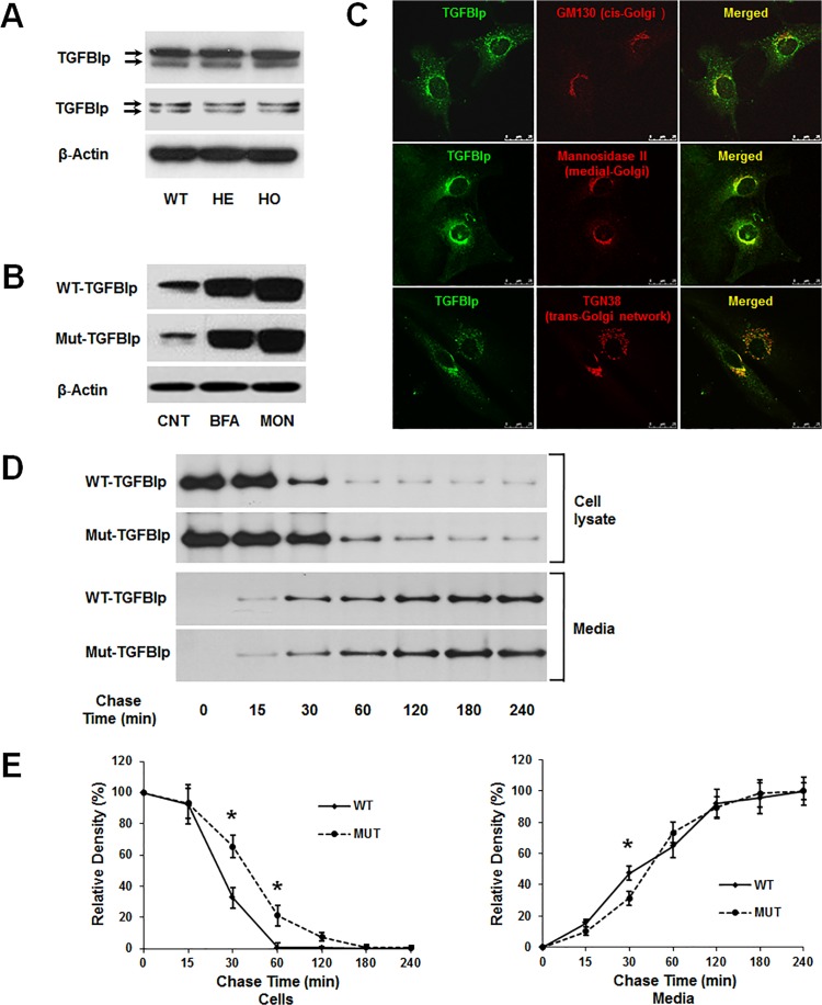
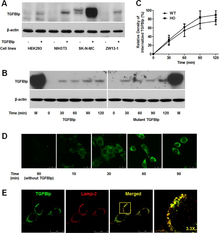
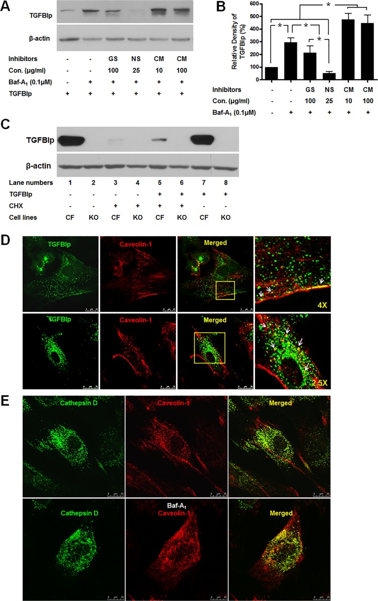
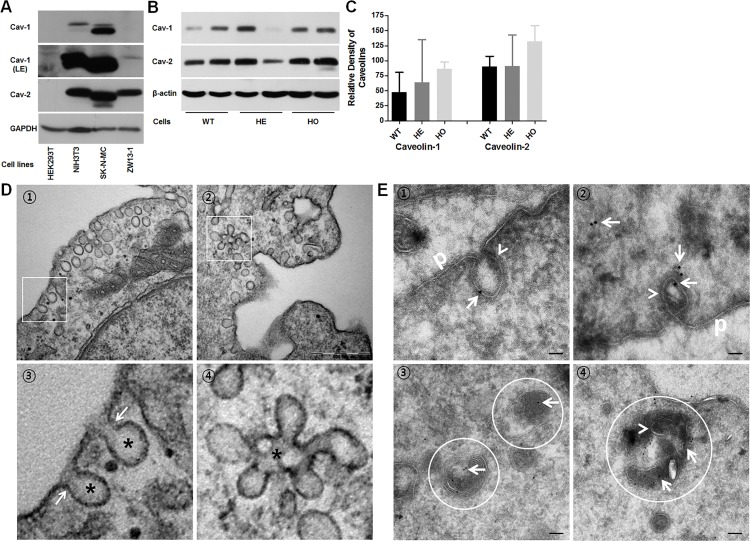
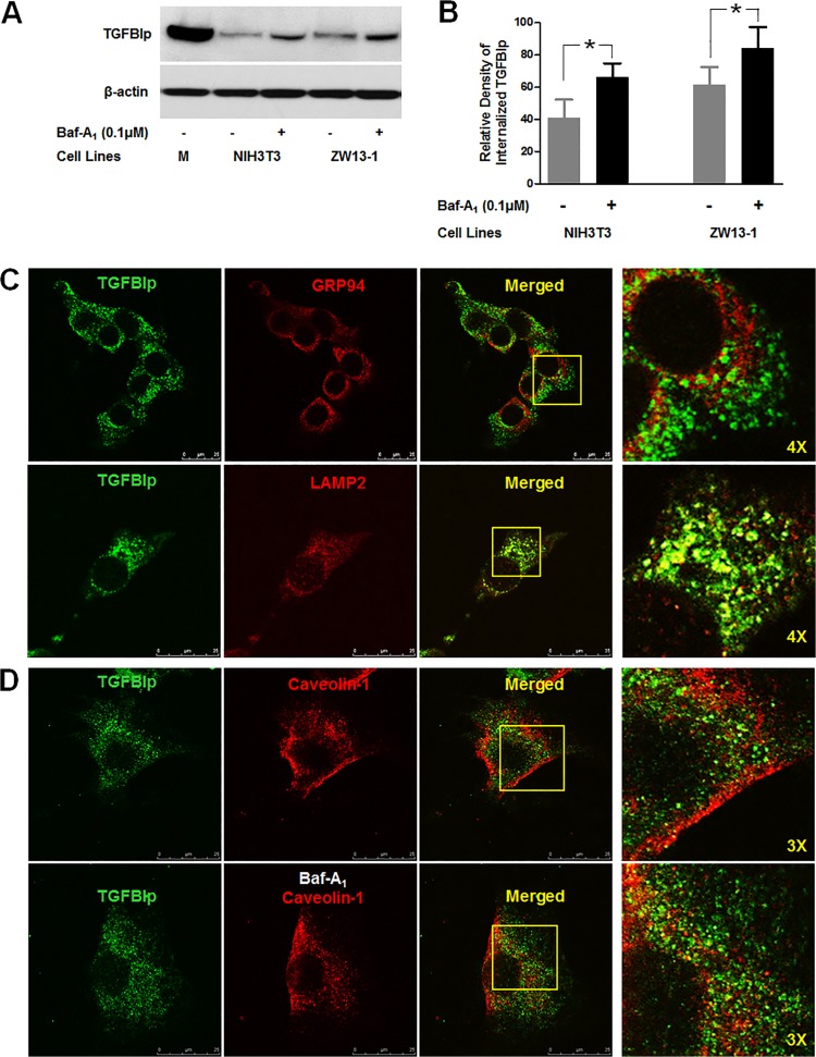
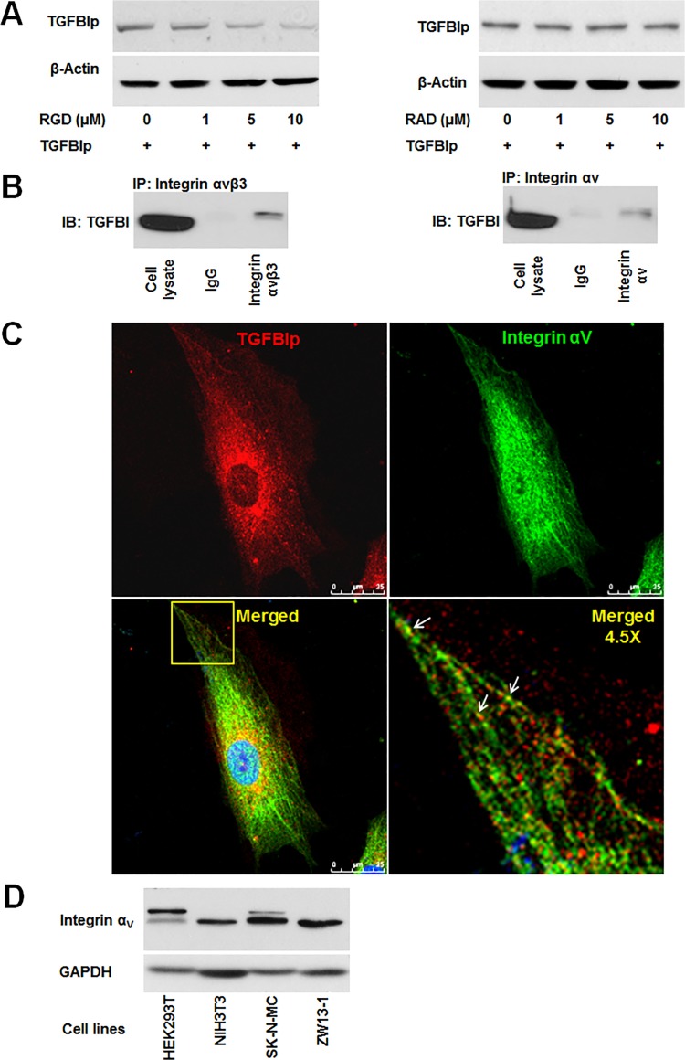
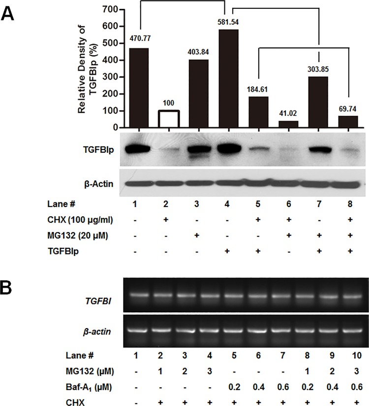
Transforming growth factor-beta-induced protein (TGFBIp) is ubiquitously expressed in the extracellular matrix (ECM) of various tissues and cell lines. Progressive accumulation of mutant TGFBIp is directly involved in the pathogenesis of TGFBI-linked corneal dystrophy. Recent studies reported that mutant TGFBIp accumulates in cells; however, the trafficking of TGFBIp is poorly understood. Therefore, we investigated TGFBIp trafficking to determine the route of its internalization and secretion and to elucidate its roles in the pathogenesis of granular corneal dystrophy type 2 (GCD2). Our data indicate that newly synthesized TGFBIp was secreted via the endoplasmic reticulum/Golgi-dependent secretory pathway, and this secretion was delayed in the corneal fibroblasts of patients with GCD2. We also found that TGFBIp was internalized by caveolae-mediated endocytosis, and the internalized TGFBIp accumulated after treatment with bafilomycin A1, an inhibitor of lysosomal degradation. In addition, the proteasome inhibitor MG132 inhibits the endocytosis of TGFBIp. Co-immunoprecipitation revealed that TGFBIp interacted with integrin αVβ3. Moreover, treatment with arginine-glycine-aspartic acid (RGD) tripeptide suppressed the internalization of TGFBIp. These insights on TGFBIp trafficking could lead to the identification of novel targets and the development of new therapies for TGFBI-linked corneal dystrophy.


Conflict of interest statement

Competing Interests: The authors have declared that no competing interests exist.

Figures

Fig 1
Fig 1. Expression and secretion of TGFBIp in wild-type (WT) and heterozygous (HE) or homozygous (HO) mutant primary corneal fibroblasts.
A. Western blot analysis of TGFBIp in cell lysates (upper panel) and conditioned media (lower panel) of WT, HE, and HO cells. β-actin was used as a loading control. Molecular weight markers (in kDa) are indicated. B. Secretion of WT and mutant TGFBIp was inhibited by treatment with brefeldin A (BFA) and monensin (MON). C. TGFBIp co-localized with markers of cis-Golgi (upper panel), medial-Golgi (middle panel), and trans-Golgi (lower panel) in cortical cells. Representative confocal images of immunofluorescence staining of TGFBIp (green) with GM130, mannosidase II, and TGN38 (all red) are shown. Overlapping areas are displayed in yellow in the merged images. Bars = 25 μm. D. Secretion of mutant TGFBIp is delayed in GCD2 corneal fibroblasts. Corneal fibroblasts from a patient with a homozygous TGFBIp mutation and a WT control were pulse-labeled for 20 min using 35S-cysteine and then incubated for 0, 15, 30, 60, 120, 180, and 240 min in unlabeled media before immunoprecipitation of TGFBIp from cell lysates and conditioned media. Phosphorimaging was performed after SDS-PAGE to detect TGFBIp. One representative experiment is shown. E. Quantitation of the experiment presented in D. Triplicate lysate samples were analyzed. *P < 0.05.
Fig 2
Fig 2. Internalization of TGFBIp in various cell lines.
A. TGFBIp was internalized in NIH3T3, SK-N-MC, and ZW13-1 cell lines but not in HEK293T. Cells were pre-incubated at 4°C for 30 min in basal medium, and then incubated for a further 120 min at 37°C in basal or TGFBIp-supplemented (~1 μg/mL) medium. Cells were washed twice with cold PBS on ice, and surface-bound TGFBIp was removed by washing three times with ice-cold acidic buffer. Cells were harvested by scraping into ice-cold PBS, pelleted by centrifugation at 1,000 × g, lysed in RIPA buffer, and 50 μg of lysate was used for western blot analysis. B. Internalization of mutant TGFBIp and WT TGFBIp was similar in NIH3T3 cells. Cells were treated as in A and analyzed by western blotting. C. Internalization of WT- and Mut-TGFBIp in the NIH3T3 cell line. Cells were incubated at 4°C for 30 min and were then shifted to 37°C in the continuous presence of TGFBIp. At the indicated time points, cells were analyzed for the amount of internalized TGFBIp. The experiment was repeated three times independently. There were no statistically significant differences between the rates of WT- and Mut-TGFBIp internalization (p > 0.05). D. Visualization of TGFBIp internalization in NIH3T3 cells by confocal microscopy. Cells were grown on glass coverslips and treated as in A, before fixation in methanol at −20°C for 3 min. Immunocytochemical staining was performed with monoclonal anti-TGFBI antibody, as described in Materials and Methods. E. TGFBIp co-localizes with Lamp-2. NIH3T3 cells grown on glass slides were subjected to immunocytochemical staining with monoclonal anti-TGFBIp and anti-Lamp-2 antibodies as described in Materials and Methods. Coverslips were mounted on the glass slides with mounting medium, and the cells were viewed using a Leica TCS SP5 confocal microscope.
Fig 3
Fig 3. Effect of inhibitors of caveolae- and clathrin-dependent endocytosis on TGFBIp internalization.
A. Inhibitors of endocytosis decreased TGFBIp levels in corneal fibroblasts. Cells were left untreated (lane 1) or treated with Baf-A1 and various endocytosis inhibitors for 60 min before incubation with TGFBIp for 120 min. TGFBIp levels were determined by western blot analysis. GS; genistein, NS; nystatin, CM; chlorpromazine. Data from one representative experiment is shown. β-actin was used as a loading control. B. Densitometric quantitation of the experiment presented in A. Data represent the TGFBIp/β-actin ratio and expressed as mean ± SD of three independently treated samples from one or two experiments. ANOVA analysis of TGFBIp levels across the treatment conditions showed no significant changes. *P≤0.05 relative to controls by Student’s t-test. C. Internalization of TGFBIp in WT corneal fibroblasts and caveolin-1-null cell line (3T3 MEF CAV-1 KO). 3T3 MEF CAV-1 KO cell does not express TGFBIp (lane 2 and 4). TGFBIp was internalized in corneal fibroblasts (lane 5) but not in 3T3 MEF CAV-1 KO cell lines (lanes 6 and 8). Cells were pre-incubated at 37°C for 60 min in basal medium or basal medium with CHX, and then incubated for a further 60 min at 37°C in basal or TGFBIp-supplemented (~1 μg/mL) medium with or without CHX. Cells were washed twice with cold PBS on ice, and surface-bound TGFBIp was removed by washing three times with ice-cold acidic buffer. Cells were harvested by scraping into ice-cold PBS, pelleted by centrifugation at 1,000 × g, lysed in RIPA buffer, and 50 μg of total protein was used for western blot analysis. D. TGFBIp co-localizes with caveolin-1 in corneal fibroblasts. Cells were grown on glass slides for 12 h and then fixed using methanol at −20°C and incubated with antibodies against TGFBIp and caveolin-1. Localization of TGFBIp (green) and caveolin-1 (red) are shown. Areas of TGFBIp and caveolin-1 co-localization appear as yellow regions in the merged image. The boxed area in the third panel was magnified and is presented as the fourth panel. Arrows in the fourth panel identify regions of TGFBIp and caveolin-1 co-localization. E. Cathepsin D co-localizes with caveolin-1 in corneal fibroblasts in the absence and presence of Baf-A1. Cells were grown on glass slides for 12 h in the absence (upper panel) or presence (lower panel) of Baf-A1 (0.1 μM) and then fixed with methanol at −20°C and incubated with antibodies against cathepsin D and caveolin-1. Localization of cathepsin D (green) and caveolin-1 (red) is shown. Areas of cathepsin D and caveolin-1 co-localization appear as yellow regions in the merged image.
Fig 4
Fig 4. Expression of caveolin-1 and -2 in established cell lines and caveolae formation in WT, HE, and HO mutant TGFBIp-expressing primary cultured corneal fibroblasts.
A. Total cellular protein (50 μg) from the specified cell lines was subjected to western blot analysis with anti-caveolin-1 (first panel), anti-caveolin-1 (second panel, LE: longer exposure), anti-caveolin-2 (third panel), and anti-Glyceraldehyde 3-phosphate dehydrogenase (GAPDH, fourth panel). GAPDH was used as a loading control. B. Western blot analysis of caveolin-1 and -2 expressions in WT, HE, and HO TGFBIp-expressing corneal fibroblasts. β-actin was used as a loading control. C. Densitometric quantitation of the experiment presented in B. D. Transmission electron microscopy (TEM) of corneal fibroblasts reveals formation of caveolae in corneal fibroblasts. Cells were grown in basal media, fixed, and then prepared for scanning TEM as described in Materials and Methods. ① Caveolae are detected on the inner surface of the plasma membrane of corneal fibroblasts. Cells contain caveolae in their apical membranes that are characterized by coat-free flask-shaped invaginations (asterisks in ③) with a diaphragm at the neck (arrows in ③). ② Note the characteristic clustering of caveolae into racemose structures, or caveosomes, on the basal side (④ asterisks). Bar = 500 nm. E. At steady state, TGFBIp is localized in the caveolae both at the plasma membrane and inside the cell. TGFBIp was immunogold-labeled on ultrathin cryosections of WT corneal fibroblasts using the TGFBIp monoclonal antibody. ① and ② plasma membrane caveolae-like structures (arrowhead) appeared as flask-shaped invaginations on the plasma membrane (p). ③ and ④ Ultrathin cryosections were labeled with anti-TGFBIp antibodies. TGFBIp-gold signal (arrow) accumulated in the lysosomes of WT corneal fibroblasts. ④ Caveolae-like structures also appeared in the lysosomes (circle). The scale bars in all panels are 100 nm.
Fig 5
Fig 5. Internalized TGFBIp is transported to lysosomes.
A. NIH3T3 and ZW13-1 cells were pre-incubated for 60 min in the absence (-) or presence (+) of Baf-A1. After incubation, cells were incubated for 120 min in normal media containing TGFBIp and western blotting was performed for TGFBIp. B. Densitometric quantitation of the experimental results presented in A. A Student’s t-test was performed to determine the significance of differences between treatments with and without Baf-A1. Data analysis showed that the p-value was less than 0.05, indicating statistical significance. The experiment was repeated three times independently. C. Co-localization of TGFBIp with GRP94, ER marker, and Lamp-2, lysosome marker, in NIH3T3 cells. Cells were subjected to immunocytochemical staining as described in Materials and Methods. Areas of co-localization appear as yellow regions in the merged image. The boxed area in the third panel was magnified and is presented as the fourth panel. D. Co-localization of TGFBIp with caveolin-1 in the absence (upper panel) and presence (lower panel) of Baf-A1. NIH3T3 cells were grown on glass slides and treated with vehicle or Baf-A1 (0.1 μM) for 60 min before incubation for 30 min at 4°C in medium containing ~1 μg/mL TGFBIp. The cells were subjected to immunocytochemical staining as described in Materials and Methods. The boxed area in the third panel was magnified and is presented as the fourth panel.
Fig 6
Fig 6. Integrin-dependent endocytosis of TGFBIp in corneal fibroblasts.
A. Endocytosis of TGFBIp was blocked by RGD peptide in a dose-dependent manner. Corneal fibroblasts were pre-incubated for 30 min in the absence (lane 1) or presence (lanes 2–4) of RGD or RAD peptides. TGFBIp (~1 μg/mL) was added to the medium and the cells were incubated for 120 min at 37°C. TGFBIp levels were measured by western blot analysis. B. TGFBIp interacts with integrin αVβ3 and αV. Cells were lysed with RIPA buffer and the lysate was immunoprecipitated with anti-integrin αVβ3 (left-hand panel) or anti-integrin αV (right-hand panel) antibody as indicated. Immunoprecipitates were resolved on 10% SDS-PAGE gels and immunoblotted with anti-TGFBIp polyclonal antibody. C. Co-localization of integrin αV with TGFBIp was visualized by confocal immunofluorescence microscopy. The merged images show TGFBIp as red, integrin αV as green, and areas of co-localization as yellow. The boxed area in the lower left-hand panel was magnified and is presented as the lower right-hand panel. Arrows identify regions of TGFBIp and integrin αV co-localization. Scale bars, 5 μm. D. Western blot analysis of HEK293T, NIH3T3, SK-N-MC, and ZW13-1 cell lines with monoclonal antibody against integrin αV. GAPDH was used as a loading control.
Fig 7
Fig 7. Effects of proteasome inhibitors on TGFBIp internalization.
A. Corneal fibroblasts were treated with CHX (to inhibit translation), and MG132 (proteasome inhibitors) or Baf-A1 (lysosomal inhibitors) for 60 min at 37°C, and then incubated with or without TGFBIp for 120 min at 37°C as indicated. The TGFBIp level was analyzed by western blotting. B. RT-PCR analysis of the effect of the specified inhibitors on the mRNA levels of TGFBI.

Similar articles

Cited by

References

    1. Munier FL, Korvatska E, Djemai A, Le Paslier D, Zografos L, Pescia G, et al. Kerato-epithelin mutations in four 5q31-linked corneal dystrophies. Nat Genet. 1997;15: 247–251. - PubMed
    1. Klintworth GK. Advances in the molecular genetics of corneal dystrophies. Am J Ophthalmol. 1999;128: 747–754. - PubMed
    1. Munier FL, Frueh BE, Othenin-Girard P, Uffer S, Cousin P, Wang MX, et al. BIGH3 mutation spectrum in corneal dystrophies. Invest Ophthalmol Vis Sci. 2002;43: 949–954. - PubMed
    1. Yuan C, Yang MC, Zins EJ, Boehlke CS, Huang AJ. Identification of the promoter region of the human betaIGH3 gene. Mol Vis. 2004;10: 351–360. - PubMed
    1. Holmbeck K, Bianco P, Caterina J, Yamada S, Kromer M, Kuznetsov SA, et al. MT1-MMP-deficient mice develop dwarfism, osteopenia, arthritis, and connective tissue disease due to inadequate collagen turnover. Cell. 1999;99: 81–92. - PubMed

Publication types

MeSH terms

LinkOut - more resources