Review
. 2015 Jul;88(1):28-34.
doi: 10.1038/ki.2015.109. Epub 2015 Apr 8.

APOL1 toxin, innate immunity, and kidney injury

Affiliations
Review

APOL1 toxin, innate immunity, and kidney injury

Sophie Limou et al. Kidney Int. 2015 Jul.

Abstract

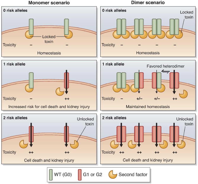
The discovery that two common APOL1 alleles were strongly associated with nondiabetic kidney diseases in African descent populations led to hope for improved diagnosis and treatment. Unfortunately, we still do not have a clear understanding of the biological function played by APOL1 in podocytes or other kidney cells, nor how the renal risk alleles initiate the development of nephropathies. Important clues for APOL1 function may be gleaned from the natural defense mechanism of APOL1 against trypanosome infections and from similar proteins (e.g., diphtheria toxin, mammalian Bcl-2 family members). This review provides an update on the biological functions for circulating (trypanosome resistance) and intracellular (emerging role for autophagy) APOL1. Further, we introduce a multimer model for APOL1 in kidney cells that reconciles the gain-of-function variants with the recessive inheritance pattern of APOL1 renal risk alleles.


Conflict of interest statement

Disclosure. All the authors declared no competing interests.

Figures

Figure 1
Figure 1. Predicted APOL1 structural domain organization
Pore-forming domain covers residues 60 to 235; BH3 domain = 154–168; membrane-addressing domain = 238–304; and SRA-interacting domain = 339–398. BH3, Bcl-2 homology domain 3.
Figure 2
Figure 2. Model of APOL1 multimers in renal cells reconciling a gain of deleterious function model with the recessive pattern of inheritance
Monomer and dimer scenarii are depicted on the left and right panels, respectively. Similarly to its trypanolytic function, we consider that APOL1 wild-type (G0, in green) toxic activity can be inhibited by a yet to be formally identified second factor (the trigger-lock system, in yellow) protecting the cell from death (locked weapon). The inhibition is lifted (actuating the trigger) by the G1 and G2 isoforms (in dark orange), enhancing the toxicity (illustrated by the black arrow) and cell death (weapon discharge) that could eventually lead to the development of glomerular injury. Only the dimer scenario offers a neutral activity for the carriers of one risk allele by limiting the number of damaging APOL1 channels (heterodimers exhibit a low or no toxicity owing to the second factor binding) and therefore reconciles with the recessive model of inheritance.
Figure 3
Figure 3. Modeling recessivity as a function of multimerization and loss of binding to the trigger-lock
We assume the following simple model: 1) There is a decrease of probability of having the trigger-lock bound to APOL1 for APOL1 risk isoforms (G1 and G2 are assumed to have equal binding) compared to wild-type (WT); 2) For carriers of 1 risk allele (WT/G1 or WT/G2 heterozygotes), multimers are formed from WT or risk molecules, drawn randomly to give a binary distribution; 3) Binding of the trigger-lock to any of the APOL1 molecules in the multimer blocks multimer activity. For this model, we plot the dominance coefficient as a function of the loss of binding probability for risk allele isoforms to the trigger-lock (expressed as a % of WT binding), from monomers to hexamers. Here, the dominance coefficient ranges from 0.5 predicting an additive model (where one risk allele carriers have half the increased risk of kidney injury of two risk alleles carriers) to 0 for a completely recessive model (where one risk allele carriers have no increased risk).

References

    1. Genovese G, Friedman DJ, Ross MD, et al. Association of trypanolytic ApoL1 variants with kidney disease in African Americans. Science. 2010;329(5993):841–5. - PMC - PubMed
    1. Duchateau PN, Pullinger CR, Cho MH, et al. Apolipoprotein L gene family: tissue-specific expression, splicing, promoter regions; discovery of a new gene. J Lipid Res. 2001;42(4):620–30. - PubMed
    1. Monajemi H, Fontijn RD, Pannekoek H, et al. The apolipoprotein L gene cluster has emerged recently in evolution and is expressed in human vascular tissue. Genomics. 2002;79(4):539–46. - PubMed
    1. Page NM, Butlin DJ, Lomthaisong K, et al. The human apolipoprotein L gene cluster: identification, classification, and sites of distribution. Genomics. 2001;74(1):71–8. - PubMed
    1. Vanhollebeke B, Pays E. The function of apolipoproteins L. Cell Mol Life Sci. 2006;63(17):1937–44. - PMC - PubMed

Publication types

LinkOut - more resources