Review
. 2015 May;13(5):269-84.
doi: 10.1038/nrmicro3432. Epub 2015 Apr 8.

Urinary tract infections: epidemiology, mechanisms of infection and treatment options

Affiliations
Review

Urinary tract infections: epidemiology, mechanisms of infection and treatment options

Ana L Flores-Mireles et al. Nat Rev Microbiol. 2015 May.

Abstract

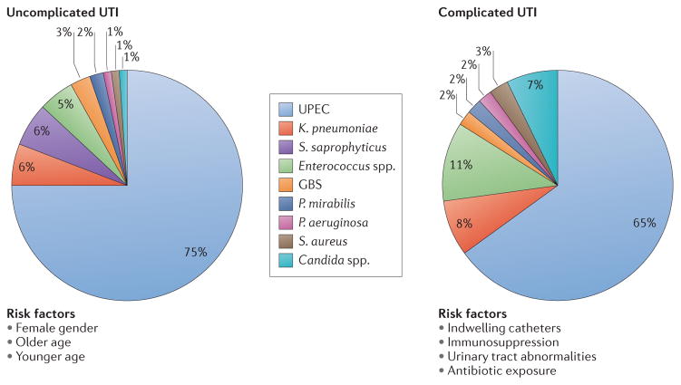
Urinary tract infections (UTIs) are a severe public health problem and are caused by a range of pathogens, but most commonly by Escherichia coli, Klebsiella pneumoniae, Proteus mirabilis, Enterococcus faecalis and Staphylococcus saprophyticus. High recurrence rates and increasing antimicrobial resistance among uropathogens threaten to greatly increase the economic burden of these infections. In this Review, we discuss how basic science studies are elucidating the molecular details of the crosstalk that occurs at the host-pathogen interface, as well as the consequences of these interactions for the pathophysiology of UTIs. We also describe current efforts to translate this knowledge into new clinical treatments for UTIs.


Conflict of interest statement

Competing interests statement

The authors declare no competing interests.

Figures

Figure 1
Figure 1. Epidemiology of urinary tract infections
Urinary tract infections (UTIs) are caused by a wide range of pathogens, including Gram-negative and Gram-positive bacteria, as well as fungi. Uncomplicated UTIs typically affect women, children and elderly patients who are otherwise healthy. Complicated UTIs are usually associated with indwelling catheters, urinary tract abnormalities, immunosuppression or exposure to antibiotics. The most common causative agent for both uncomplicated and complicated UTIs is uropathogenic Escherichia coli (UPEC). For uncomplicated UTIs, other causative agents are (in order of prevalence) Klebsiella pneumoniae, Staphylococcus saprophyticus, Enterococcus faecalis, group B Streptococcus (GBS), Proteus mirabilis, Pseudomonas aeruginosa, Staphylococcus aureus and Candida spp. For complicated UTIs, the other causative agents are (in order of prevalence) Enterococcus spp., K. pneumoniae, Candida spp., S. aureus, P. mirabilis, P. aeruginosa and GBS.
Figure 2
Figure 2. Pathogenesis of urinary tract infections
a | Uncomplicated urinary tract infections (UTIs) begin when uropathogens that reside in the gut contaminate the periurethral area (step 1) and are able to colonize the urethra. Subsequent migration to the bladder (step 2) and expression of pili and adhesins results in colonization and invasion of the superficial umbrella cells (step 3). Host inflammatory responses, including neutrophil infiltration (step 4), begin to clear extracellular bacteria. Some bacteria evade the immune system, either through host cell invasion or through morphological changes that result in resistance to neutrophils, and these bacteria undergo multiplication (step 5) and biofilm formation (step 6). These bacteria produce toxins and proteases that induce host cell damage (step 7), releasing essential nutrients that promote bacterial survival and ascension to the kidneys (step 8). Kidney colonization (step 9) results in bacterial toxin production and host tissue damage (step 10). If left untreated, UTIs can ultimately progress to bacteraemia if the pathogen crosses the tubular epithelial barrier in the kidneys (step 11). b | Uropathogens that cause complicated UTIs follow the same initial steps as those described for uncomplicated infections, including periurethral colonization (step 1), progression to the urethra and migration to the bladder (step 2). However, in order for the pathogens to cause infection, the bladder must be compromised. The most common cause of a compromised bladder is catheterization. Owing to the robust immune response induced by catheterization (step 3), fibrinogen accumulates on the catheter, providing an ideal environment for the attachment of uropathogens that express fibrinogen-binding proteins. Infection induces neutrophil infiltration (step 4), but after their initial attachment to the fibrinogen-coated catheters, the bacteria multiply (step 5), form biofilms (step 6), promote epithelial damage (step 7) and can seed infection of the kidneys (steps 8 and 9), where toxin production induces tissue damage (step 10). If left untreated, uropathogens that cause complicated UTIs can also progress to bacteraemia by crossing the tubular epithelial cell barrier (step 11).
Figure 3
Figure 3. Virulence factors of uropathogenic Escherichia coli that contribute to urinary tract infections
a | In the bladder, uropathogenic Escherichia coli (UPEC) expression of type 1 pili is essential for colonization, invasion and persistence. The type 1 pilus adhesin, FimH, binds mannosylated uroplakins and integrins that coat the surface of umbrella cells. Uroplakin binding by FimH induces actin rearrangement and bacterial internalization via unknown mechanisms. FimH–α3β1 integrin interactions induce actin rearrangement via activation of RHO-family GTPases (such as RAC proteins), resulting in bacterial invasion. Inside the host cell, UPEC can subvert host defences and resist antibiotic treatment. However, lipopolysaccharide (LPS) released by UPEC is sensed by Toll-like receptor 4 (TLR4), which induces cyclic AMP (cAMP) production via adenylyl cyclase 3 (AC3) activation, resulting in exocytosis of vesicular UPEC across the apical plasma membrane. UPEC subverts this innate defence mechanism by escaping into the cytoplasm, where it then multiplies to form intracellular bacterial communities (IBCs). Maturation of IBCs causes bacterial dispersal and allows the invasion of other host cells, which enables UPEC to re-enter the IBC cycle. Alternatively, UPEC can establish quiescent intracellular reservoirs (QIRs) in the underlying transitional cells. QIRs consist of 4–10 non-replicating bacteria within membrane-bound compartments encased in F-actin and can remain viable for months. In addition, UPEC survives within the harsh bladder environment by secreting several factors that are important for nutrient acquisition. The toxin α-haemolysin (HlyA) promotes host cell lysis through pore formation, facilitating iron release and nutrient acquisition. The siderophores expressed by UPEC allow the bacterium to scavenge iron and thus promote survival during a urinary tract infection (UTI). HlyA also triggers epithelial exfoliation to promote the spread of UPEC to other hosts following urine expulsion or to expose deeper layers of the uroepithelium for QIRs. Cytotoxic necrotizing factor 1 (CNF1) is also important for host cell remodelling and functions by binding to the receptor basal cell adhesion molecule (BCAM) on host cells to induce constitutive activation of the RHO GTPases RAC1, RHOA and cell division control 42 (CDC42), resulting in actin cytoskeletal rearrangements and membrane ruffling. Activation of RAC1 also induces the host cell anti-apoptotic and pro-survival pathways, preventing apoptosis of colonized epithelial cells and allowing the UPEC population to expand. The extracellular survival of UPEC also requires evasion of the innate immune system by the adoption of a filamentous morphology, which renders the bacterium more resistant to neutrophil killing than their bacillary form. b | UPEC colonization of the kidneys is dependent on expression of pyelonephritis-associated (P) pili, which bind globoside-containing glycolipids lining the renal tissue. The P pilus adhesin, PapG, also interacts with TLR4, reducing the expression of polymeric immunoglobulin receptor (PIGR). This results in impaired immunoglobulin A (IgA) transport across the epithelium, thereby modulating the local secretory antibody immune response and preventing UPEC opsonization and clearance.
Figure 4
Figure 4. Mechanisms of pathogenesis during catheter-associated urinary tract infections
a | Catheter-associated urinary tract infections (CAUTIs) mediated by Proteus mirabilis depend on the expression of mannose-resistant Proteus-like (MR/P) pili for initial attachment, and for biofilm formation on the catheter and in the bladder. Subsequent urease production induces the formation of calcium crystals and magnesium ammonium phosphate precipitates in the urine through the hydrolysis of urea to carbon dioxide and ammonia, resulting in a high pH. The production of extracellular polymeric substances by bacteria attached to the catheter traps these crystals, allowing the formation of a crystalline biofilm, which protects the community from the host immune system and from antibiotics. In addition, these structures prevent proper urine drainage, resulting in reflux and promoting the progression to pyelonephritis, septicaemia and shock. Finally, production of the bacterial toxins haemolysin (HpmA) and Proteus toxic agglutinin (Pta) is important for tissue destruction and bacterial dissemination to the kidneys. HpmA induces pore formation by inserting itself into the cell membrane and destabilizing the host cell, causing tissue damage, exfoliation and nutrient release. Pta punctures the host cell membrane, causing cytosol leakage and resulting in osmotic stress and depolymerization of actin filaments, thus compromising the structural integrity of the cell. The release of nutrients via these toxins also allows the bacteria to scavenge iron using siderophores. b | Enterococcus faecalis pathogenesis during CAUTIs depends on catheter implantation, which results in bladder inflammation and causes fibrinogen release, deposition onto the catheter, and accumulation. E. faecalis takes advantage of the presence of fibrinogen and uses it as a food source through the production of proteases. E. faecalis also binds fibrinogen through the endocarditis- and biofilm-associated (Ebp) pilus, allowing the formation of biofilms that protect the bacteria against the immune system.

References

    1. Stamm WE, Norrby SR. Urinary tract infections: disease panorama and challenges. J Infect Dis. 2001;183 (Suppl 1):S1–S4. - PubMed
    1. Schappert SM, Rechtsteiner EA. Ambulatory medical care utilization estimates for 2007. Vital Health Stat. 2011;13:1–38. - PubMed
    1. Foxman B. Urinary tract infection syndromes: occurrence, recurrence, bacteriology, risk factors, and disease burden. Infect Dis Clin North Am. 2014;28:1–13. This paper presents the most recent information about UTIs and their socioeconomic impact. - PubMed
    1. Foxman B. The epidemiology of urinary tract infection. Nature Rev Urol. 2010;7:653–660. - PubMed
    1. Hooton TM. Uncomplicated urinary tract infection. New Engl J Med. 2012;366:1028–1037. - PubMed

Publication types

MeSH terms

Substances