. 2015 May 15;4(5):644-54.
doi: 10.1021/acssynbio.5b00002. Epub 2015 Apr 24.

Engineering Escherichia coli into a protein delivery system for mammalian cells

Affiliations

Engineering Escherichia coli into a protein delivery system for mammalian cells

Analise Z Reeves et al. ACS Synth Biol. .

Abstract

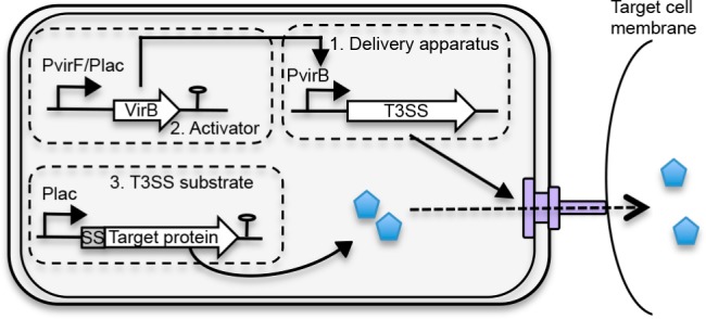
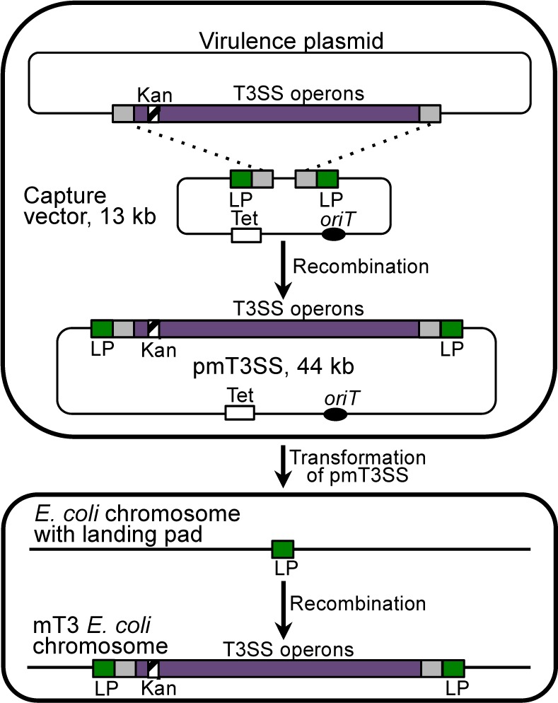
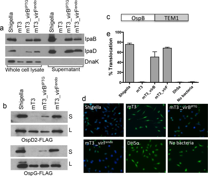
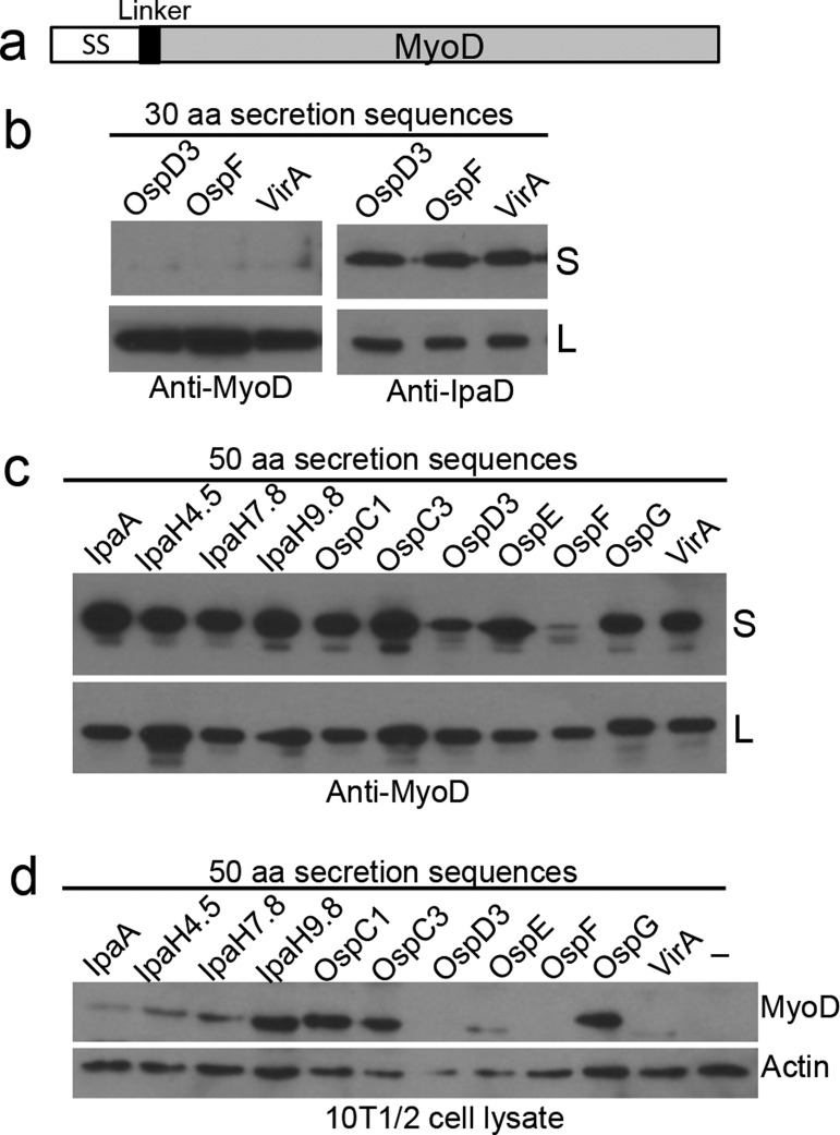
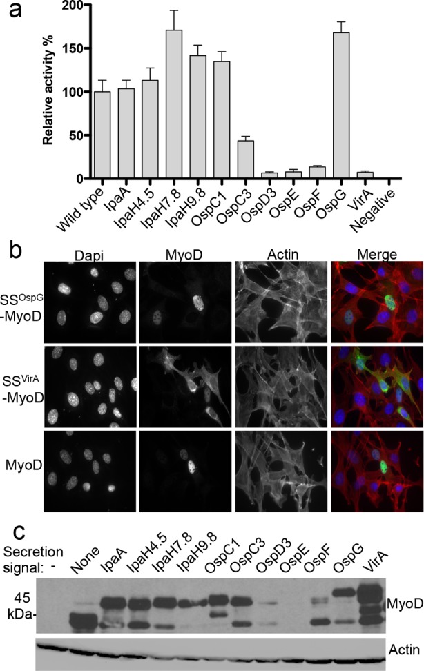
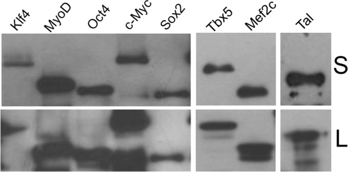
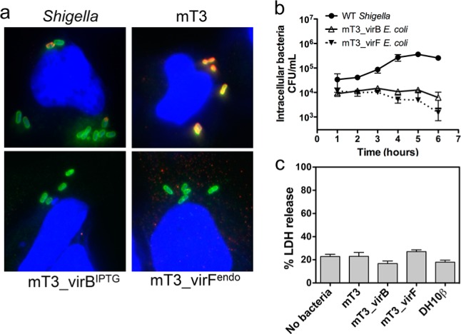
Many Gram-negative pathogens encode type 3 secretion systems, sophisticated nanomachines that deliver proteins directly into the cytoplasm of mammalian cells. These systems present attractive opportunities for therapeutic protein delivery applications; however, their utility has been limited by their inherent pathogenicity. Here, we report the reengineering of a laboratory strain of Escherichia coli with a tunable type 3 secretion system that can efficiently deliver heterologous proteins into mammalian cells, thereby circumventing the need for virulence attenuation. We first introduced a 31 kB region of Shigella flexneri DNA that encodes all of the information needed to form the secretion nanomachine onto a plasmid that can be directly propagated within E. coli or integrated into the E. coli chromosome. To provide flexible control over type 3 secretion and protein delivery, we generated plasmids expressing master regulators of the type 3 system from either constitutive or inducible promoters. We then constructed a Gateway-compatible plasmid library of type 3 secretion sequences to enable rapid screening and identification of sequences that do not perturb function when fused to heterologous protein substrates and optimized their delivery into mammalian cells. Combining these elements, we found that coordinated expression of the type 3 secretion system and modified target protein substrates produces a nonpathogenic strain that expresses, secretes, and delivers heterologous proteins into mammalian cells. This reengineered system thus provides a highly flexible protein delivery platform with potential for future therapeutic applications.

Keywords: bacterial engineering; protein delivery; synthetic biology; type 3 secretion system.


Figures

Figure 1
Figure 1
Components of the bacterial protein delivery system in Escherichia coli. (1) The delivery apparatus encodes the genes required to assemble a functional type 3 secretion system (T3SS) from Shigella flexneri. When T3SS genes are expressed, the secretion system assembles in the bacterial inner and outer membranes. Upon contact with a eukaryotic target cell, the secretion system forms a conduit between the bacterial and target cell that allows for protein delivery directly into the target cell cytoplasm. (2) The type 3 genes are activated by the VirB transcription factor, whose expression is induced either from a lac promoter by the addition of IPTG or from its native promoter, which is, in turn, activated by the expression of the VirB transcriptional activator, VirF. (3) The type 3 secreted substrates are target protein(s) fused to type 3 secretion sequence (SS) at their N-termini. Expression of the target protein is induced by the presence of IPTG and can be coordinated with expression of the type 3 secretion apparatus genes.
Figure 2
Figure 2
Generation of mT3 Escherichia coli, the protein delivery strain. A kanamycin-resistance cassette (striped box) was inserted into a nonessential region of the Shigella flexneri virulence plasmid to assist in selection of proper recombination events with the capture vector. A capture vector was constructed that contains regions of homology to the regions of the Shigella virulence plasmid flanking the type 3 secretion genes, which are represented as gray boxes. Landing pad (LP) sequence, denoted as a green box, flanks the pieces of T3SS gene homology to facilitate downstream integration into the E. coli chromosome. An origin of transfer (oriT), which can mobilize the plasmid between bacterial host strains by conjugation, is represented by a black oval. λ-Red recombination was then used to introduce the region of the Shigella virulence plasmid that contains the T3SS genes onto the capture vector. The resulting 44 kb plasmid (pmT3SS) contains the entire T3SS. When pmT3SS is introduced into a strain of E. coli harboring an engineered landing pad sequence, recombination leads to integration of the intervening sequence, in this case the T3SS operons, into the chromosome, generating the strain mT3 E. coli.
Figure 3
Figure 3
mT3 Escherichia coli secretes and delivers proteins into mammalian cells. Shigella and mT3 E. coli strains were grown under conditions that induce type 3 secretion system expression. Secretion was induced by exposure to Congo Red dye, and delivery was induced by bacterial contact with mammalian cells. (a) Western blot analysis of T3SS apparatus proteins in mT3 E. coli. Whole cell lysate and supernatant proteins were separated by SDS-PAGE and immunoblotted with anti-IpaB or anti-IpaD antibodies. DnaK is a cytoplasmic protein unrelated to type 3 secretion and serves as a loading and bacterial cell lysis control. (b) Plasmids expressing FLAG-tagged versions of native Shigella effectors were introduced into each strain background, and cell lysate (L) and secreted proteins (S) were probed with anti-FLAG antibodies. The blots shown are representative of at least three experiments. Each strain was transformed with a target protein (substrate) plasmid that expresses an IPTG-inducible construct of an OspB–TEM-1 fusion protein illustrated in (c). (d) Images of HeLa cells loaded with CCF4/AM exposed to wild-type Shigella or mT3 E. coli strains expressing OspB–TEM-1. (e) Translocation was quantified by measuring the percentage of cells that fluoresce blue (cleaved CCF4/AM). Data are expressed as the mean of three independent experiments performed in triplicate. Error bars represent the standard error of the mean (SEM). At least 600 cells were counted for each sample.
Figure 4
Figure 4
Secretion sequence–MyoD fusion proteins are recognized as type 3 secreted substrates and directly delivered into mammalian cells. (a) Schematic of MyoD fused to a 30 or 50 amino acid secretion sequence and separated by a flexible glycine linker. (b) Secretion assay of the set of 30 amino acid secretion sequence fusion proteins to MyoD (SS–MyoD) in mT3_virFendo. Blots were probed with an anti-MyoD or anti-IpaD antibody. (c) Secretion assay of the library of 50 amino acid SS–MyoD fusion proteins in mT3_virFendo. Blots were probed with an anti-MyoD antibody. (d) Delivery of SS–MyoD into MEFs exposed to mT3_virFendoE. coli expressing each of the designated SS–MyoD proteins. After 1 h, MEF cell lysates were collected and probed with anti-MyoD and anti-actin antibodies. Actin serves as a loading control for cell lysate. S, supernatant; L, whole cell lysate. Supporting Information Figure S5 demonstrates that E. coli DH10β does not secrete these proteins in the absence of the Shigella type 3 secretion system operons.
Figure 5
Figure 5
Fusion to type 3 secretion sequences (SS) can affect heterologous protein activity by altering protein stability or localization. MEFs were transfected with equal amounts of mammalian expression plasmids that express wild-type or the designated SS–MyoD proteins. Cells were fixed after 7 days and stained for myosin heavy and light chain expression. Myosin positive cells were enumerated. (a) Relative myogenic activity was determined by dividing the number of myosin positive cells produced by transfection with SS–MyoD by the average amount generated by wild-type MyoD, with wild type set to 100%. Data are expressed as the mean plus the standard error of the mean (SEM) from four independent transfections. (b) Localization of representative SS–MyoD derivatives. Cells were fixed and stained with anti-MyoD antibody 24 h post-transfection. Nuclei and actin were stained with DAPI and phalloidin, respectively. (c) Stability of SS–MyoD fusion proteins. Lysates from 10T1/2 cells transfected with wild-type or SS–MyoD fusion proteins were probed with anti-MyoD antibody 24 h after transfection. Actin was used as a loading control.
Figure 6
Figure 6
mT3_virFendoE. coli expresses and secretes a variety of target proteins modified by the Shigella OspG type 3 secretion sequence. Plasmids expressing FLAG-tagged versions of target proteins were introduced into mT3_virFendoE. coli cell lysate, and type 3 secreted proteins were probed with anti-FLAG antibodies. Included are iPS reprogramming factors, MyoD, two cardiac reprogramming factors, and a TALE–activator fusion protein. The blots shown are representative of at least three experimental repeats. S, supernatant; L, whole cell lysates.
Figure 7
Figure 7
Type 3 secretion genes in mT3 E. coli induce invasion but not replication or cytotoxicity in HeLa cells. (a) HeLa cells were differentially stained following a 1 h exposure to bacteria. To distinguish internal vs external bacteria (Methods), prior to permeabilization, HeLa cells were fixed and stained with anti-E. coli antibodies followed by Alexa-Fluor 568 (red) conjugated secondary antibodies. After this initial staining, HeLa cells were permeabilized, followed by another round of staining with primary anti-E. coli antibodies and Alexa-Fluor 488 (green) conjugated secondary antibodies. This procedure results in internalized bacteria staining green, whereas external bacteria stain both red and green, appearing yellow. Nuclei were stained with DAPI (blue). (b) mT3_virBIPTG and mT3_virFendoE. coli are able to invade, but they grow very poorly in HeLa cells compared to that of wild-type Shigella. HeLa cells were infected at an MOI of 100:1, and intracellular bacteria were enumerated for 6 h postinfection in a gentamicin protection assay. Values represent the means of measurements for triplicate samples from a representative experiment. Error bars represent the SEM. (c) HeLa cells were exposed to bacteria for 4 h, and supernatants were analyzed for cytotoxicity by lactate dehydrogenase (LDH) release assay. Data are expressed as the mean + standard error of the mean (SEM) from four independent experiments.

Similar articles

Cited by

References

    1. Bichsel C.; Neeld D.; Hamazaki T.; Chang L. J.; Yang L. J.; Terada N.; Jin S. (2013) Direct reprogramming of fibroblasts to myocytes via bacterial injection of MyoD protein. Cell. Reprogramming 15, 117–125. - PMC - PubMed
    1. Jia J.; Jin Y.; Bian T.; Wu D.; Yang L.; Terada N.; Wu W.; Jin S. (2014) Bacterial delivery of TALEN proteins for human genome editing. PLoS One 9, e91547. - PMC - PubMed
    1. Carleton H. A.; Lara-Tejero M.; Liu X.; Galan J. E. (2013) Engineering the type III secretion system in non-replicating bacterial minicells for antigen delivery. Nat. Commun. 4, 1590. - PMC - PubMed
    1. Chen L. M.; Briones G.; Donis R. O.; Galan J. E. (2006) Optimization of the delivery of heterologous proteins by the Salmonella enterica serovar Typhimurium type III secretion system for vaccine development. Infect. Immun. 74, 5826–5833. - PMC - PubMed
    1. Akeda Y.; Kimura T.; Yamasaki A.; Kodama T.; Iida T.; Honda T.; Oishi K. (2012) Functional cloning of Vibrio parahaemolyticus type III secretion system 1 in Escherichia coli K-12 strain as a molecular syringe. Biochem. Biophys. Res. Commun. 427, 242–247. - PubMed

Publication types

Substances