. 2015 Apr 8;10(4):e0121427.
doi: 10.1371/journal.pone.0121427. eCollection 2015.

Crotoxin from Crotalus durissus terrificus is able to down-modulate the acute intestinal inflammation in mice

Affiliations

Crotoxin from Crotalus durissus terrificus is able to down-modulate the acute intestinal inflammation in mice

Caroline de Souza Almeida et al. PLoS One. .

Abstract

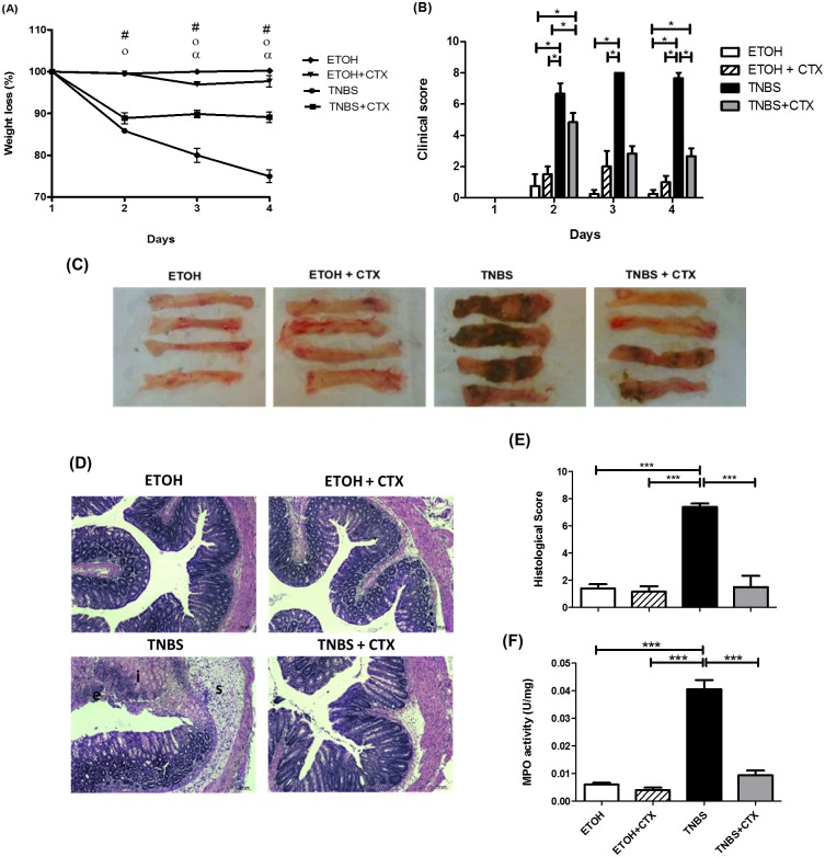
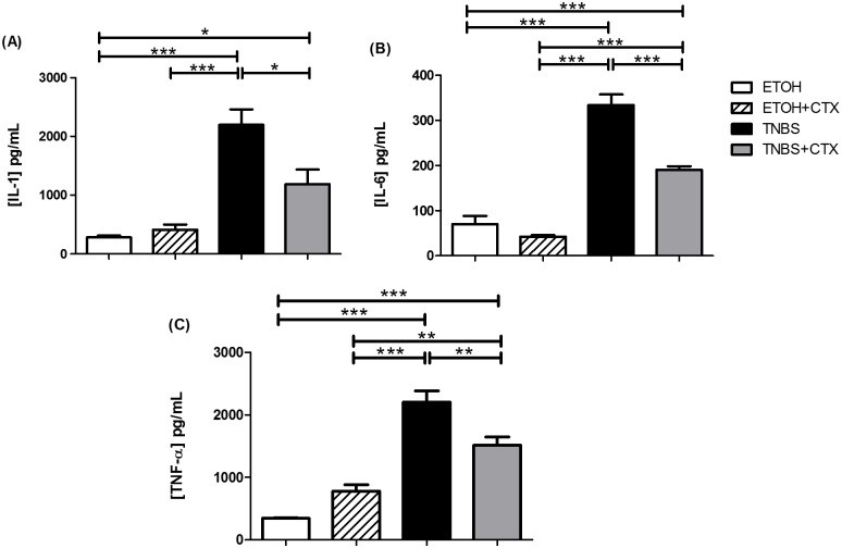
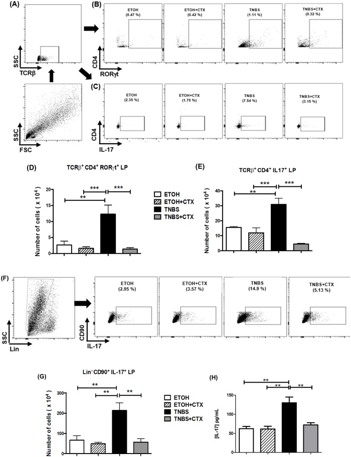
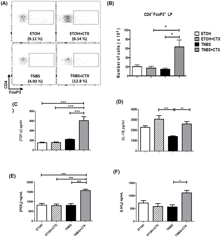
Inflammatory bowel diseases (IBD) is the result of dysregulation of mucosal innate and adaptive immune responses. Factors such as genetic, microbial and environmental are involved in the development of these disorders. Accordingly, animal models that mimic human diseases are tools for the understanding the immunological processes of the IBD as well as to evaluate new therapeutic strategies. Crotoxin (CTX) is the main component of Crotalus durissus terrificus snake venom and has an immunomodulatory effect. Thus, we aimed to evaluate the modulatory effect of CTX in a murine model of colitis induced by 2,4,6- trinitrobenzene sulfonic acid (TNBS). The CTX was administered intraperitoneally 18 hours after the TNBS intrarectal instillation in BALB/c mice. The CTX administration resulted in decreased weight loss, disease activity index (DAI), macroscopic tissue damage, histopathological score and myeloperoxidase (MPO) activity analyzed after 4 days of acute TNBS colitis. Furthermore, the levels of TNF-α, IL-1β and IL-6 were lower in colon tissue homogenates of TNBS-mice that received the CTX when compared with untreated TNBS mice. The analysis of distinct cell populations obtained from the intestinal lamina propria showed that CTX reduced the number of group 3 innate lymphoid cells (ILC3) and Th17 population; CTX decreased IL-17 secretion but did not alter the frequency of CD4+Tbet+ T cells induced by TNBS instillation in mice. In contrast, increased CD4+FoxP3+ cell population as well as secretion of TGF-β, prostaglandin E2 (PGE2) and lipoxin A4 (LXA4) was observed in TNBS-colitis mice treated with CTX compared with untreated TNBS-colitis mice. In conclusion, the CTX is able to modulate the intestinal acute inflammatory response induced by TNBS, resulting in the improvement of clinical status of the mice. This effect of CTX is complex and involves the suppression of the pro-inflammatory environment elicited by intrarectal instillation of TNBS due to the induction of a local anti-inflammatory profile in mice.


Conflict of interest statement

Competing Interests: The authors have declared that no competing interests exist.

Figures

Fig 1
Fig 1. Effect of CTX treatment on the colitis induced by TNBS instillation in BALB/c mice.
(A) Body weight changes (%) of BALB/c mice during four days after the intrarectal instillation of TNBS (2.5 mg/animal) in 45% ethanol solution. The control mice received the 45% of ethanol solution. CTX (0.035 mg/kg) was administered i.p. 18 h after TNBS-induced colitis, and saline solution was administered as control. # (p<0.05) ETOH versus TNBS and TNBS+CTX; o (p<0.05) ETOH+CTX versus TNBS and TNBS+CTX; α (p<0.05) TNBS versus TNBS+CTX (n = 4–6 mice/group). (B) Disease activity index calculated as described in material and methods. The results were expressed as mean ± SEM (n = 4–6 mice/group); (C) Macroscopic appearance of colonic portion (4 cm) obtained from each mice/group at 4 days after TNBS-induced colitis; (D) Histological analysis of perirectal segment from mice of distinct experimental groups stained with H&E (Structures: (e) epithelial damage, (i) inflammatory infiltrate and (s) submucosa edema) obtained after 4 days of TNBS-colitis; (E) Histological score of inflammatory reaction perirectal segment of each experimental group of mice (n = 4–5 mice/group); (F) MPO activity of colonic tissue of each experimental mice-group. Groups: ETOH (control- 45% ETOH); ETOH+CTX (45% ethanol group treated with CTX); TNBS (TNBS instillation in 45% ETOH- inflammatory bowel disease) and TNBS+CTX (TNBS-instillation in 45% ETOH that received the CTX) (n = 4–5 animals/group). * p<0.05; *** p<0.001.
Fig 2
Fig 2. Secretion of pro-inflammatory cytokines in colonic tissue homogenates of TNBS-mice treated or not with CTX.
Production of IL-1β (A), IL-6 (B) and TNF-α (C) was measured in homogenates of colonic segments by ELISA. The results represent the mean of the cytokine secretion in individual mice/group ± SEM. * p<0.05, ** p<0.01 and *** p<0.001 (n = 4–5 animals/group).
Fig 3
Fig 3. Effect of CTX on Th17 cells, ILC3 and IL-17 secretion of mice with acute colitis induced by TNBS.
Cell suspensions were prepared from lamina propria of distinct experimental groups after 4 days of TNBS-induced colitis and that received or not the CTX. The TCRß+CD4+RORγt+, TCRß+CD4+IL-17+ and Lin-CD90+IL-17+ cells were analyzed by flow cytometry. (A, B and C) Strategy for the analysis of TCRß+CD4+RORγt+ and TCRß+CD4+IL-17+ cells in the lamina propria obtained from each group of mice. The results of TCRß+CD4+RORγt+ (D) and TCRß+CD4+IL-17+ cells (E) expressed as the mean of the absolute number ± SEM. The samples were prepared from a pool of cells from 4–5 animals/group performed in duplicate. The results are from 2–3 independent experiments. (F) Strategy for the analysis of the Lin-CD90+IL-17+ cells. (G) The results of Lin-CD90+IL-17+ cells were expressed as a mean of the absolute number ± SEM. The samples were prepared from a pool of cells from 4–5 animals/group performed in duplicate. The results are from 2 independent experiments. (H) Secretion of IL-17 in colonic tissue homogenates determined by ELISA. The results represent the mean obtained in the individual samples/group ± SEM. * p<0.05, ** p<0.01 and *** p<0.001 (n = 4–5 animals/group).
Fig 4
Fig 4. CD4+Tbet+ cell population and IFN-γ secretion of mice with acute colitis induced by TNBS treated or not with CTX.
(A) Representative dot plots of CD4+Tbet+ cells in the lamina propria of distinct group of mice. (B) CD4+Tbet+ cells were expressed as a mean of the absolute number of cells ± SEM. The samples were prepared from a pool of cells from 4–5 animals/group performed in duplicate. The results are from 3 independent experiments. (C) Secretion of IFN-γ in colonic tissue homogenates determined by ELISA. The results represent the mean obtained in the individual samples/group ± SEM. * p<0.05, ** p<0.01 and *** p<0.001; (n = 4–5 animals/group).
Fig 5
Fig 5. Analysis of CD4+FoxP3+ cells and anti-inflammatory mediators in TNBS-mice treated or not with CTX.
Cell suspensions were prepared from lamina propria of distinct experimental groups after 4 days of TNBS-induced colitis for the analysis of CD4+FoxP3+ cells by flow cytometry. The samples were prepared from a pool of cells from 4–5 animals/group performed in duplicate. The results are from 2 independent experiments. (A) Representative dot plots of CD4+FoxP3+ cells in the lamina propria obtained from distinct experimental groups. (B) Results of CD4+FoxP3+ cells expressed as a mean of the absolute number of cells in duplicate of 2 independent experiments ± SEM. Secretion of TGF-β (C) and IL-10 (D) in colonic tissue homogenates determined by ELISA. Production of PGE2 (E) and LXA4 (F) was performed by ELISA in colonic tissue homogenates. The results represent the mean obtained in individual mice/group ± SEM. * p<0.05, ** p<0.01 and *** p<0.001; (n = 4–5 animals/group).

Similar articles

Cited by

References

    1. Geboes K. From inflammation to lesion. Acta Gastroenterol Belg. 1994;57: 273–284. - PubMed
    1. Sartor RB Current concepts of the etiology and pathogenesis of ulcerative colitis and Crohn’s disease. Gastroenterol Clin North Am. 1995;24: 475–507. - PubMed
    1. Kraus TA, Mayer L. Oral tolerance and inflammatory bowel disease. Curr Opin Gastroenterol. 2005;21: 692–696. - PubMed
    1. Sartor RB. Pathogenesis and immune mechanisms of chronic inflammatory bowel diseases. Am J Gastroenterol. 1997;92: 5S–11S. - PubMed
    1. Brandtzaeg P. Inflammatory bowel disease: clinics and pathology. Do inflammatory bowel disease and periodontal disease have similar immunopathogeneses? Acta OdontolScand, 2001;59: 235–243. - PubMed

Publication types