. 2015 Apr 8;10(4):e0123027.
doi: 10.1371/journal.pone.0123027. eCollection 2015.

SaeRS-dependent inhibition of biofilm formation in Staphylococcus aureus Newman

Affiliations

SaeRS-dependent inhibition of biofilm formation in Staphylococcus aureus Newman

David Cue et al. PLoS One. .

Abstract

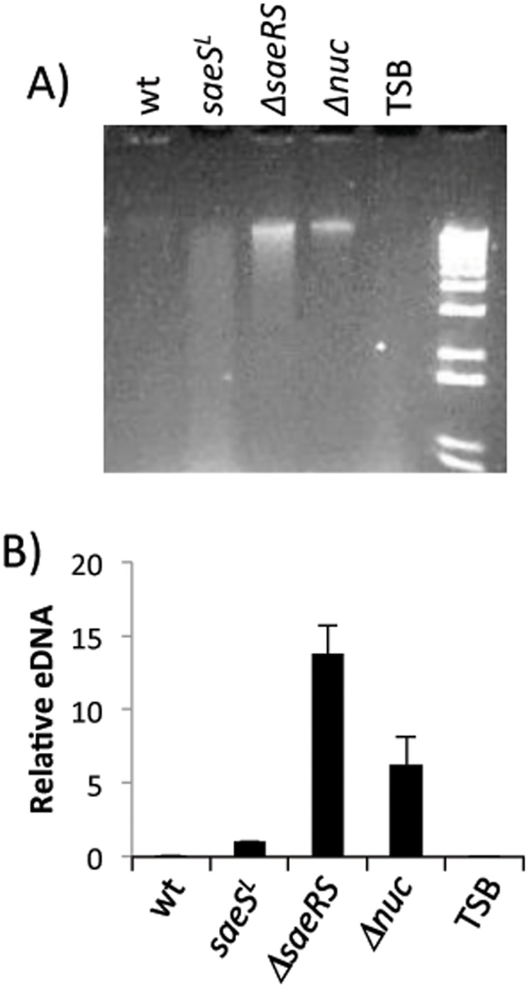
The SaeRS two-component regulatory system of Staphylococcus aureus is known to affect the expression of many genes. The SaeS protein is the histidine kinase responsible for phosphorylation of the response regulator SaeR. In S. aureus Newman, the sae system is constitutively expressed due to a point mutation in saeS, relative to other S. aureus strains, which results in substitution of proline for leucine at amino acid 18. Strain Newman is unable to form a robust biofilm and we report here that the biofilm-deficient phenotype is due to the saeSP allele. Replacement of the Newman saeSP with saeSL, or deletion of saeRS, resulted in a biofilm-proficient phenotype. Newman culture supernatants were observed to inhibit biofilm formation by other S. aureus strains, but did not affect biofilm formation by S. epidermidis. Culture supernatants of Newman saeSL or Newman ΔsaeRS had no significant effect on biofilm formation. The inhibitory factor was inactivated by incubation with proteinase K, but survived heating, indicating that the inhibitory protein is heat-stable. The inhibitory protein was found to affect the attachment step in biofilm formation, but had no effect on preformed biofilms. Replacement of saeSL with saeSP in the biofilm-proficient S. aureus USA300 FPR3757 resulted in the loss of biofilm formation. Culture supernatants of USA300 FPR3757 saeSP, did not inhibit biofilm formation by other staphylococci, suggesting that the inhibitory factor is produced but not secreted in the mutant strain. A number of biochemical methods were utilized to isolate the inhibitory protein. Although a number of candidate proteins were identified, none were found to be the actual inhibitor. In an effort to reduce the number of potential inhibitory genes, RNA-Seq analyses were done with wild-type strain Newman and the saeSL and ΔsaeRS mutants. RNA-Seq results indicated that sae regulates many genes that may affect biofilm formation by Newman.


Conflict of interest statement

Competing Interests: The authors have declared that no competing interests exist.

Figures

Fig 1
Fig 1. SaePQRS regulates biofilm formation by S. aureus strain Newman.
(A) Stationary phase cultures of the indicated strains were diluted to an OD660 of 0.05 and inoculated into wells of a microtiter plate. The microtiter plate wells had been precoated with human plasma. After 16 h, biofilms were washed, fixed, and stained with crystal violet. The wells labeled “medium” indicates wells that contained sterile biofilm medium. (B) Inhibition of UAMS-1 biofilm formation by culture supernatants. Stationary phase culture supernatants of strain Newman and its derivatives were harvested, filter sterilized and added to microtiter plate wells preinoculated with S. aureus UAMS-1 suspended in biofilm medium. The source of each culture supernatant is listed to the right of the picture. The wells labeled “medium” indicates sterile TSB-0G medium was added in place of an exogenous culture supernatant. Plates were incubated and processed as described for A.
Fig 2
Fig 2. The biofilm inhibitory factor is a heat stable protein.
Stationary phase culture supernatants of strain Newman (wt) and Newman ΔsaeRS were harvested, filter sterilized, and diluted into biofilm medium. Anti-biofilm activity of supernatants was tested against S. aureus strain UAMS-1. (A) Proteinase K treatment of culture supernatants. Supernatants from the indicated strains were incubated with proteinase K and Tris buffer, or buffer only (mock prtK), at 42°C for 1 hr and then 95°C for 10 minutes, before adding to biofilm medium. “None” indicates wells containing UAMS-1 but no culture supernatant. Wells labeled “medium” contained sterile biofilm medium. (B) Supernatants were incubated at the indicated temperatures for the indicated time periods before adding to wells containing UAMS-1 suspended in biofilm medium.
Fig 3
Fig 3. saeRS regulates attachment but not dispersal.
(A) Stationary phase cultures of Newman and the ΔsaePQRS::kan strain were diluted into biofilm medium, inoculated into a 24-well plate and allowed to attach for 1 h at 37°C before washing and staining with crystal violet. (B) Supernatants from Newman and Newman ΔsaePQRS::kan were diluted into biofilm media and the attachment phenotype of strain UAMS-1 was tested with and without supernatant supplementation. (C) Newman (row 2) and Newman ΔsaePQRS::kan (row 3) culture supernatants were added at the indicated times following inoculation with UAMS-1. Wells in row 1 were supplemented with sterile medium rather than culture supernatant.
Fig 4
Fig 4. Strain-dependent production of the biofilm inhibitory protein.
(A) Biofilm formation by strain USA300 FPR 3757 and derivatives. Wells were inoculated with the indicated USA 300 FBR 3757 derivatives. +saePQRS L and +saePQRS P indicate the presence of plasmids pCWSAE50 or pCWSAE51, respectively. (B) Anti-biofilm activity of USA300 derivatives. Culture supernatants from the strains listed to the right of the figure were tested for inhibition of biofilm formation by strain UAMS-1.
Fig 5
Fig 5. The biofilm inhibitory protein limits biofilm in S. aureus but not S. epidermidis strains.
Culture supernatants from strain Newman or the ΔsaePQRS::kan mutant were added to biofilm medium and anti-biofilm activity was tested against the bacterial strains listed to the left of the figure. “Medium” indicates that sterile medium was added in place of culture supernatant.
Fig 6
Fig 6. Confirmation of RNA-Seq data using RT-qPCR.
RNA was isolated from cultures of strain Newman (saeS P), CYL11481 (saeS L) and CYL11771 (ΔsaeRS) for use in RT-qPCR assays. Values for the saeS L (open bars) and ΔsaeRS (grey bars) strains are expressed relative to wild type Newman, which was assigned an arbitrary value of 1. All assays were performed with at least two RNA preparations obtained from separate cultures.
Fig 7
Fig 7. Zymograms of S. aureus murein hydrolase activity.
Cell free extracts of strains Newman (wt), saeS L (CYL11481) and ΔsaeRS (CYL11771) were run on 10% acrylamide-SDS gels containing heat killed S. aureus RN4220 cells (leftmost panel) or heat killed Micrococcus luteus cells (middle panel). Numbers to the left of each gel indicate the molecular weights of size standards. Dark bands indicate regions of murein hydrolase activity. The large clear band migrating at approximately 60 kDa is presumably the Map protein, which is highly expressed in strain Newman. The rightmost panel is a Coomassie Blue-stained gel showing the same extracts used for the other panels. The large band migrating at approximately 60 kDa is presumably the Map protein.
Fig 8
Fig 8. Extracellular (e) DNA in culture supernatants.
Culture supernatants from strain Newman and its derivatives were filter sterilized, extracted with phenol-chloroform and ethanol precipitated. Precipitates were suspended in H2O. The volumes of water were varied to compensate for slight differences in OD660 of the initial cultures. (A) Agarose gel of isolated eDNAs. (B) qPCR results. qPCR was performed with isolated eDNAs using oligonucleotide primers specific for the hu gene. Data are expressed relative to the amount of eDNA in the saeS L strain supernatant, which was arbitrarily assigned a value of 1. Data are means from two independent cultures of each strain. Δnuc indicates a nuclease deficient mutant of Newman. TSB indicates material isolated from sterile culture medium (TSB-0G).

References

    1. Boles BR, Horswill AR. Staphylococcal biofilm disassembly. Trends Microbiol. 2011;19: 449–455. 10.1016/j.tim.2011.06.004 - DOI - PMC - PubMed
    1. Gotz F. Staphylococcus and biofilms. Mol Microbiol. 2002;43: 1367–1378. - PubMed
    1. O’Gara JP. Ica and beyond. Biofilm mechanisms and regulation in Staphylococcus epidermidis and Staphylococcus aureus . FEMS Microbiol Lett. 2007;270: 179–188. - PubMed
    1. Otto M. Staphylococcal biofilms. Curr Top Microbiol Immunol 2008;322: 207–228. - PMC - PubMed
    1. Otto M. Staphylococcus epidermidis pathogenesis. Methods Mol Biol 2014;1106:17–31. 10.1007/978-1-62703-736-5_2 - DOI - PubMed

Publication types

MeSH terms

Substances

Associated data