. 2015 Apr 8;10(4):e0122919.
doi: 10.1371/journal.pone.0122919. eCollection 2015.

MYB elongation is regulated by the nucleic acid binding of NFκB p50 to the intronic stem-loop region

Affiliations

MYB elongation is regulated by the nucleic acid binding of NFκB p50 to the intronic stem-loop region

Lloyd A Pereira et al. PLoS One. .

Abstract

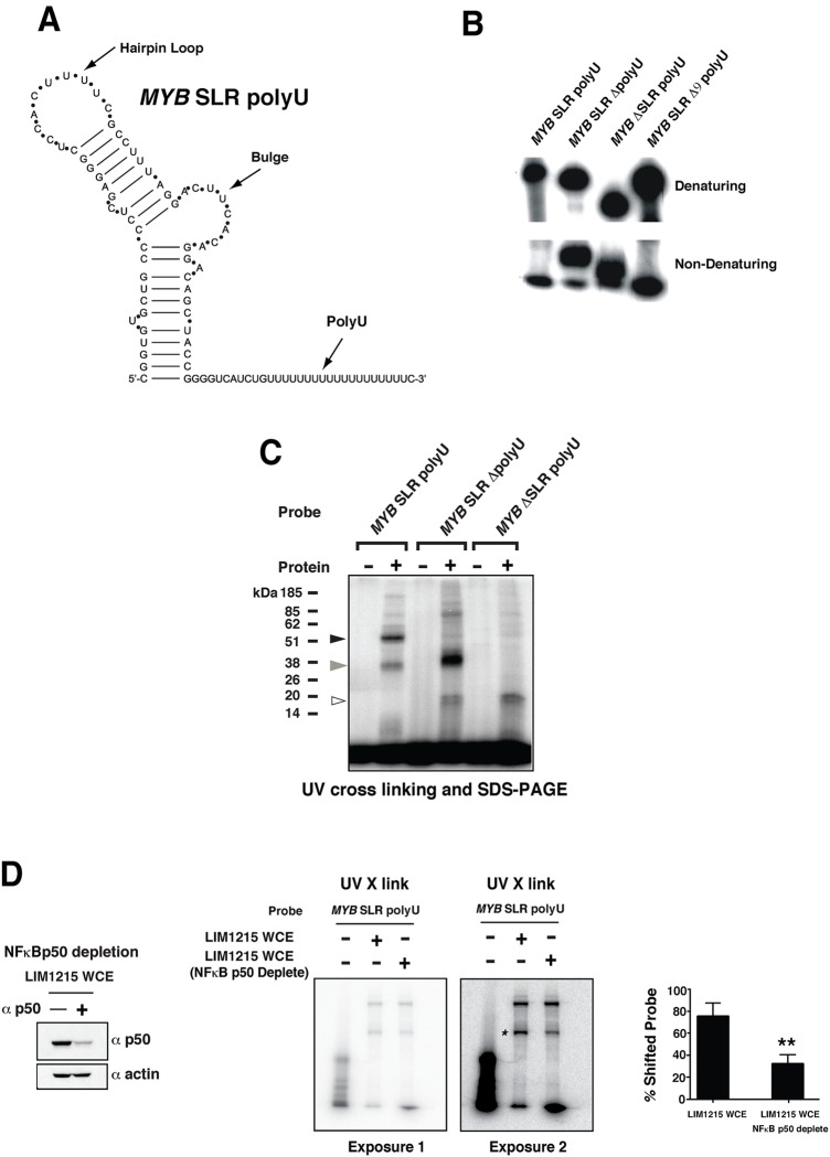
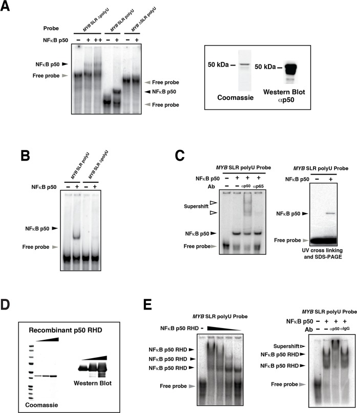
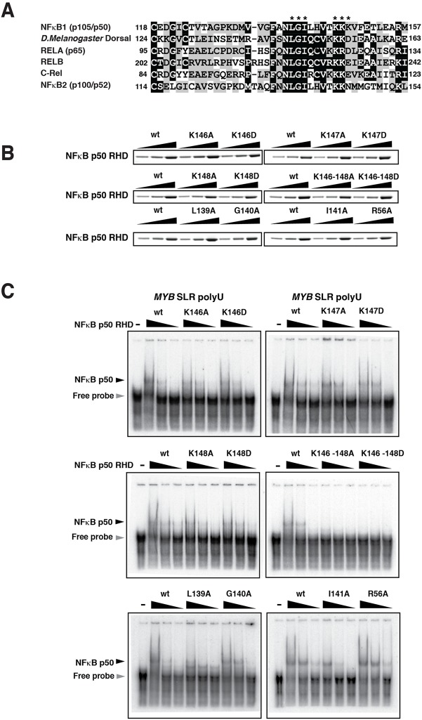
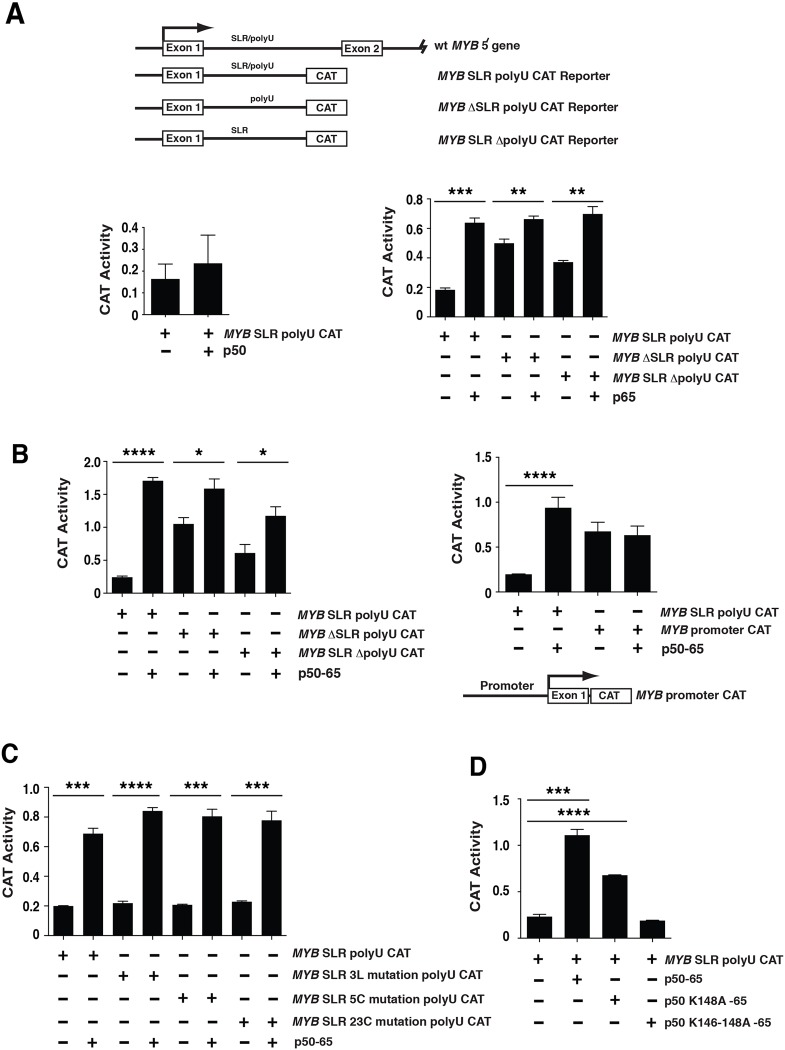
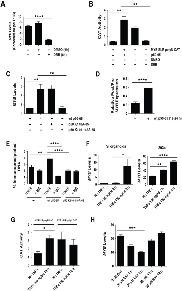
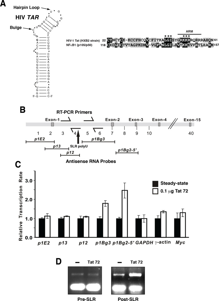
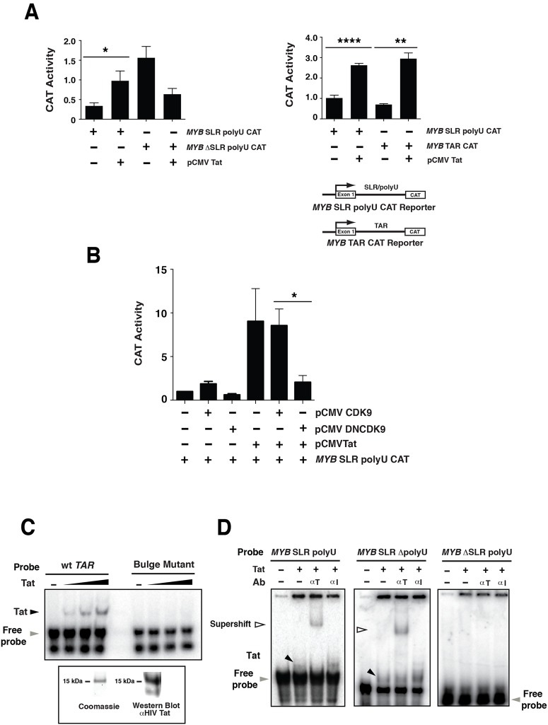
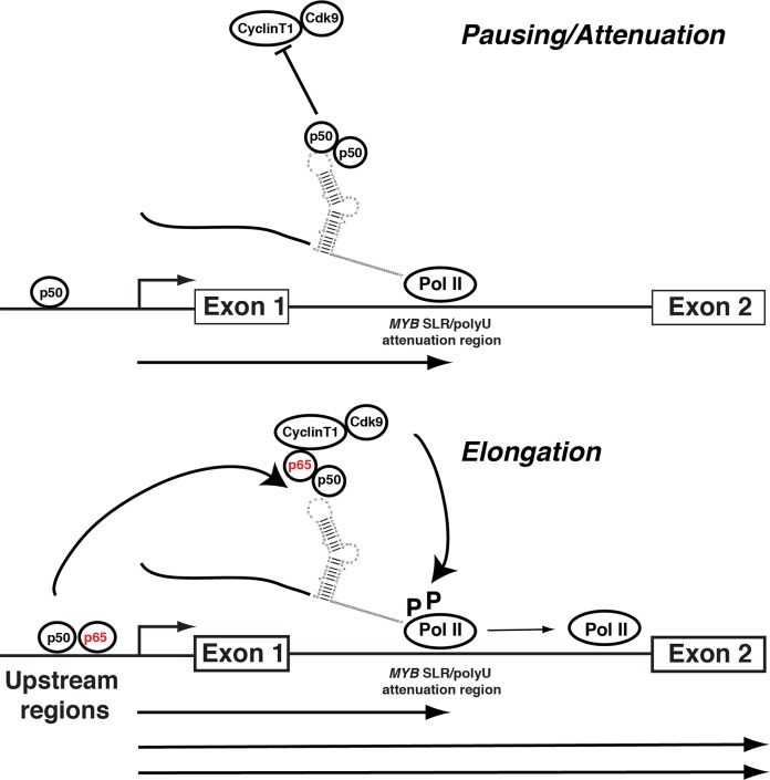
MYB transcriptional elongation is regulated by an attenuator sequence within intron 1 that has been proposed to encode a RNA stem loop (SLR) followed by a polyU tract. We report that NFκBp50 can bind the SLR polyU RNA and promote MYB transcriptional elongation together with NFκBp65. We identified a conserved lysine-rich motif within the Rel homology domain (RHD) of NFκBp50, mutation of which abrogated the interaction of NFκBp50 with the SLR polyU and impaired NFκBp50 mediated MYB elongation. We observed that the TAR RNA-binding region of Tat is homologous to the NFκBp50 RHD lysine-rich motif, a finding consistent with HIV Tat acting as an effector of MYB transcriptional elongation in an SLR dependent manner. Furthermore, we identify the DNA binding activity of NFκBp50 as a key component required for the SLR polyU mediated regulation of MYB. Collectively these results suggest that the MYB SLR polyU provides a platform for proteins to regulate MYB and reveals novel nucleic acid binding properties of NFκBp50 required for MYB regulation.


Conflict of interest statement

Competing Interests: The authors have declared that no competing interests exist.

Figures

Fig 1
Fig 1. Cellular proteins bind to the MYB SLR polyU RNA.
(A) MYB SLR polyU structure analysis. The mfold predicted structure of the MYB SLR polyU is shown. (B) Electrophoretic mobility of radiolabeled MYB SLR polyU RNA transcripts (MYB SLR polyU; 310 bases), polyU tract-deleted (MYB SLR ΔpolyU; 291 bases), stem-loop deleted (MYB ΔSLR polyU; 249 bases) and 9 of the 19 polyU residues deleted (MYB SLR Δ9 polyU; 300 bases). RNAs were subjected to electrophoresis in a denaturing 4% acrylamide gel where probes migrate according to size and a 4% non-denaturing acrylamide gel where secondary structure is maintained. (C) LIM1215 whole cell extracts were incubated with radiolabeled RNA probes generated from pBluescript II KS MYB SLR polyU, polyU tract deleted (MYB SLR ΔpolyU) and stem-loop region deleted (MYB ΔSLR polyU) templates and subjected to UV-cross linking followed by SDS-PAGE. Two species (~50 kDa, black arrow and ~ 38 kDa, grey arrow) were observed with the MYB SLR polyU probe, and one species (~ 38 kDa, grey arrow) with the MYB SLR ΔpolyU probe. A ~20 kDa doublet is also evident with the MYB ΔSLR polyU and MYB SLR ΔpolyU probes, white arrow. (D) LIM1215 whole cell extract was depleted of NFκBp50 by incubation with NFκBp50 antibody. Depletion of NFκBp50 was confirmed by Western blot analysis. Depleted extracts were subsequently incubated with radiolabeled MYB SLR polyU RNA probe and subjected to UV-cross linking followed by SDS-PAGE. Two phosphorimaging exposures of the same SDS-PAGE gel are shown to highlight the reduction in the 50kDa signal as indicated by the asterisk. Phosphorimaging quantitation confirmed reduction of the 50kDa signal. Error bars represent mean ± SEM, ** P <0.01.
Fig 2
Fig 2. NFκBp50 binds directly to the MYB SLR polyU.
(A) Radiolabeled RNA probes generated from pBluescript II KS MYB SLR polyU, polyU tract-deleted (MYB SLR ΔpolyU) or stem-loop region deleted (MYB ΔSLR polyU) templates were incubated with 50 ng of NFκBp50 or in the case of the MYB SLR ΔpolyU probe with 25 ng and 50 ng NFκB p50 and the reactions resolved on a 5% Tris-glycine gel. Coomassie gel and Western blot analysis of the NFκBp50 protein are shown. (B) Radiolabeled RNA probes generated from pGEM-MYB SLR polyU or MYB SLR ΔpolyU were incubated with 50 ng of NFκBp50 and the reactions resolved on a 5% Tris-glycine gel. (C) Left; an RNA probe generated from pGEM-MYB SLR polyU was incubated with 50 ng of NFκBp50 and NFκBp50-MYB SLR polyU RNA-protein complexes were supershifted by the addition of anti-NFκBp50 antibody. Anti-NFκBp65 antibody was used as a control. Right; RNA shifts were performed as above and the reactions subjected to UV-cross linking and SDS-PAGE. (D) NFκBp50 RHD analysed by Coomassie staining and Western blot analysis. (E) Left; an RNA probe generated from pBluescript II KS MYB SLR polyU was incubated with increasing amounts of recombinant NFκBp50 RHD and the reactions resolved on a 5% Tris-glycine gel. Right; an RNA probe generated from pBluescript II KS MYB SLR polyU was incubated with recombinant NFκBp50 RHD and NFκBp50-MYB SLR polyU RNA-protein complexes were super-shifted by the addition of anti-NFκBp50 antibody. Anti-rabbit IgG was used as a control. In (A-E) the black arrows indicate the position of the NFκBp50-MYB polyU SLR or NFκBp50-MYB SLR ΔpolyU RNA complexes; white arrows show the position of the complexes in the presence of the anti-p50 antibody; grey arrow indicates free MYB polyU SLR or MYB SLR ΔpolyU RNA probes.
Fig 3
Fig 3. The NFκBp50 RHD binds the MYB SLR polyU RNA via a lysine-rich region.
(A) Sequence alignment of the RHD of NFκB proteins. Black boxes indicate amino acid identity and grey boxes indicate similarity. The numbers refer to the first position of the segments within the respective proteins. The asterisk refers to residues that were analyzed by mutagenesis. (B) Wild type (wt) and mutant NFκBp50 RHDs analyzed by Coomassie staining. (C) Radiolabeled RNA probe generated from pBluescript II KS MYB SLR polyU template was incubated with 12.5 ng of recombinant wt or mutant NFκBp50 RHD as indicated and the reactions resolved on a 5% Tris-glycine gel. The black arrow indicates the position of the NFκBp50 RHD-MYB SLR polyU RNA complex; the grey arrow indicates free probe.
Fig 4
Fig 4. NFκBp50 and NFκBp65 induce MYB elongation via the MYB SLR polyU.
(A) Top panel: The 5´ genomic structure of MYB and the CAT reporter constructs is depicted. MYB ΔSLR polyU CAT has a 76 bp deletion of the SLR sequence up to the 19 nucleotide polyU stretch. MYB SLR ΔpolyU CAT contains a deletion of the 19 nucleotide polyU stretch. Bottom left panel: Transactivation studies in 293 cells using 2 μg of the MYB SLR polyU CAT reporter and 0.5 μg of pcDNA NFκBp50. Bottom right panel: Transactivation studies in 293 cells using 2 μg of the MYB SLR polyU CAT, MYB ΔSLR polyU CAT or MYB SLR ΔpolyU CAT reporters and 0.5 μg of pcDNA NFκBp65. (B) Left panel: Transactivation studies in 293 cells using 2 μg of the MYB SLR polyU CAT, MYB ΔSLR polyU CAT or MYB SLR ΔpolyU CAT reporters and 0.25 μg of pcDNA NFκBp50-p65; Right panel: Transactivation studies in 293 cells using 2 μg of the MYB SLR polyU CAT or MYB Promoter CAT reporters and 0.25 μg of pcDNA NFκBp50-p65. (C) Transactivation studies in 293 cells using 2 μg of the MYB SLR polyU CAT, MYB SLR 3L mutation polyU CAT, MYB SLR 5C mutation polyU CAT or MYB SLR 23C mutation polyU CAT reporters with 0.25 μg of pcDNA NFκBp50-p65. (D) Transactivation studies in 293 cells using 2 μg of the MYB SLR polyU CAT reporter with 0.25 μg of pcDNA NFκBp50-p65, 0.25 μg of pcDNA NFκBp50 K148A-p65 or 1 μg of pcDNA NFκBp50 K146-148A-p65. Error bars represent mean ± SEM, * P <0.05, ** P <0.01, *** P <0.001, **** P <0.0001.
Fig 5
Fig 5. NFκBp50-p65, P-TEFb and TNFα influence MYB elongation.
(A) 293 cells were treated with the P-TEFb inhibitor DRB for 6 h and endogenous MYB expression assessed by QPCR. (B) Transactivation studies in 293 cells using 2 μg of the MYB SLR polyU CAT reporter and 0.125 μg of pcDNA NFκBp50-p65. At 12 h post transfection cells were treated with DRB and incubated for a further 24 h. (C) NFκBp50-p65 induces endogenous MYB. Total RNA was isolated from 293 cells transfected with; 1 μg of pcDNA NFκB p50-p65, 1 μg of pcDNA NFκBp50 K148A-p65 or 4 μg of pcDNA NFκBp50 K146-148A-p65 and analyzed by Q-PCR to measure MYB expression levels and (D) 1 μg of pcDNA NFκB p50-p65 and analyzed by Q-PCR to measure intronic pre-mRNA MYB transcript upstream (preSLR) and downstream (post SLR) of the MYB SLR polyU. Data are expressed as a ratio “post/pre”, a measure of the amount of transcription through the SLR. (E) ChIP analysis of RNA polymerase II levels at the MYB SLR polyU. 293 cells were transfected with pcDNA, NFκB p50-p65 or pcDNA NFκBp50 K146-148A-p65. Cross-linked chromatin extracts were prepared at 48h post transfection and RNA polymerase II was detected by anti-pol II followed by Q-PCR. (F) SI organoids cultures and 293 cells were exposed to TNFα (20–100 ng/ml) for the times indicated and total RNA was isolated and analyzed by Q-PCR to measure MYB expression levels. (G) 293 cells were transfected with 2 μg of the MYB SLR polyU CAT or MYB ΔSLR polyU CAT reporter. At 24 h post transfection cells were exposed to 100 ng/ml TNFα for 10 h and CAT reporter activity assessed. (H) 293 cells were exposed to BAY inhibitor for the times indicated and total RNA was isolated and analyzed by Q-PCR to measure MYB expression levels. Error bars represent mean ± SEM, * P <0.05, ** P <0.01, *** P <0.001, **** P <0.0001.
Fig 6
Fig 6. HIV-Tat induces MYB expression and transcriptional elongation.
(A) The RNA stem loop structure of the HIV TAR [26]. Sequence alignment of the TAR binding region of HIV-1 Tat with the RHD of NFκBp50. Black boxes indicate amino acid identity and grey boxes indicate similarity. The Tat RNA binding domain (ARM motif) is indicated by the black line. The numbers refer to the first position of the segments within the respective proteins. The asterisk refers to NFκBp50 residues that were analyzed by mutagenesis. (B) The genomic arrangement of the MYB locus. The anti-sense RNA probes, the location of the SLR region within intron 1 and the location of RT-PCR primers used to examine RNA elongation across this region by nuclear run-on transcription are shown. (C) Transcription of the MYB gene in 293 cells as assessed by nuclear run-on transcription. Nuclear run on assays were performed as described [20, 21]. Transcriptional activity was normalized to GAPDH signal and the steady state rate of transcription for each transcript length. Relative transcription between untransfected cells and cell transfected with Tat is shown. Densitometric analysis of the radioactivity bound to the filters was performed using Imagequant software and represents the mean values obtained from duplicate filters. (D) Nuclear RNA was isolated from 293 cells transfected with 100 ng pCMVTat (72) and subjected to RT-PCR to detect intron 1 RNA pre- and post-MYB SLR attenuator region.
Fig 7
Fig 7. HIV-Tat increases MYB reporter activity via the intron 1 MYB SLR.
(A) Transactivation studies in 293 cells using: Left panel, 2 μg of MYB SLR polyU CAT or MYB ΔSLR polyU CAT and 2 μg of pCMV Tat (101). Right panel, 2 μg of MYB SLR polyU CAT or MYB TAR CAT and 2 μg of pCMV Tat (101). (B) Transactivation studies in 293 cells using 2 μg of MYB SLR polyU CAT reporter, 1.5 μg pCMV Tat (72) and 1.5 μg of pCMVCDK9 or pCMVDNCDK9. (C) TAR or bulge mutant (T+23A) TAR RNA probes were incubated with 25, 50 and 100 ng of Tat and reactions resolved on a 5% Tris-glycine gel. The black arrow indicates the position of the Tat-TAR RNA complex; the grey arrow indicates free TAR RNA probe. Coomassie gel and Western blot analysis confirming the expression and integrity of Tat is shown. (D) Binding of Tat to the MYB SLR polyU. RNA probes were generated from pGEM-3Zf MYB SLR polyU, MYB SLR ΔpolyU or MYB ΔSLR polyU templates, incubated with 50 ng of Tat and reactions resolved on a 5% Tris-glycine gel. Tat-MYB SLR polyU or Tat-MYB SLR RNA-protein complexes were super-shifted by the addition of anti-Tat antibody (100 ng). A mouse isotype control IgG (100 ng) was used as a control. The black arrows indicate the position of the Tat-MYB SLR polyU or Tat-MYB SLR RNA-protein complexes; the white arrows show the position of this complex in the presence of an anti-Tat antibody; the grey arrow indicates free MYB SLR polyU, MYB SLR ΔpolyU or MYB ΔSLR polyU probe. Error bars represent mean ± SEM, * P <0.05, ** P <0.01, **** P <0.0001.
Fig 8
Fig 8. Model for the NFκBp50 and p65 regulation of MYB elongation through the intron 1 MYB SLR polyU region and upstream sequences.
NFκBp50 binding at upstream sequences stimulates transcription from the multiple start sites. Transcripts are then paused/attenuated at the downstream MYB SLR polyU. In the absence of NFκBp65, NFκBp50 occupies the MYB SLR polyU region and MYB transcription is paused. In contrast the formation of a NFκBp50-p65 heterodimer on the MYB SLR polyU contributes to the stimulation of MYB transcription elongation by the NFκBp65-mediated recruitment of P-TEFb and the subsequent Ser2 phosphorylation of Pol II CTD (elongating form) by CDK9. This model is consistent with i) previous findings that NFκBp65 mediates transcriptional elongation through the direct recruitment of P-TEFb [50] and ii) our recent observations that ERα recruits P-TEFb to a region near the MYB SLR polyU and that this interaction is concordant with the accumulation of Ser2 CTD phosphorylated pol II bound to this region and the relief of MYB transcriptional attenuation [22]. This model also raises the possibility that the regulation of MYB may also involve the interaction of upstream bound NFκBp50/65 with the downstream intron 1 MYB SLR polyU. Recent data indicate that enhancer elements loop towards murine Myb intron 1 to bring the transcription apparatus to the vicinity of the pausing region and regulate Myb attenuation/elongation [19] and we have shown that the upstream regions of MYB lie in proximity to the MYB SLR polyU S5 Fig Furthermore, recent structure based analyses of HIV-1 LTR interactions suggest that pre-formed NFκBp50-DNA complexes can interact with downstream HIV TAR RNA [48].

Similar articles

Cited by

References

    1. Ramsay RG, Gonda TJ. MYB function in normal and cancer cells. Nat Rev Cancer. 2008;8: 523–534. 10.1038/nrc2439 - DOI - PubMed
    1. Drabsch Y, Hugo H, Zhang R, Dowhan DH, Miao YR, Gewirtz AM, et al. Mechanism of and requirement for estrogen-regulated MYB expression in estrogen-receptor-positive breast cancer cells. Proc Natl Acad Sci U S A. 2007;104: 13762–13767. - PMC - PubMed
    1. Miao RY, Drabsch Y, Cross RS, Cheasley D, Carpinteri S, Pereira L, et al. MYB is essential for mammary tumorigenesis. Cancer Res. 2011;71: 7029–37. 10.1158/0008-5472.CAN-11-1015 - DOI - PubMed
    1. Biroccio A, Benassi B, D'Agnano I, D'Angelo C, Buglioni S, Mottolese M, et al. c-Myb and Bcl-x overexpression predicts poor prognosis in colorectal cancer: clinical and experimental findings. Am J Pathol. 2001;158: 1289–1299. - PMC - PubMed
    1. Ciznadija D, Tothill R, Waterman ML, Zhao L, Huynh D, Yu RM, et al. Intestinal adenoma formation and MYC activation are regulated by cooperation between MYB and Wnt signaling. Cell Death Differ. 2009;16: 1530–1538. 10.1038/cdd.2009.94 - DOI - PubMed

Publication types