

Review
doi: 10.1038/jcbfm.2015.60.
Epub 2015 Apr 8.
Diverse functions of pericytes in cerebral blood flow regulation and ischemia
Affiliations
- PMID: 25853910
- PMCID: PMC4640260
- DOI: 10.1038/jcbfm.2015.60
Item in Clipboard
Review
Diverse functions of pericytes in cerebral blood flow regulation and ischemia
J Cereb Blood Flow Metab.
2015 Jun.
Abstract
Pericytes are mural cells with contractile properties. Here, we provide evidence that microvascular pericytes modulate cerebral blood flow in response to neuronal activity ('functional hyperemia'). Besides their role in neurovascular coupling, pericytes are responsive to brain damage. Cerebral ischemia is associated with constrictions and death of capillary pericytes, followed by fibrotic reorganization of the ischemic tissue. The data suggest that precapillary arterioles and capillaries are major sites of hemodynamic regulation in the brain.
Figures
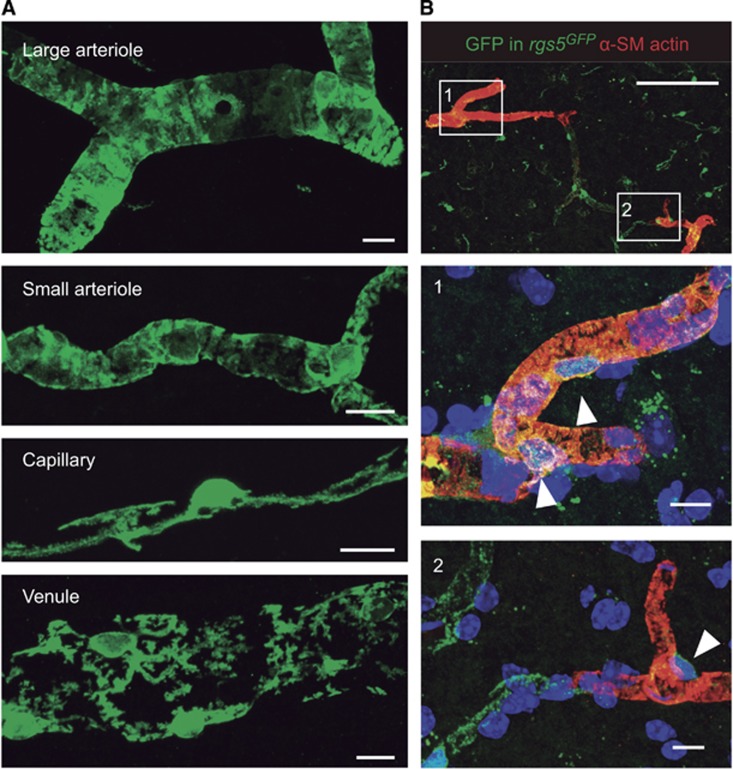
Different mural cell phenotypes in the brain microcirculation. (A) In Sox10-iCreERT2 mice crossed to a green fluorescent protein (GFP) Cre-reporter strain, neural crest-derived vascular mural cells, including smooth muscle cells and pericytes, are genetically labeled with GFP. Large arterioles, including penetrating vessels, and their smaller branches show a complete coverage of the vessel lumen with robust circumferential mural cell processes. In contrast, capillary pericytes extend longitudinal processes that do not completely cover the endothelial tube. Pericytes in venules show a distinct stellate morphology. (B) In rgs5GFP mice, pericytes express GFP. Double staining for GFP (green) and α-smooth muscle (SM) actin (red) reveals the circumferential contractile machinery of pericytes (arrowheads) in penetrating vessels (1). An abrupt decrease in α-SM actin immunoreactivity is found in downstream vessels, including capillaries (2). Scale bars: 10 μm (A, B) middle and bottom panels), 100 μm (B, top panel).
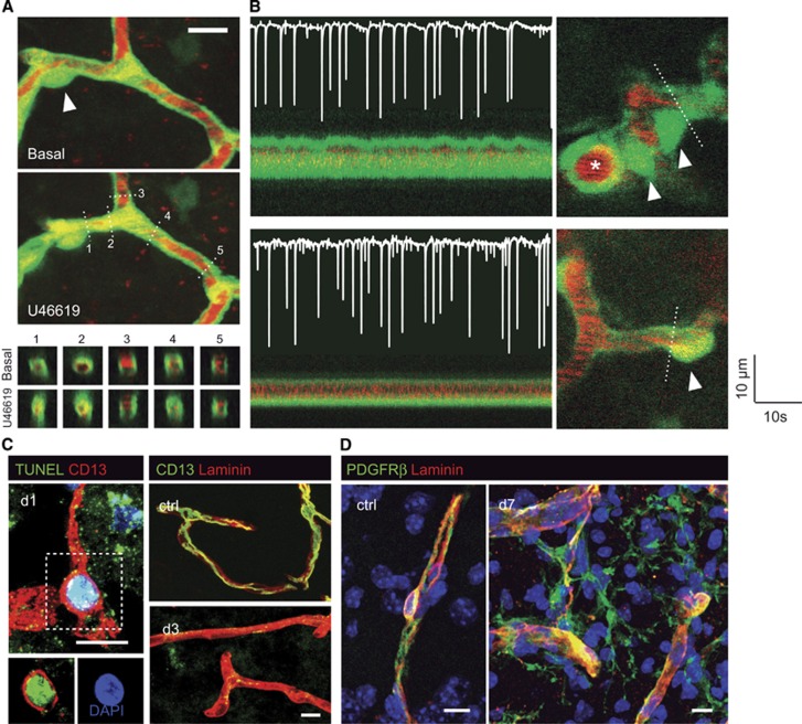
Contractility of the deep brain microcirculation. (A) Two-photon images showing the constriction of a capillary by the thromboxane A2 receptor agonist, U46619, in GFP transgenic mice in vivo. The plasma is labeled with TRITC (red). Note the collapse of the capillary segment in the vicinity of the pericyte body (arrowhead), while some blood flow is maintained in the capillary to the right. The bottom panels represent orthogonal reconstructions showing vessel cross-sections at the planes indicated by dashed lines. (B) Responses to neuronal activity in the first branches of a penetrating arteriole (top panel) and a downstream capillary (bottom panel). The images on the left trace the oscillations of vascular diameter observed at the planes indicated by dashed lines in the images on the right. Arrowheads point at pericyte bodies. The superimposed electrocorticogram recordings on the left show neuronal spikes elicited by the microinfusion of bicuculline into the cortex. Dilatations are present in the branches of the penetrating arteriole (top panel, marked with an asterisk), but not in capillaries (bottom panel). (C) Pericyte loss and stromal proliferation after cerebral ischemia. Double CD13+ TUNEL+ pericyte in the lesioned tissue 1 day after ischemia, indicative of DNA damage (left panel). Compared with nonischemic tissue, laminin-immunoreactive vascular segments lack CD13+ immunoreactivity (right panel). (D) PDGFRβ+ pericyte in nonischemic parenchyma (left panel). Proliferation of vessel-associated PDGFRβ+ stromal cells in the ischemic parenchyma 7 days after the insult (right panel). Scale bars: 10 μm.
References
-
- 2Woolsey T, Rovainen C, Cox S, Henegar M, Liang G, Liu D et al. Neuronal units linked to microvascular modules in cerebral cortex: response elements for imaging the brain. Cereb Cortex 1996; 6: 647–660. - PubMed
-
- 5Armulik A, Genové G, Betsholtz C. Pericytes: developmental, physiological, and pathological perspectives, problems, and promises. Dev Cell 2011; 21: 193–215. - PubMed
Publication types
MeSH terms
LinkOut - more resources
Full Text Sources
Other Literature Sources
