. 2015 Apr 9:6:6775.
doi: 10.1038/ncomms7775.

Borealin dimerization mediates optimal CPC checkpoint function by enhancing localization to centromeres and kinetochores

Affiliations

Borealin dimerization mediates optimal CPC checkpoint function by enhancing localization to centromeres and kinetochores

Michael E Bekier et al. Nat Commun. .

Abstract

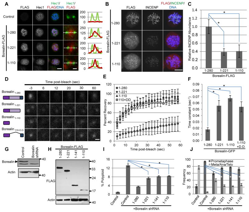
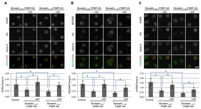
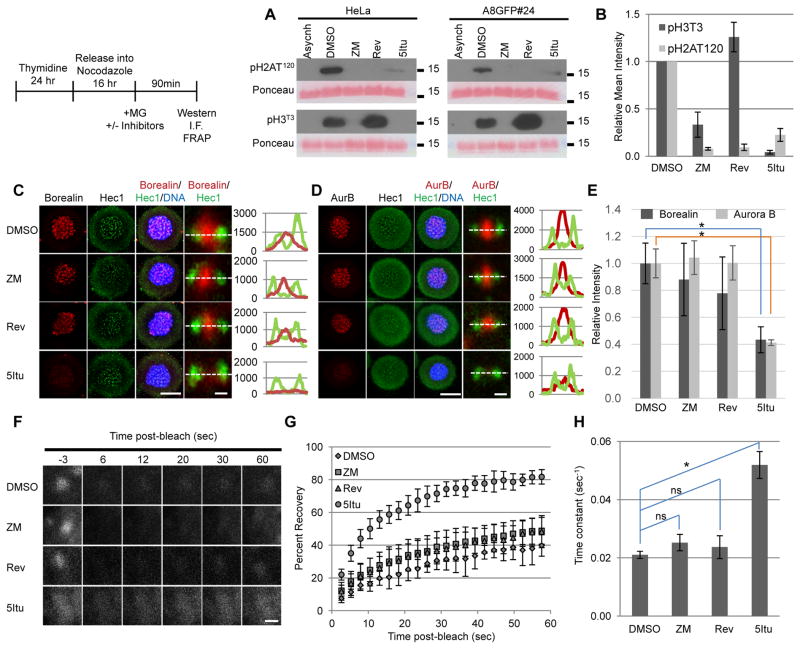
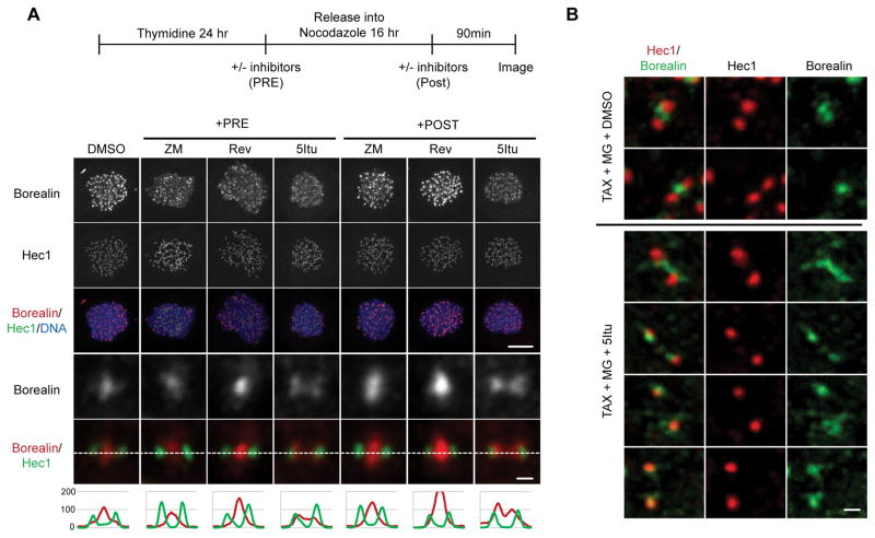
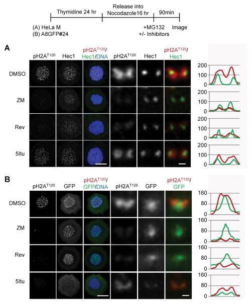
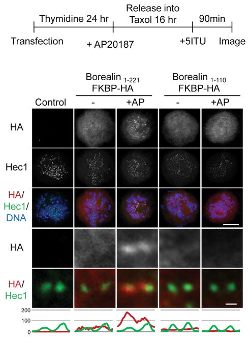
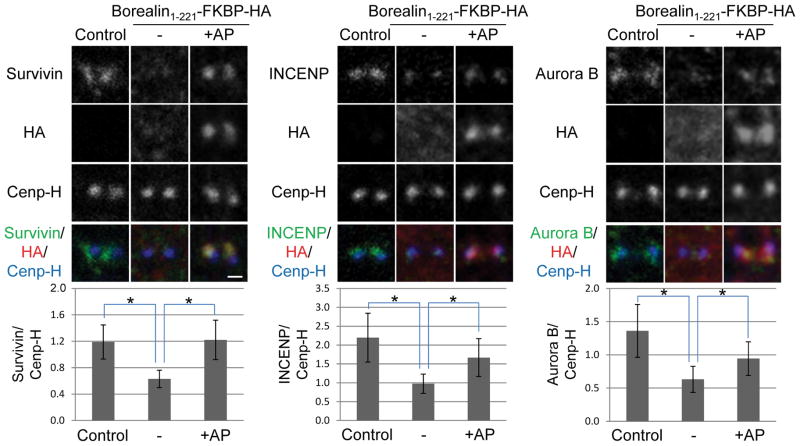
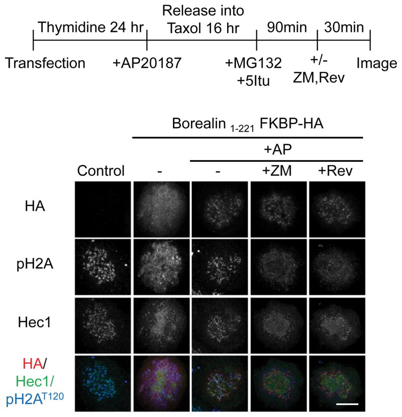
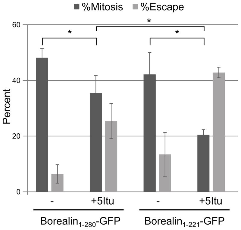
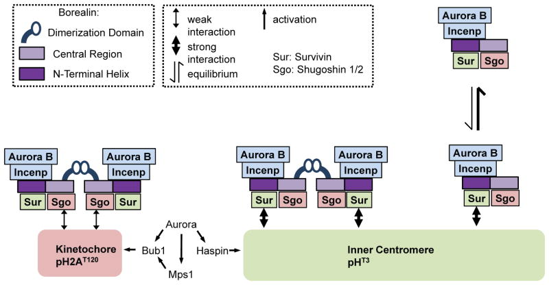
The chromosomal passenger complex (CPC) localizes to centromeres where it activates the mitotic checkpoint in response to inappropriate inter-kinetochore tension. This error correction function is essential for proper chromosome segregation. Here we define several critical features of CPC localization and function. First, the Borealin dimerization domain suppresses dynamic exchange at the centromere to allow optimal CPC function. Second, Borealin dimerization is essential to target a subpopulation of CPC proximal to the kinetochore when the mitotic spindle is disrupted. This subpopulation is also needed for full CPC checkpoint function. The existence of a pool of CPC at the kinetochore suggests that error correction is more complicated than predicted from the Aurora B phosphorylation gradient model. Finally, Haspin kinase plays a key role in maintaining the slowly exchanging centromere Borealin pool, while Aurora B and Mps1 play minimal roles in maintaining CPC localization once cells are in mitosis.


Conflict of interest statement

Competing financial interests

The authors declare no competing financial interests.

Figures

Figure 1
Figure 1. Borealin structure-function analysis
A.) Immunofluorescence using antibodies to FLAG and Hec1 in HeLa M cells transfected with FLAG-tagged Borealin and then blocked in taxol. Whole cell maximum projections (scale bar = 10μm), individual sister kinetochores (scale bar = 0.5μm) and line scans of Hec1 and Flag intensity in kinetochore images are shown. B.) Maximum projections from cells treated as in A and stained with antibodies against FLAG and INCENP (scale bar = 10μm). C.) Intensity of INCENP in the DNA area of greater than 30 cells. Pixel intensities in INCENP images are relative to the average level of INCENP in untransfected samples from the same experiment. Bars represent standard deviation. D.) Representative still images of GFP foci from FRAP experiments in nocodazole-arrested HeLa M cells after transient transfection with GFP-tagged Borealin truncations (scale bar = 1μm). E.) Post-bleach recovery of GFP-Borealin measured by FRAP. Averages and standard deviations of more than eight centromeres per condition are shown. F.) Average time-constant values of FRAP curves from E (see methods). G.) Borealin knockdown in HeLa M cells after transient transfection with pBabePURO, and either empty pSUPER (Control) or pSUPER-Borealin shRNA. Cell lysates obtained after puromycin selection to remove untransfected cells were analyzed by western blotting with antibodies against Borealin and Actin. H.) Expression of FLAG-tagged Borealin truncations. HeLa M cells were transfected with pSUPER-Borealin shRNA, pBABEpuro along with FLAG-tagged forms of Borealin. FLAG and Actin were detected by western analysis. I.) Percentage of cells with greater than 4N DNA content (Polyploid). HeLa M cells were transfected with pSUPER-Borealin shRNA, pBABEpuro and FLAG-tagged Borealin, exposed to puromycin and analyzed by flow cytometry for DNA content 4 days post-transfection. J.) Frequency of mitotic phases in Borealin-knockdown cells expressing truncated forms of Borealin. HeLa M cells were transfected with pSUPER-Borealin shRNA, a plasmid encoding H2BGFP and Borealin truncations. Three days post-transfection, mitotic phases of GFP-positive cells were determined. Scoring was carried out in a blinded manner (n≥100 for each condition). Bars from I and J represent standard error from three independent transfections. In sections C, F, I and J the asterisk (*) indicates p<0.05 using a student’s t-test.
Figure 2
Figure 2. Borealin dimerization regulates CPC abundance at centromeres
HeLa M cells were transfected with the indicated constructs and then exposed to thymidine for 24 hours to synchronize in S phase. At the time of thymidine removal, taxol was added in combination with AP20187 (+AP) or DMSO (−). Cells were fixed 16 hours later and analyzed by immunofluorescence. Intensity of centromere staining of Survivin (A), INCENP (B) and Aurora B (C) was quantified relative to the kinetochore marker CenpH (scale bar = 20μm). Bars represent standard deviation from averages of at least 50 centromere signals from 10 different cells per condition. *p<0.05 using a student’s t-test.
Figure 3
Figure 3. Inner centromere localization of Borealin is regulated by Haspin kinase activity
HeLa M cells were synchronized and treated as shown in the schematic. Briefly, cells released from a thymidine block in the presence of nocodazole to synchronously blocked them in mitosis. Mitotic cells were then exposed to MG132 to prevent mitotic exit and either DMSO, ZM447439 (ZM), reversine (Rev), or 5Itu. Ninety minutes after adding MG132 with or without inhibitors, cells were analyzed by western blotting (A, B), immunofluorescence (“I.F.” C, D, E) or FRAP (F, G, H). A.) Western blot analysis of histone extracts. Histones were extracted and analyzed by western blotting with the antibodies against pH2AT120 and pH3T3. Ponceau S staining served as loading control. Asynchronously growing cells (Asynch) were used as a negative control. B.) Quantification of band intensities from three independent western blots prepared as in A. C&D.) Maximum projections of HeLa M cells treated as described in the schematic. Immunofluorescence was performed using antibodies to either Borealin or Aurora B and Hec1. Hoechst was used to visualize DNA. Whole cell maximum projections (scale bar = 10μm) are shown with close-up images and line scans of individual sister kinetochores/centromeres from a single plane to the right (scale bar = 0.5μm). E.) Quantification of the relative level of Borealin or Aurora B intensity in DNA area of max projections from C and D, respectively. Greater than 30 cells were quantified. F.) Representative still images of single centromeres from A8GFP#24 cells stably expressing Borealin-GFP treated as shown in the schematic (scale bar = 1μm). G.) Post-recovery kinetics of Borealin-GFP under the indicated conditions. More than eight centromeres per condition were averaged in a single experiment. H.) The average time-constant values of FRAP curves from G (see methods). In sections B, E and H, bars represent standard deviations and the asterisk (*) indicates p<0.05 using a student’s t-test.
Figure 4
Figure 4. Kinetochore-proximal Borealin pool observed after Haspin inhibition
A.) Borealin localization compared to Hec1. Cells were prepared as shown in the schematic. Maximum projection images of nocodazole-arrested HeLa M cells treated with the indicated inhibitors either before (+PRE) or after (+POST) mitotic entry are shown. Immunofluorescence was performed using antibodies against endogenous Borealin and Hec1. Examples of whole cell maximum projections (scale bar = 10μm) are shown. Close-up images and line scans of individual sister kinetochores/centromeres from a single plane are at the bottom (scale bar = 0.5μm). B.) Co-localization of Borealin and Hec1 in taxol arrested cells exposed to 5Itu. HeLa M cells were exposed to taxol for 6 hours, and the with MG132 with or without 5Itu. Immunofluorescence carried out 90 minutes post-5Itu addition. Confocal images were obtained with a 0.1μm z-step followed by deconvolution using AutoQuant X3 software. Projection images are shown (scale bar = 0.5μm).
Figure 5
Figure 5. Kinetochore localization of Borealin overlaps with pH2AT120
HeLa M cells or a stable clone of HeLa M expressing Borealin-GFP (A8GFP#24) were treated as indicated in the schematic. Briefly, cells were released from a thymidine block in the presence of nocodazole. Sixteen hours later when most cells were blocked in mitosis, DMSO, ZM447439 (ZM), reversine (Rev) or 5Itu was added. MG132 was added under all conditions to block mitotic exit. Cells were fixed 90 minutes after inhibitors were added, stained with the indicated antibodies and analyzed by confocal microscopy. A.) Partial overlap between pH2AT120 with Hec1 in HeLa M cells exposed to 5Itu. Examples of maximum projections are shown (scale bar = 10μm). Close-up images and line scans of individual sister kinetochores/centromeres from a single plane are to the right (scale bar = 0.5μm). B.) Partial co-localization of Borealin-GFP with pH2AT120 in cells blocked in 5Itu. A8GFP#24 cells were treated as indicated in the schematic and stained with antibodies to GFP to enhance detection as well as pH2AT120. Examples of maximum projections (scale bar = 10μm) and kinetochore pairs (scale bar = 0.5μm) are shown.
Figure 6
Figure 6. Borealin dimerization is essential for localization near the kinetochore
HeLa M cells were transiently transfected with Borealin-FKBP fusions, synchronized as shown and exposed to the homo-dimerizing agent AP20187 (+AP) or DMSO (−). All samples include 5Itu to reveal the kinetochore pool. Immunofluorescence was carried out using antibodies to HA to detect the Borealin-FKBP fusions, as well as Hec1 to stain the kinetochore. Examples of maximum projection images are show in the top three rows (scale bar = 10μm). Close-up images and line scans of individual sister kinetochores/centromeres from a single plane are also shown (scale bar = 0.5μm).
Figure 7
Figure 7. Monomeric but not dimeric Borealin1-221-FKBP reduces CPC subunits at the kinetochore
HeLa M cells were synchronized and treated as shown in the schematic to figure 5. However, when fixed, the cells were stained with antibodies to Survivin, INCENP, Aurora B, CenpH to mark the kinetochore, and HA to detect the Borealin-FKBP fusions. As in figure 5, all cells were exposed to 5Itu to remove the inner centromere pool and reveal the kinetochore pool. Examples of close-up confocal images of individual sister kinetochores/centromeres are shown for all conditions (scale bar = 0.5μm). Intensity of CPC family members was quantified and is shown relative to CenpH at individual kinetochores. At least 50 kinetochores from 10 different cells were analyzed. Bars represent standard deviation. *p<0.05 using a student’s t-test.
Figure 8
Figure 8. Kinetochore Borealin can be uncoupled from pH2AT120
HeLa M cells were synchronized and treated as shown in the schematic. Briefly, cells were transfected with the indicated Borealin-FKBP and synchronously released into a taxol block. All cells received AP20187 to induce dimerization. Once cells were in mitosis, 5Itu was added for 90 minutes. Then, either ZM447439 (ZM) or reversine (Rev) was added for an additional 30 minutes before cells were fixed. Immunofluorescence was carried out with antibodies to pH2AT120, Hec1, or HA to detect the Borealin-FKBP fusion. Maximum projection images are shown (scale bar = 10μm).
Figure 9
Figure 9. Checkpoint function of a kinetochore-proximal CPC pool
HeLa M cells were transfected and synchronized into S-phase by 24-hour treatment with 2mM thymidine. Following synchronization, cells were released into taxol for 16 hours and time-lapse imaging was performed immediately after adding either DMSO (−) or 5Itu. The percentage of Borealin-GFP expressing cells remaining in mitosis or having escaped mitosis by 15-hours after 5Itu treatment is shown. Bars represent standard error from three independent transfections. *p<0.05 from a student’s t-test.
Figure 10
Figure 10. Model for the role of dimerization in CPC interactions with the centromere
Dimeric CPC stably binds at inner centromeres possibly via two Survivin-pH3T3 interactions mediated by Borealin dimerization. Removing the dimerization domain of Borealin results in rapid exchange at inner centromeres since monomeric CPC might only contact a single pH3T3 molecule. When endogenous Borealin is knocked-down, Borealin mutants lacking dimerization transiently associate with the centromere to provide partial function. However, when endogenous Borealin is present, dimerization-defective mutants compete for other CPC family members to increase their exchange and act as dominant negatives. At the kinetochore, dimeric CPC binds with a high rate of turnover potentially via dual pH2AT120-Sgo1/2 interactions. These kinetochore-interactions are too weak to allow association in the context of a monomeric CPC. According to the prevailing model of CPC targeting, CPC interacts simultaneously with pH3T3 via Survivin and pH2AT120 via Sgo1/2. Our results do not preclude simultaneous binding, but do suggest that pH3T3 would contribute most of the binding energy if this binding mode occurs. Theoretically, CPC monomers or dimers could simultaneously interact with single pH3T3 and pH2AT120.

Similar articles

Cited by

References

    1. Ricke RM, van Deursen JM. Aneuploidy in health, disease, and aging. The Journal of cell biology. 2013;201:11–21. - PMC - PubMed
    1. Duesberg P, Rausch C, Rasnick D, Hehlmann R. Genetic instability of cancer cells is proportional to their degree of aneuploidy. Proceedings of the National Academy of Sciences of the United States of America. 1998;95:13692–13697. - PMC - PubMed
    1. Ricke RM, Jeganathan KB, van Deursen JM. Bub1 overexpression induces aneuploidy and tumor formation through Aurora B kinase hyperactivation. The Journal of cell biology. 2011;193:1049–1064. - PMC - PubMed
    1. Forment JV, Kaidi A, Jackson SP. Chromothripsis and cancer: causes and consequences of chromosome shattering. Nature reviews Cancer. 2012;12:663–670. - PubMed
    1. Jones MJ, Jallepalli PV. Chromothripsis: chromosomes in crisis. Developmental cell. 2012;23:908–917. - PMC - PubMed

Publication types

MeSH terms