. 2015 May;64(5):609-20.
doi: 10.1007/s00262-015-1664-x. Epub 2015 Feb 18.

Broadening the repertoire of melanoma-associated T-cell epitopes

Affiliations

Broadening the repertoire of melanoma-associated T-cell epitopes

Thomas Mørch Frøsig et al. Cancer Immunol Immunother. 2015 May.

Abstract

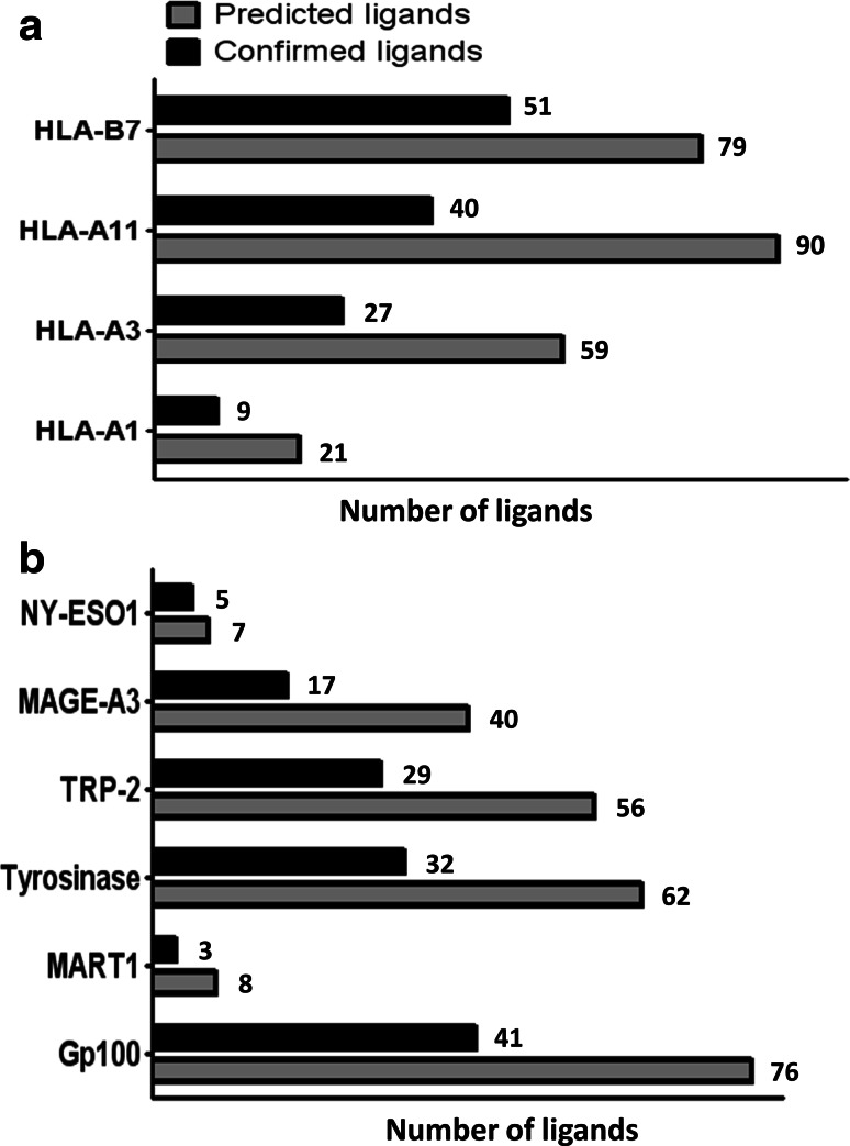
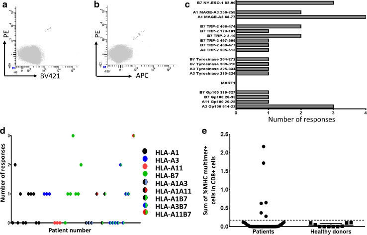
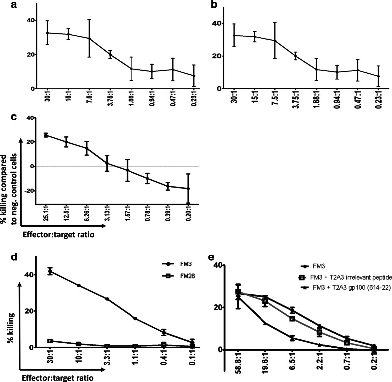
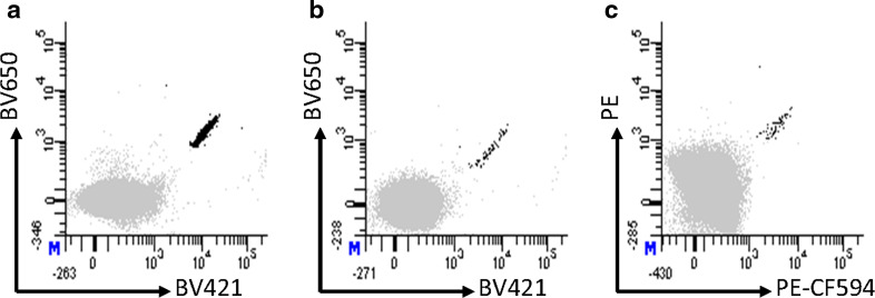
Immune therapy has provided a significant breakthrough in the treatment of metastatic melanoma. Despite the remarkable clinical efficacy and established involvement of effector CD8 T cells, the knowledge of the exact peptide-MHC complexes recognized by T cells on the tumor cell surface is limited. Many melanoma-associated T-cell epitopes have been described, but this knowledge remains largely restricted to HLA-A2, and we lack understanding of the T-cell recognition in the context of other HLA molecules. We selected six melanoma-associated antigens (MAGE-A3, NY-ESO-1, gp100, Mart1, tyrosinase and TRP-2) that are frequently recognized in patients with the aim of identifying novel T-cell epitopes restricted to HLA-A1, -A3, -A11 and -B7. Using in silico prediction and in vitro confirmation, we identified 127 MHC ligands and analyzed the T-cell responses against these ligands via the MHC multimer-based enrichment of peripheral blood from 39 melanoma patients and 10 healthy donors. To dissect the T-cell reactivity against this large peptide library, we used combinatorial-encoded MHC multimers and observed the T-cell responses against 17 different peptide-MHC complexes in the patient group and four in the healthy donor group. We confirmed the processing and presentation of HLA-A3-restricted T-cell epitopes from tyrosinase (TQYESGSMDK) and gp100 (LIYRRRLMK) and an HLA-A11-restricted T-cell epitope from gp100 (AVGATKVPR) via the cytolytic T-cell recognition of melanoma cell lines and/or K562 cells expressing the appropriate antigen and HLA molecule. We further found T-cell reactivity against two of the identified sequences among tumor-infiltrating lymphocytes from melanoma patients, suggesting a potential clinical relevance of these sequences.


Conflict of interest statement

No conflict of interest to declare.

Figures

Fig. 1
Fig. 1
MHC ELISA affinity testing of in silico predicted ligands. Peptides were selected according to the measured absorbance related to the rescue of the specific peptide-MHC complex after UV light-mediated peptide exchange. The absorbance level was calculated relative to a positive control virus-derived peptide and peptides were selected if this normalization value was higher than a given threshold in one of two independent experiments. a Shows the number of predicted and confirmed ligands distributed according to HLA restriction, and b shows the number of predicted and confirmed ligands distributed according to target the proteins
Fig. 2
Fig. 2
Detection of immune responses after MHC multimer-based enrichment. a, b Example of a confirmed response found with combinatorial encoding of MHC multimers detecting HLA-A3 Gp100614-22 reactive T cells, after a PE-MHC multimer-based enrichment of PBMCs. a 0.004 % of 270,000 CD8 cells detected in the PE/BV421 MHC multimer combination. b 0.004 % of 510,000 CD8 cells detected in the PE/APC MHC multimer combination. Only CD8 cells negative for all MHC multimer (gray) or specific for exactly two MHC multimers (black) are shown, the gating strategy is provided in Supplementary Figure 2. c The total number of T-cell responses detected per peptide sequence, grouped according to their protein origin. d The total number of responses detected per patient, when plotted according to patients’ HLA type. Each dot represents one patient. e The sum of melanoma-associated MHC multimer-specific T cells detected for each patient/healthy donor. The dotted line represents the mean frequency of responses found in the heathy donor cohort+ 3*standard deviation. Responses with higher frequency in the patient cohort are: HLA-B7/gp100319-327, HLA-B7/gp10026-35 and HLA-B7/NY-ESO82-90; HLA-A1/MAGE-A368-77 (2 responses); and HLA-A3/gp100614-22
Fig. 3
Fig. 3
Recognition of processed antigen in K562 cells or melanoma cell lines. a Lysis of K562/HLA-A3/gp100 cells compared with K562/HLA-A3 cells when co-cultured with increasing numbers of HLA-A3 gp100614-622-specific T cells (60.0 % of CD8 cells) in the given specific effector/target cell ratio, determined as the ratio between MHC multimer positive T cells and K562/HLA-A3/gp100 cells. b Lysis of K562/HLA-A3/tyrosinase cells compared with K562/HLA-A3 cells when co-cultured with increasing numbers of HLA-A3/tyrosinase325-334-specific T cells (81.7 % of CD8 cells) in the given effector:target cell ratio. c Lysis of K562/HLA-A11/gp100 cells compared with K562/HLA-A11 cells when co-cultured with increasing amounts of an HLA-A1/gp10020-28-specific T-cell culture (51.8 % of CD8 cells) in the given effector/target cell ratio. d Chromium-release cytotoxicity testing of the HLA-A3/gp100614-622 culture (60.0 % of CD8 cells) against gp100 positive and HLA-A3-positive (FM3) and HLA-A3-negative (FM28) melanoma cell lines. e Cytolytic recognition of the FM3 melanoma cell line and cold target inhibition of gp100614-22-specific T cell-mediated killing with HLA-A3-transduced T2 pulsed with either gp100614-22 or an irrelevant tyrosinase-derived peptide. Data shown in (ac) were measured with the VITAL-Far Red assay after 48 h of co-culturing of effector and target cells at 37 °C, and data shown in (d, e) were obtained with a standard Cr51-release assay after 4 h of co-culturing of effector and target cells at 37 °C. Shown in all plots is the relative lysis of positive target cells. The effector/target ratio is calculated based on MHC multimer-specific effector T cells. Bars represent the standard deviation of two independent experiments
Fig. 4
Fig. 4
T-cell responses in melanoma TILs detected by dual-color-coded MHC multimers. a 0.41 % HLA-B7/TRP-2466-474-specific T cells of 609.000 CD8 cells detected with the BV421:BV650 MHC multimer combination. b 0.037 % HLA-B7/TRP-2466-474-specific T cells of 176.000 CD8 cells detected with the BV421:BV650 MHC multimer combination, c 0.017 % HLA-B7/TRP-2497-506-specific T cells of 366.000 CD8 cells detected with the PE:PE-CF594 MHC multimer combination. All responses were additionally confirmed using another two-color combination

Similar articles

Cited by

References

    1. Hodi FS, O’Day SJ, McDermott DF, et al. Improved survival with ipilimumab in patients with metastatic melanoma. N Engl J Med. 2010;363:711–723. doi: 10.1056/NEJMoa1003466. - DOI - PMC - PubMed
    1. Topalian SL, Hodi FS, Brahmer JR, et al. Safety, activity, and immune correlates of anti-PD-1 antibody in cancer. N Engl J Med. 2012;366:2443–2454. doi: 10.1056/NEJMoa1200690. - DOI - PMC - PubMed
    1. Hamid O, Robert C, Daud A, et al. Safety and tumor responses with lambrolizumab (anti-PD-1) in melanoma. N Engl J Med. 2013;369:134–144. doi: 10.1056/NEJMoa1305133. - DOI - PMC - PubMed
    1. Brahmer JR, Tykodi SS, Chow LQM, et al. Safety and activity of anti-PD-L1 antibody in patients with advanced cancer. N Engl J Med. 2012;366:2455–2465. doi: 10.1056/NEJMoa1200694. - DOI - PMC - PubMed
    1. Wolchok JD, Kluger H, Callahan MK, et al. Nivolumab plus ipilimumab in advanced melanoma. N Engl J Med. 2013;369:122–133. doi: 10.1056/NEJMoa1302369. - DOI - PMC - PubMed

Publication types

MeSH terms