Review
. 2015 Jun;72(11):2135-56.
doi: 10.1007/s00018-015-1857-7. Epub 2015 Feb 18.

Fat deposition and accumulation in the damaged and inflamed skeletal muscle: cellular and molecular players

Affiliations
Review

Fat deposition and accumulation in the damaged and inflamed skeletal muscle: cellular and molecular players

Clara Sciorati et al. Cell Mol Life Sci. 2015 Jun.

Abstract

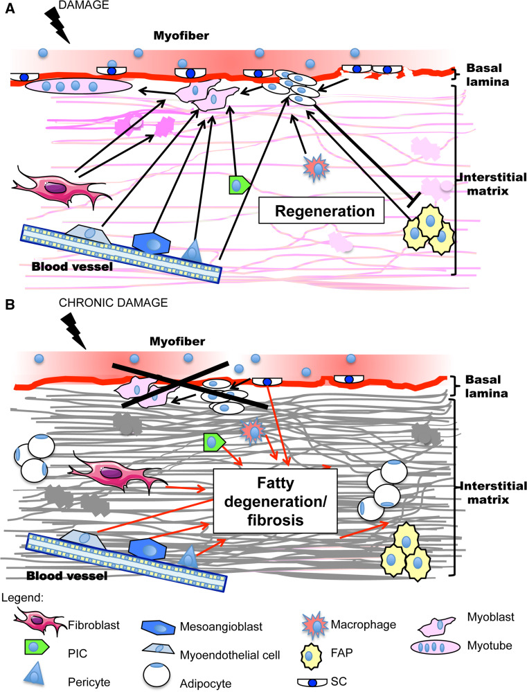
The skeletal muscle has the capacity to repair damage by the activation and differentiation of fiber sub-laminar satellite cells. Regeneration impairment due to reduced satellite cells number and/or functional capacity leads to fiber substitution with ectopic tissues including fat and fibrous tissue and to the loss of muscle functions. Muscle mesenchymal cells that in physiological conditions sustain or directly contribute to regeneration differentiate in adipocytes in patients with persistent damage and inflammation of the skeletal muscle. These cells comprise the fibro-adipogenic precursors, the PW1-expressing cells and some interstitial cells associated with vessels (pericytes, mesoangioblasts and myoendothelial cells). Resident fibroblasts that are responsible for collagen deposition and extracellular matrix remodeling during regeneration yield fibrotic tissue and can differentiate into adipose cells. Some authors have also proposed that satellite cells themselves could transdifferentiate into adipocytes, although recent results by lineage tracing techniques seem to put this theory to discussion. This review summarizes findings about muscle resident mesenchymal cell differentiation in adipocytes and recapitulates the molecular mediators involved in intramuscular adipose tissue deposition.


Figures

Fig. 1
Fig. 1
Schematic representation of muscle resident cells involved in regeneration or in adipo-fibrous tissue formation and of their interactions. a In physiological conditions, fiber damage induces satellite cells (SC) activation after basal lamina breakage, proliferation and differentiation into myoblasts that fuse to form new myotubes. Fibro-adipogenic precursors (FAP), located in the interstitial matrix, sustain SC cell regeneration by soluble factors and conversely SC inhibit their differentiation into adipocytes. Interstitial cells (PW1-expressing cells or PIC, pericytes, mesoangioblasts and myoendothelial cells) differentiate into myoblasts and contribute to regeneration. Fibroblasts, endothelium and infiltrating macrophages sustain myogenesis. Fibroblasts are also responsible of ECM remodeling, which in addition favors myoblast migration and fusion. b In a situation of persistent damage regeneration fails because of SC number reduction and/or functional impairment. In this case, ectopic fibro-adipose tissue deposition comes into view: it is sustained by interstitial mesenchymal cells, including FAP, as well as by fibroblasts and chronic inflammation. SC transdifferentiation along the adipocyte program also occurs
Fig. 2
Fig. 2
Nitric oxide inhibits adipose tissue deposition and FAP adipocyte differentiation in dystrophic muscle. a Nitric oxide effect on adipose tissue deposition (upper panels) and on FAP differentiation in adipocytes (lower panels) for tibialis anterior muscles, cells isolated from untreated mdx mice (untreated) and from mice that received an NO donor drug (NO-treated). Adipose tissue in section was revealed using an anti-perilipin antibody (upper panels) while O-red oil was used to count adipocytes in culture (lower panels). Hoechst was employed for nuclei staining. Quantification of perilipin-positive area (upper) and adipocytes number (lower) are reported in the right graphs (mean ± S.D). **p < 0.001 and ****p < 0.0001 vs. untreated, scale bars are 20 μm. b Expression of PPARγ mRNA and mir27b (left and right, respectively) in tibialis anterior muscles obtained from untreated mdx mice (untreated) and from mice that received an NO donor drug (NO treated); mean ± S.D. *p < 0.05 vs. untreated. c Percentage of collagen-positive cells in FAPs cultured in the absence (untreated) or in the presence of an NO donor (NO treated) and stimulated with TGFβ; mean ± S.D

References

    1. Charge SB, Rudnicki MA. Cellular and molecular regulation of muscle regeneration. Physiol Rev. 2004;84:209–238. - PubMed
    1. Kuang S, Rudnicki MA. The emerging biology of satellite cells and their therapeutic potential. Trends Mol Med. 2008;14:82–91. - PubMed
    1. Relaix F, Zammit PS. Satellite cells are essential for skeletal muscle regeneration: the cell on the edge returns centre stage. Development. 2012;139:2845–2856. - PubMed
    1. Yin H, Price F, Rudnicki MA. Satellite cells and the muscle stem cell niche. Physiol Rev. 2013;93:23–67. - PMC - PubMed
    1. Seale P, Sabourin LA, Girgis-Gabardo A, Mansouri A, Gruss P, Rudnicki MA. Pax7 is required for the specification of myogenic satellite cells. Cell. 2000;102:777–786. - PubMed

Publication types

MeSH terms

LinkOut - more resources