. 2015 Sep;72(17):3387-99.
doi: 10.1007/s00018-015-1894-2. Epub 2015 Apr 9.

Spinocerebellar ataxia type 19/22 mutations alter heterocomplex Kv4.3 channel function and gating in a dominant manner

Affiliations

Spinocerebellar ataxia type 19/22 mutations alter heterocomplex Kv4.3 channel function and gating in a dominant manner

Anna Duarri et al. Cell Mol Life Sci. 2015 Sep.

Abstract

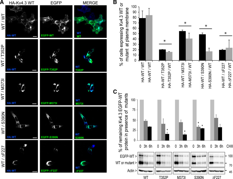
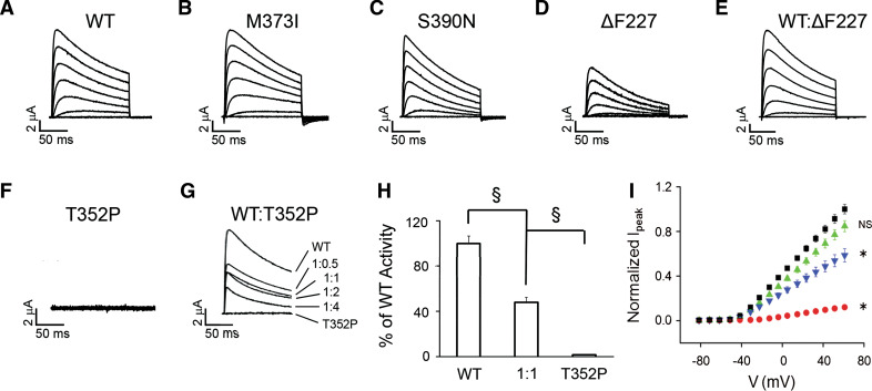
The dominantly inherited cerebellar ataxias are a heterogeneous group of neurodegenerative disorders caused by Purkinje cell loss in the cerebellum. Recently, we identified loss-of-function mutations in the KCND3 gene as the cause of spinocerebellar ataxia type 19/22 (SCA19/22), revealing a previously unknown role for the voltage-gated potassium channel, Kv4.3, in Purkinje cell survival. However, how mutant Kv4.3 affects wild-type Kv4.3 channel functioning remains unknown. We provide evidence that SCA19/22-mutant Kv4.3 exerts a dominant negative effect on the trafficking and surface expression of wild-type Kv4.3 in the absence of its regulatory subunit, KChIP2. Notably, this dominant negative effect can be rescued by the presence of KChIP2. We also found that all SCA19/22-mutant subunits either suppress wild-type Kv4.3 current amplitude or alter channel gating in a dominant manner. Our findings suggest that altered Kv4.3 channel localization and/or functioning resulting from SCA19/22 mutations may lead to Purkinje cell loss, neurodegeneration and ataxia.


Conflict of interest statement

The authors declare that they do not have any competing or financial interests.

Figures

Fig. 1
Fig. 1
WT/mutant Kv4.3 heterocomplexes are intracellulary retained and less stable than WT/WT Kv4.3 homocomplexes. a The confocal images show the anti-HA immuno-staining of permeabilized and fixed HeLa cells co-expressing HA–Kv4.3 WT (blue) and EGFP-fused WT, -T352P, -M373I, -S390N or -ΔF227 Kv4.3 (green). Scale bar 20 µm. b Flow cytometry was used to quantify the extracellular HA-tagged Kv4.3 WT (black bars) or mutant Kv4.3 (gray bars) at the plasma membrane of non-permeabilized HeLa cells expressing either WT/WT homocomplexes or WT/mutant heterocomplexes. The cells expressing Kv4.3 WT on the plasma membrane in the presence of any of the mutant subunits were strongly reduced compared with that in the presence of another WT subunit (black bars; WT/WT 79 % vs. WT/T352P: 21 %, WT/M373I: 55 %, WT/S390N: 49 % and WT/ΔF227: 20 %). Likewise, markedly reduced levels of all HA-tagged Kv4.3 mutant subunits were observed in the presence of WT (gray bars; WT/WT 84 % vs. T352P/WT: 16 %, M373I/WT: 41 %, S390N/WT: 17 % and ΔF227/WT: 24 %). c After 6 h of cycloheximide (CHX) treatment, the WT/mutant heterocomplexes were more rapidly degraded than the WT/WT homocomplexes (remaining protein WT/WT: 34 ± 1.1 %; WT/T352P: 16.1 ± 4.4 %; WT/M373I: 14.7 ± 1.9 %; WT/S390N: 29.7 ± 0.6 %, but not for WT/ΔF227: 32.5 ± 7.4 %). The bars represent the normalized expression of WT EGFP-Kv4.3 at t = 0 shown in percentages. Data in b and c represent the average of three independent experiments and the error bars represent the mean ± SEM, t test and ANOVA *p < 0.05 vs. WT in b and p < 0.005 vs. WT in c
Fig. 2
Fig. 2
Kv4.3 mutant subunits exhibit a temperature-sensitive folding defect. a Images of HeLa cells expressing HA–Kv4.3 WT or HA–T352P, –M373I, –S390N and –ΔF227 incubated at 37 °C or at 30 °C for 24 h. Non-permeabilized cells were stained using anti-HA antibody to detect Kv4.3 at the plasma membrane. Scale bar 20 µm. b Flow cytometry analysis is used to quantify the level of HA–Kv4.3 at the cell surface in HeLa cells cultured at 37 °C or at 30 °C. The graphs show a significant increase in the number of cells expressing the mutant subunits at the plasma membrane cultured at 30 °C (black bars; WT: 89 % ± 1.8; T352P: 86.9 % ± 1.2; M373I: 89.5 % ± 1.4; S390N: 77.8 % ± 3.3; and ΔF227: 67.8 % ± 10.1) compared to cells cultured at 37 °C (gray bars; WT: 96.5 % ± 2.1; T352P: 55.4 % ± 5.5; M373I: 71.1 % ± 3.4; S390N: 33.4 % ± 3; and ΔF227: 13.1 % ± 0.6). Bars show the average of three independent experiments (mean ± SEM, *p < 0.01, **p < 0.001)
Fig. 3
Fig. 3
KChIP2 drives the formation of stable WT/mutant Kv4.3 heterocomplexes at plasma membrane. a Confocal images of permeabilized and fixed HeLa cells expressing KChIP2 together with HA–WT/EGFP–WT homocomplexes or HA–WT/EGFP–mutant heterocomplexes (green) stained with anti-HA (blue) and anti-KChIP2 (red) antibodies. In the presence of KChIP2, all WT/mutant heterocomplexes were detected at the plasma membrane (merged in violet). Scale bar 20 µm. b Flow cytometry was used to quantify the percentage of cells expressing the extracellular HA-tagged Kv4.3 WT (black bars) or mutant subunits (gray bars) in the presence of KChIP2 at the plasma membrane of non-permeabilized HeLa cells. Similar levels of HA–Kv4.3 WT were detected at the plasma membrane for WT/WT homocomplexes vs. WT/mutant heterocomplexes (WT/WT: 79.5 % vs. WT/T352P: 77.8 %, WT/M373I: 71.8 %, WT/S390N: 75.1 % and WT/ΔF227: 75 %). Similarly, the levels of HA-tagged Kv4.3 mutants at plasma membrane for mutant/WT heterocomplexes were similar to WT/WT homocomplexes (WT/WT: 87.5 % vs. WT/T352P: 71.6 %, WT/M373I: 71 %, WT/S390N: 80.3 % and WT/ΔF227: 69.6 %). c Time course cycloheximide (CHX) experiments were performed in HeLa cells expressing the EGFP-WT/WT homocomplexes or EFGP–WT/mutant heterocomplexes in the presence (black lines) or absence (red lines) of KChIP2, and the remaining protein was analyzed by Western blot and quantified. Notably, in the presence of KChIP2, all WT/mutant heterocomplexes were significantly more stable (black lines) than the heterocomplexes without KChIP2 (red lines). Data in b and c represent the average of three independent experiments and the error bars represent the mean ± SEM, t test in b showed no significant differences and *p < 0.00001 vs. KChIP2 presence in c. In c the graphs represent the Western blot protein densitometries normalized by actin, showing the percentage of the remaining Kv4.3 protein
Fig. 4
Fig. 4
T352P-mutant Kv4.3 suppresses Kv4.3 WT activity by a dominant mechanism. af Representative current traces, recorded in oocytes at ~18 °C with a two-electrode voltage clamp 1–3 days after RNA injection, are shown for a WT, b M373I, c S390N, d ΔF227, e a 1:1 mixture of WT and ΔF227 and f T352P. Currents were evoked by pulsing from a holding potential of −100 mV to voltages ranging from −80 to +70 mV in 10 mV increments. For clarity, every other trace has been omitted. g Representative current traces evoked at +60 mV are shown for oocytes injected with WT, T352P or indicated ratios of WT:T352P RNA have been overlaid. h WT or T352P were expressed separately or at a 1:1 ratio keeping the amount of WT RNA constant. Normalized peak current amplitudes measured at +60 mV were WT alone, 1.00 ± 0.06 (n = 24); WT:T352P at a 1:1 ratio, 0.48 ± 0.09 (n = 30); and T352P alone, 0.02 ± 0.01 (n = 7). The 1:1 ratio differed significantly from WT alone, evaluated using a one-way ANOVA followed by Student’s t test (§ p < 0.00001). i Current density plot. Normalized peak current amplitude as a function of voltage is shown for wild type (black squares), ΔF227 (red circles), wild type:ΔF227 expressed at a 1:1 molar ratio (green triangles) or wild type:T352P expressed at a 1:1 molar ratio (blue inverted triangles). RNA encoding KChIP2 was co-injected at an equimolar ratio with the total amount of Kv4.3 RNA. Peak current amplitudes were measured as a function of voltage and normalized to that for wild-type Kv4.3 expressed in parallel in the same batch of oocytes. Data from different batches of oocytes were then averaged (n = 12). Data are provided as mean ± SEM. Statistical significance was assessed using data obtained at +50 mV: *significantly different from wild type (*p < 0.05) by ANOVA followed by Tukey’s post hoc test; NS not significantly different from wild type
Fig. 5
Fig. 5
SCA19/22 mutations alter the functional properties of Kv4.3 homocomplexes and heterocomplexes. a Conductance values were calculated from peak current amplitudes assuming a linear open channel current–voltage relationship and normalized to the maximum value obtained in the experiment. Normalized conductance values were plotted as a function of test voltage for WT (black squares), ΔF227 (open circles), a 1:1 mixture of WT and ΔF227 (red circles), a 1:1 mixture of WT and T352P (green triangles), M373I (blue inverted triangles) and S390N (diamond symbols) and data were fitted with a single Boltzmann function to obtain values for V ½,act and the slope factor (see Table 1). b Representative current traces evoked by pulsing from −100 to +60 mV have been scaled and overlaid for WT (black), ΔF227 (magenta), a 1:1 mixture of WT and ΔF227 (red), a 1:1 mixture of WT and T352P (green), M373I, (blue) and S390N (cyan). c The box plot shows the time to reach peak current amplitude at +60 mV. d The box plot shows the I 150ms/I peak ratio, calculated by dividing the current amplitude remaining at the end of a 150 ms pulse by the peak current amplitude. e Steady-state inactivation was evaluated using a two-pulse protocol. Peak amplitudes during the test pulse were normalized to the peak amplitude in the absence of a prepulse (I/I max) and plotted versus prepulse voltage. Data were fitted with a single Boltzmann function (solid curves) to obtain values for V ½,inact and the slope factor (see Table 1). f To measure the rate of recovery from inactivation, currents were evoked by pulsing from −80 to +60 mV for 400 ms (pulse 1). The voltage was then returned to −80 mV for variable durations ranging from 500 to 1100 ms, in 40 ms steps, prior to a second pulse to +60 mV for 400 ms (pulse 2). The fractional recovery was calculated as the peak amplitude during pulse 2 divided by the peak amplitude during pulse 1 (I/I max) and plotted versus the interpulse duration. Each data set was fitted with a single exponential function to obtain the time constant for recovery (τ rec) (Table 1). Data are shown as mean ± SEM. In b and c, statistical significance compared to WT alone was evaluated by one-way ANOVA followed by Student’s t test: p < 0.005; # p < 0.0005; § p < 0.00001. Mean values ± SEM of the time to peak and the I 150ms/I peak ratio are provided in Table 1. Results obtained with 1:1 mixtures of WT:M373I and WT:S390N are provided in Table 1

References

    1. Lee YC, Durr A, Majczenko K, Huang YH, Liu YC, Lien CC, Tsai PC, Ichikawa Y, Goto J, Monin ML, Li JZ, Chung MY, Mundwiller E, Shakkottai V, Liu TT, Tesson C, Lu YC, Brice A, Tsuji S, Burmeister M, Stevanin G, Soong BW. Mutations in KCND3 cause spinocerebellar ataxia type 22. Ann Neurol. 2012;72:859–869. doi: 10.1002/ana.23701. - DOI - PMC - PubMed
    1. Duarri A, Nibbeling E, Fokkens MR, Meijer M, Boddeke E, Lagrange E, Stevanin G, Brice A, Durr A, Verbeek DS. The L450P mutation in KCND3 brings spinocerebellar ataxia and Brugada syndrome closer together. Neurogenetics. 2013;14:257–258. doi: 10.1007/s10048-013-0370-0. - DOI - PubMed
    1. Duarri A, Jezierska J, Fokkens M, Meijer M, Schelhaas HJ, den Dunnen WF, van Dijk F, Verschuuren-Bemelmans C, Hageman G, van de Vlies P, Kusters B, van de Warrenburg BP, Kremer B, Wijmenga C, Sinke RJ, Swertz MA, Kampinga HH, Boddeke E, Verbeek DS. Mutations in potassium channel kcnd3 cause spinocerebellar ataxia type 19. Ann Neurol. 2012;72:870–880. doi: 10.1002/ana.23700. - DOI - PubMed
    1. Bahring R, Dannenberg J, Peters HC, Leicher T, Pongs O, Isbrandt D. Conserved Kv4N-terminal domain critical for effects of Kv channel-interacting protein 2.2 on channel expression and gating. J Biol Chem. 2001;276:23888–23894. doi: 10.1074/jbc.M101320200. - DOI - PubMed
    1. Shibata R, Misonou H, Campomanes CR, Anderson AE, Schrader LA, Doliveira LC, Carroll KI, Sweatt JD, Rhodes KJ, Trimmer JS. A fundamental role for KChIPs in determining the molecular properties and trafficking of Kv4.2 potassium channels. J Biol Chem. 2003;278:36445–36454. doi: 10.1074/jbc.M306142200. - DOI - PubMed

Publication types

Supplementary concepts

LinkOut - more resources