. 2015 Apr 8:5:9623.
doi: 10.1038/srep09623.

Plasticity of listeriolysin O pores and its regulation by pH and unique histidine [corrected]

Affiliations

Plasticity of listeriolysin O pores and its regulation by pH and unique histidine [corrected]

Marjetka Podobnik et al. Sci Rep. .

Erratum in

Abstract

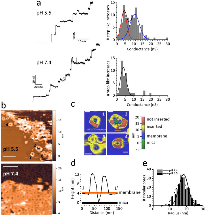
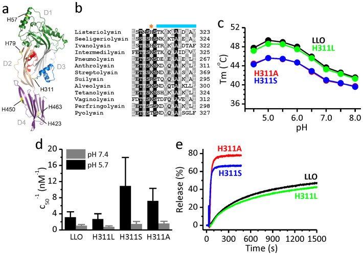
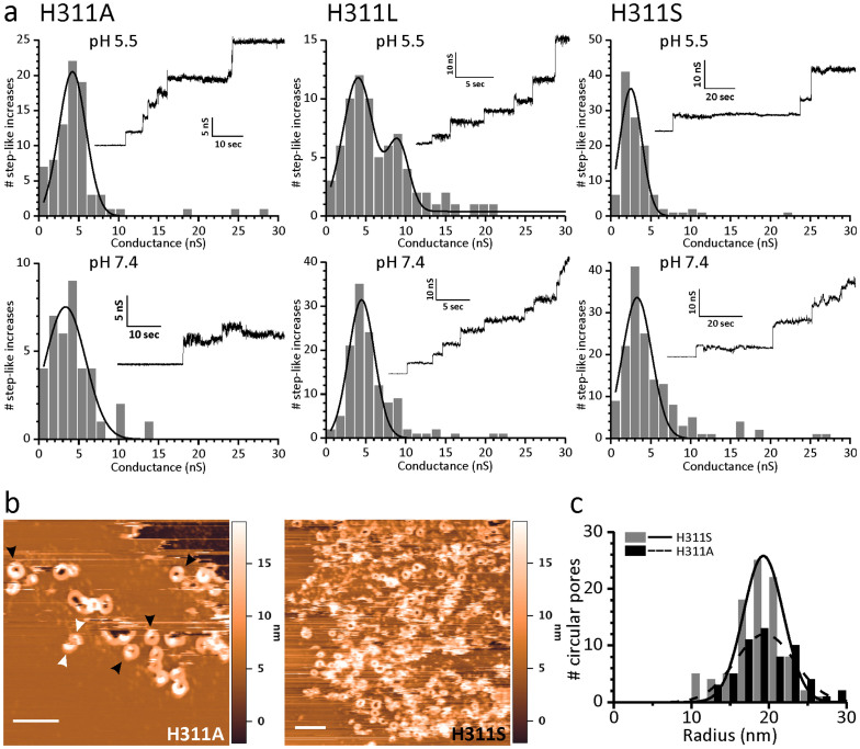
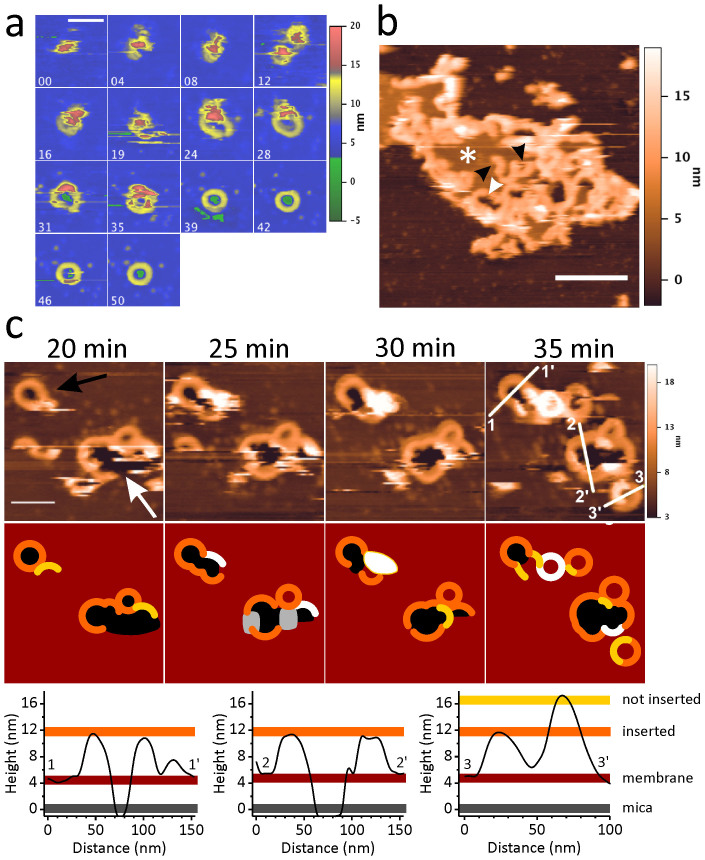
Pore formation of cellular membranes is an ancient mechanism of bacterial pathogenesis that allows efficient damaging of target cells. Several mechanisms have been described, however, relatively little is known about the assembly and properties of pores. Listeriolysin O (LLO) is a pH-regulated cholesterol-dependent cytolysin from the intracellular pathogen Listeria monocytogenes, which forms transmembrane β-barrel pores. Here we report that the assembly of LLO pores is rapid and efficient irrespective of pH. While pore diameters at the membrane surface are comparable at either pH 5.5 or 7.4, the distribution of pore conductances is significantly pH-dependent. This is directed by the unique residue H311, which is also important for the conformational stability of the LLO monomer and the rate of pore formation. The functional pores exhibit variations in height profiles and can reconfigure significantly by merging to other full pores or arcs. Our results indicate significant plasticity of large β-barrel pores, controlled by environmental cues like pH.


Conflict of interest statement

The authors declare no competing financial interests.

Figures

Figure 1
Figure 1. Properties of LLO pores at pH 5.5 and 7.4.
(a) Conductance traces (left) of pores as measured by PLM. The protein concentration used was 5 nM or 1 nM for pH 5.5 and 7.4, respectively; the recorded current traces were obtained by applying +40 mV. Histograms of single channel conductance distribution are reported to the right of each trace. Two populations of pores that are apparent at pH 5.5 are shown by red and blue line. (b) Images of representative AFM scans of LLO oligomers formed on SLB at two designated pH values. 200 nm scale bars (white lines) are shown. Numbers (1–4) denote aggregates of various shapes not yet completely inserted and are shown enlarged in panel (c). Scale bar in (c) is 30 nm. (d) Height profiles of protein aggregates were traced along the black line shown in the AFM image in (b). (e) Distribution of radii estimated from AFM images in (b).
Figure 2
Figure 2. Position of the unique H311 in the structure of LLO and properties of LLO mutants.
(a) Crystal structure of LLO (PDB-ID 4CDB) is shown in ribbons. Domain 1 (D1) is in green, domain 2 (D2) in light brown, domain 3 (D3) in gray and domain 4 (D4) in purple. TMH1 region is in red and TMH2 is in blue. Side chains of all histidines are shown as sticks. (b) Multiple sequence alignment of some of the CDC family members with position of H311 and part of TMH2 indicated by orange asterisk and blue line, respectively. (c) Thermal stability of LLO and mutants as described by Tm values measured by DSF at different pH values. All measurements were performed twice. (d) Hemolytic activity at pH 5.7 or pH 7.4. At least 4 independent measurements for each protein were performed. Hemolytic buffer and erythrocytes were either at pH 5.7 or 7.4. T = 25°C. Hemolytic activity is expressed as inverse values of protein concentration at which half maximal rate of hemolysis is achieved (c50). (e) Kinetics of calcein release at pH 5.5 at room temperature. Representative traces of two independent experiments are reported. Final protein concentration was 0.39 nM.
Figure 3
Figure 3. Pore properties of LLO mutants at pH 5.5 and 7.4.
(a) Conductance of pores as measured by PLM. The protein concentration was 5 nM or 1 nM at pH 5.5 and 7.4, respectively. Histograms of single channel conductance distribution are reported together (inset) with the recorded current traces that were obtained applying +40 mV. (b) AFM images of pores formed by LLO mutants on SLB at pH 5.5. 200 nm scale bars (white lines) are shown. Black arrowheads denote parts of rings not yet completely inserted, while white arrowheads denote arcs that also are not inserted. (c) Distribution of the radii estimated from AFM images shown in (b).
Figure 4
Figure 4. Dynamics of LLO pore formation as imaged by AFM.
(a) Time course of pore formation by the wild type LLO on SLB at pH 5.5. Numbers at the upper left side of each image show the time in minutes. The color code on the height scale is the same as in Fig. 1C; scale bar length corresponds to 100 nm. (b) Scan of the wild type LLO oligomers on SLB at pH 5.5. White asterisk marks a lipid bilayer patch. Black arrows indicate arc-like structures. A large porous structure formed by arcs facing each other is marked by white arrow. Scale bar length corresponds to 500 nm. (c) Time evolution of oligomers formed by the H311A mutant on SLB at pH 5.5. Upper part: Four AFM scans, acquired with a 5 minutes time interval, show the growth and fusion of new structures with an already formed ring (black arrow) and the closure of a large structure, mainly consisting of or being contoured with joined arcs (white arrow). The time indicated above the panels was counted after injection of the protein across SLB. Below: sketch reconstruction of AFM scans in the upper panel to clearly depict the shape and height of various protein aggregates. Height profiles of protein aggregates along the white lines shown in the AFM image in (c) after 35 min are shown below the image.

References

    1. Hamon M. A., Ribet D., Stavru F. & Cossart P. Listeriolysin O: the Swiss army knife of Listeria. Trends Microbiol. 20, 360–368 (2012). - PubMed
    1. Swaminathan B. & Gerner-Smidt P. The epidemiology of human listeriosis. Microbes Infect. 9, 1236–1243 (2007). - PubMed
    1. Gaillard J. L., Berche P., Mounier J., Richard S. & Sansonetti P. In vitro model of penetration and intracellular growth of Listeria monocytogenes in the human enterocyte-like cell line Caco-2. Infect. Immun. 55, 2822–2829 (1987). - PMC - PubMed
    1. Portnoy D. A., Jacks P. S. & Hinrichs D. J. Role of hemolysin for the intracellular growth of Listeria monocytogenes. J. Exp. Med. 167, 1459–1471 (1988). - PMC - PubMed
    1. Cossart P. et al. Listeriolysin O is essential for virulence of Listeria monocytogenes: direct evidence obtained by gene complementation. Infect. Immun. 57, 3629–3636 (1989). - PMC - PubMed

Publication types

MeSH terms

LinkOut - more resources