. 2015 Apr 15;29(8):791-802.
doi: 10.1101/gad.259341.115. Epub 2015 Apr 8.

Senescent cells communicate via intercellular protein transfer

Affiliations

Senescent cells communicate via intercellular protein transfer

Anat Biran et al. Genes Dev. .

Abstract

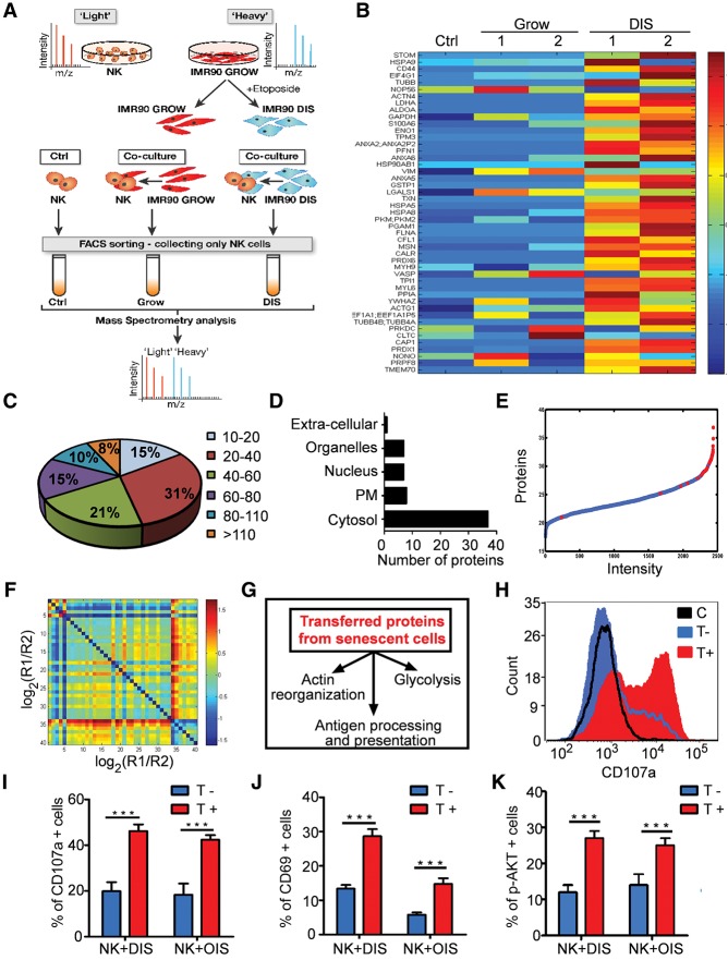
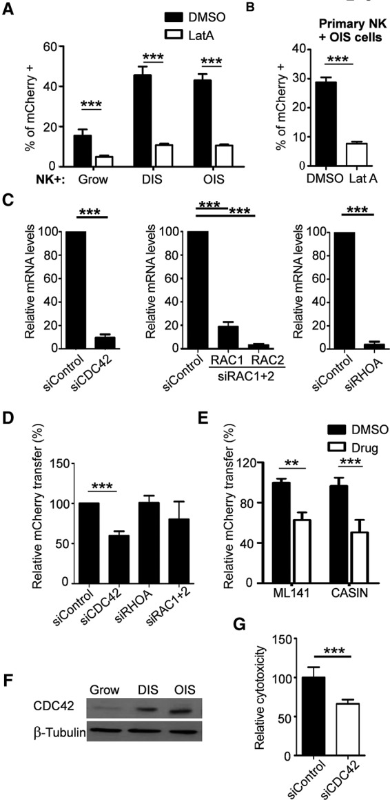
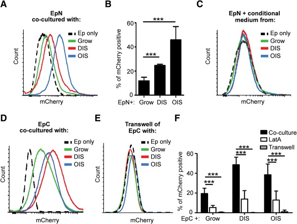
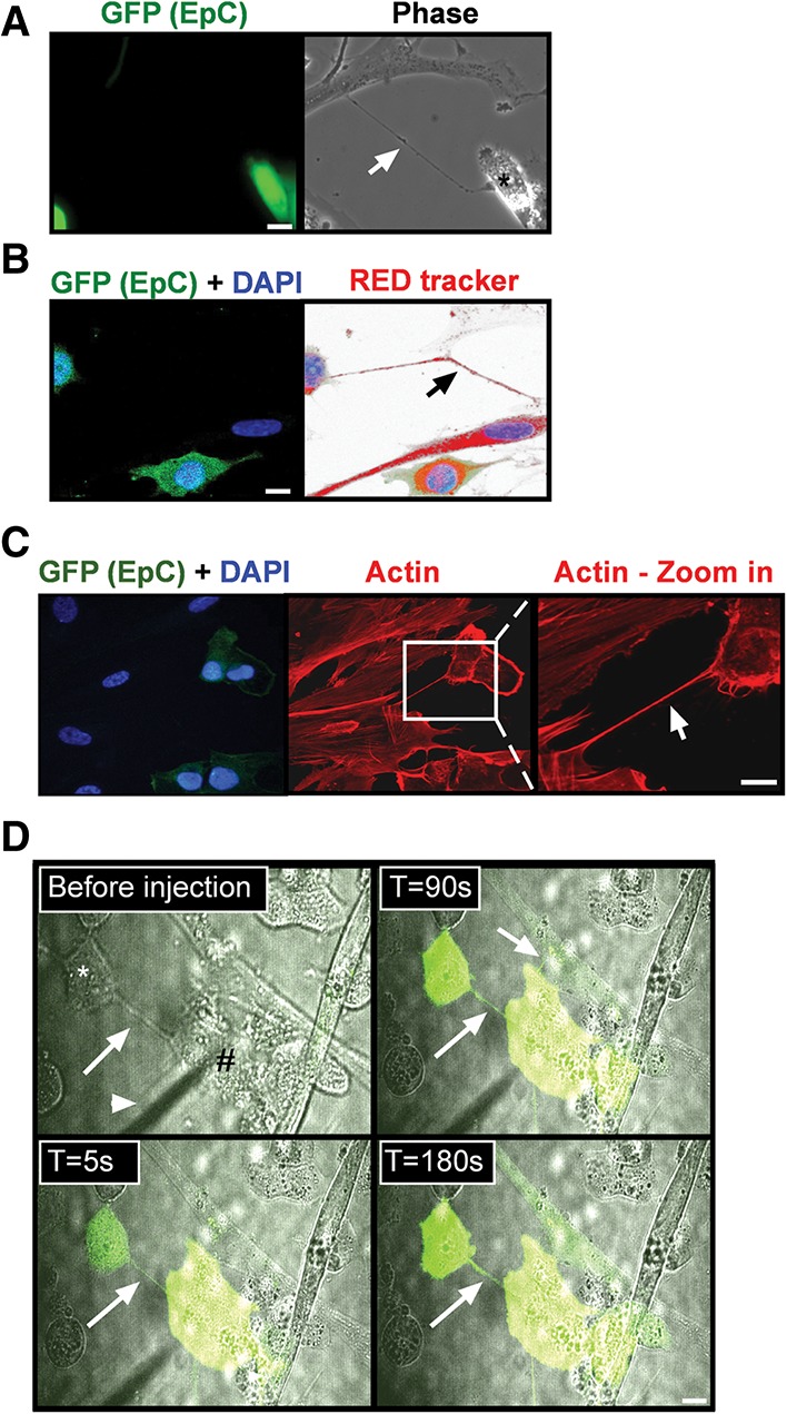
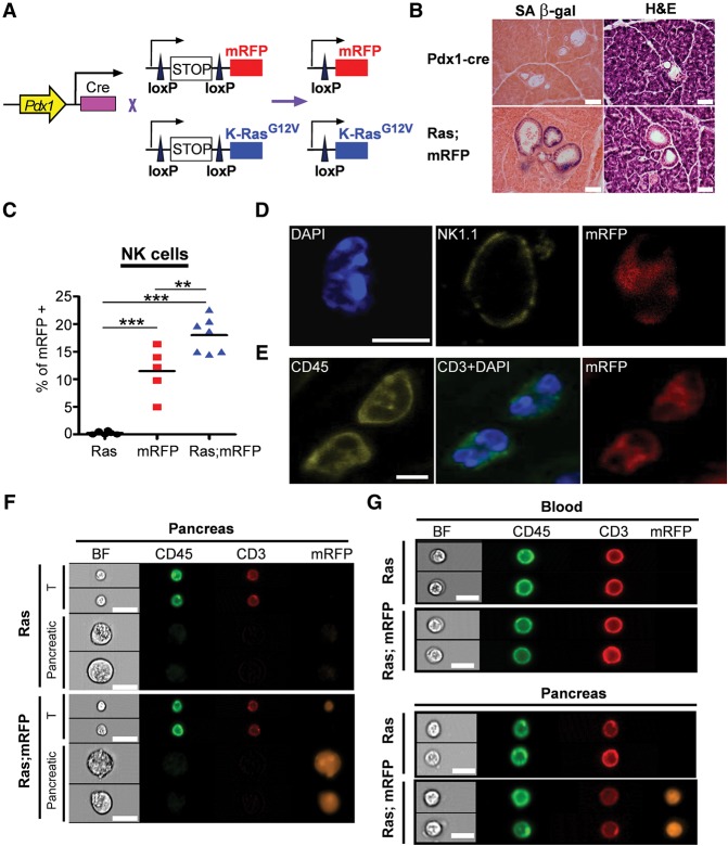
Mammalian cells mostly rely on extracellular molecules to transfer signals to other cells. However, in stress conditions, more robust mechanisms might be necessary to facilitate cell-cell communications. Cellular senescence, a stress response associated with permanent exit from the cell cycle and the development of an immunogenic phenotype, limits both tumorigenesis and tissue damage. Paradoxically, the long-term presence of senescent cells can promote tissue damage and aging within their microenvironment. Soluble factors secreted from senescent cells mediate some of these cell-nonautonomous effects. However, it is unknown whether senescent cells impact neighboring cells by other mechanisms. Here we show that senescent cells directly transfer proteins to neighboring cells and that this process facilitates immune surveillance of senescent cells by natural killer (NK) cells. We found that transfer of proteins to NK and T cells is increased in the murine preneoplastic pancreas, a site where senescent cells are present in vivo. Proteomic analysis and functional studies of the transferred proteins revealed that the transfer is strictly dependent on cell-cell contact and CDC42-regulated actin polymerization and is mediated at least partially by cytoplasmic bridges. These findings reveal a novel mode of intercellular communication by which senescent cells regulate their immune surveillance and might impact tumorigenesis and tissue aging.

Keywords: actin polymerization; cellular senescence; cytoplasmic bridges; natural killer cells.


Figures

Figure 1.
Figure 1.
mCherry is preferentially transferred from senescent cells to NK cells by a contact-dependent mechanism. Primary human NK cells or NK92 cells were cocultured with senescent IMR-90 cells expressing the mCherry-H-Ras12V fusion protein (OIS cells) for 2 h. (A) The cells were fixed and stained for CD56 (green). Bar, 10 µM. (B) NK92 cells were collected, stained for CD56 and with DAPI, and analyzed by FACS compared with NK92 cells without coculture (control [C]). NK92 cells that received mCherry were designated as T+. (C) The percentage of T+ NK92 cells was determined following 2 h of coculture with OIS cells. (D) The percentage of T+ human primary NK cells was determined following 1 h of coculture with OIS cells. (E) NK92 cells were analyzed by ImageStream following coculture with OIS cells. (F) Following coculture with OIS cells, NK92 cells were FACS-sorted for T+ and T populations and analyzed by Western blot for the presence of mCherry-H-Ras12V (using anti-pan-Ras antibodies). (G) IPT to NK92 cells from OIS, DNA damage-induced senescence (DIS), or growing IMR-90 cells was determined by FACS analysis after 2 h of coculture. (H) Representative analysis of IPT from growing, quiescent, apoptotic, or DIS cells, all expressing mCherry, to NK92 cells after 2 h of coculture. (I) NK92 cells were cocultured with growing, OIS, or DIS cells or separated within the same well by a transwell filter (0.4-µm pore) and then analyzed by FACS. (J) Primary human NK cells were cocultured for 1 h with OIS cells or separated by a transwell filter and analyzed for IPT. All graphs are expressed as means ± SEM from at least three independent experiments. (***) P < 0.001.
Figure 2.
Figure 2.
Transferred proteins identified by proteomic analysis lead to NK activation and cytotoxicity. (A) Diagram of the trans-SILAC experiment. (B) SPIN-ordered expression matrix. Colors depict relative levels of transfer (centered and normalized), where red denotes high levels of transfer, and blue denotes low levels of transfer. Rows represent proteins, and columns represent samples: NK92 cells without coculture (Ctrl) or cocultured with growing control (Grow) or DIS cells. (C) Size distribution (in kilodaltons) of the identified transferred proteins. (D) Subcellular localization of the identified transferred proteins. (E) Quantitative proteomic analysis of DIS cells. Proteins from senescent cells were ranked according to their relative intensity levels, reflecting protein abundance. Transferred proteins from DIS to NK92 cells are marked in red. (F) A matrix of protein ratios R1/R2, where the transferred protein abundance is denoted by R1 and protein abundance in DIS cells is denoted by R2. (G) Kyoto Encyclopedia of Genes and Genomes (KEGG) pathway analysis identified the pathways affected by the transferred proteins. (HK) Primary human NK cells were cocultured for 1 h with OIS or DIS cells, collected, and gated by FACS as T or T+ compared with control (C) NK cells. (H) Representative FACS analysis of CD107a levels in control, T, and T+ populations following IPT from DIS cells. (I) Average percentage of CD107a-positive cells in T and T+ populations. (J) Average percentage of CD69-positive cells in T and T+ populations. (K) Average percentage of phosphorylated AKT (p-AKT)-positive cells between T and T+ populations. Results are expressed as means ± SEM from at least three independent experiments. (***) P < 0.001.
Figure 3.
Figure 3.
IPT is dependent on actin polymerization. NK cells were cocultured with growing, DIS, or OIS cells. NK cells were then collected, stained for CD56 and with DAPI, and analyzed by FACS for the presence of mCherry. (A) The percentage of T+ NK92 cells following 2 h of coculture in the presence of 200 nM LatA or vehicle (DMSO). (B) The percentage of T+ primary human NK cells following 1 h of coculture in the presence of 200 nM LatA or vehicle. (C) CDC42, RAC1, RAC2, and RHOA siRNAs induce efficient knockdown 3 d after transfection. (D) DIS cells with knockdown of CDC42, RHOA, or RAC1 and RAC2 were cocultured for 2 h with NK92 cells. (E) DIS cells were treated with ML141, CASIN (CDC42 inhibitors), or DMSO (control); washed; and then cocultured with NK92 cells. (F) Western blot analysis of CDC42 levels in growing, DIS, and OIS IMR-90 cells. (G) NK-mediated cytotoxicity toward DIS cells following CDC42 knockdown (siCDC42). Results are expressed as means ± SEM from at least three independent experiments. (***) P < 0.001.
Figure 4.
Figure 4.
Senescent cells transfer proteins to epithelial cells. (AC) MCF10A cells expressing GFP (EpN) were cocultured with growing, DIS, or OIS cell for 24 h. Transfer of mCherry to MCF10A was analyzed by FACS. (A) Representative FACS analysis of mCherry levels in EpN cells compared with MCF10A alone (Ep only). (B) The percentage of mCherry-positive EpN cells following the coculture. (C) EpN cells were cultured with medium collected from growing, DIS, or OIS cells. (DF) H1299 cells expressing GFP (EpC) were cocultured for 24 h with growing, DIS, and OIS cells and analyzed by FACS. (D) Representative FACS analysis of mCherry levels in EpC cells. (E) EpC cells were separated by a transwell chamber from growing, DIS, or OIS cells. (F) The percentage of mCherry-positive EpC cells following coculture in the presence of LatA or in a transwell chamber. Results are expressed as means ± SEM from at least three independent experiments. (***) P < 0.001.
Figure 5.
Figure 5.
Senescent cells form CBs with cancer cells. (AC) GFP-expressing EpC cells were cocultured with DIS cells, stained, and analyzed by confocal microscope. (A) The CB (arrow) was detected in the coculture. (B) A CB (arrow) was detected when the cells were stained with RedTracker dye and DAPI. (C) Cells were stained for F-actin (red) and DAPI. (D) Using a microinjection needle (arrowhead), a DIS fibroblast (#) was injected with calcein fluorescent dye. Transfer of the dye via a CB (arrow) from the senescent cell to the cancer cells (asterisk) was monitored over time. Bars, 10 µM.
Figure 6.
Figure 6.
mRFP is transferred to immune cells in the premalignant pancreas in vivo. (A) Schematic representation of the Ras;mRFP mouse model. (B) H&E- and SA-β-gal-stained sections from Ras;mRFP mice. Bar, 50 µm. (C) FACS quantification of mRFP-positive NK cells extracted from the pancreata. n ≥ 5 mice for each genotype; (***) P < 0.001; (**) P < 0.01. (D) Sections of a pancreas from Ras;mRFP mice were stained for NK1.1 (yellow) and with DAPI (blue) and imaged by confocal microscope. Bar, 5 µm. (E) Sections of a pancreas from Ras;mRFP mice were stained for CD45 (yellow) and CD3 (green) and with DAPI (blue). Bar, 5 µm. (F,G) ImageStream analysis of cells derived from the blood or pancreata of mice of the indicated genotype. (F) T cells (CD45+;CD3+) and pancreatic resident cells have different characteristics and are clearly distinguishable by ImageStream analysis. Bar, 20 μm. (G) Representative images of mRFP-positive and mRFP-negative T cells acquired using ImageStream. Bar, 10 µm.

References

    1. Abounit S, Zurzolo C. 2012. Wiring through tunneling nanotubes—from electrical signals to organelle transfer. J Cell Sci 125: 1089–1098. - PubMed
    1. Acosta JC, O'Loghlen A, Banito A, Guijarro MV, Augert A, Raguz S, Fumagalli M, Da Costa M, Brown C, Popov N, et al. 2008. Chemokine signaling via the CXCR2 receptor reinforces senescence. Cell 133: 1006–1018. - PubMed
    1. Acosta JC, Banito A, Wuestefeld T, Georgilis A, Janich P, Morton JP, Athineos D, Kang TW, Lasitschka F, Andrulis M, et al. 2013. A complex secretory program orchestrated by the inflammasome controls paracrine senescence. Nat Cell Biol 15: 978–990. - PMC - PubMed
    1. Arkwright PD, Luchetti F, Tour J, Roberts C, Ayub R, Morales AP, Rodriguez JJ, Gilmore A, Canonico B, Papa S, et al. 2010. Fas stimulation of T lymphocytes promotes rapid intercellular exchange of death signals via membrane nanotubes. Cell Res 20: 72–88. - PMC - PubMed
    1. Baker DJ, Wijshake T, Tchkonia T, LeBrasseur NK, Childs BG, van de Sluis B, Kirkland JL, van Deursen JM. 2011. Clearance of p16Ink4a-positive senescent cells delays ageing-associated disorders. Nature 479: 232–236. - PMC - PubMed

Publication types