. 2015 Apr 8;35(14):5435-41.
doi: 10.1523/JNEUROSCI.5269-14.2015.

Optogenetic evidence for inhibitory signaling from orexin to MCH neurons via local microcircuits

Affiliations

Optogenetic evidence for inhibitory signaling from orexin to MCH neurons via local microcircuits

John Apergis-Schoute et al. J Neurosci. .

Abstract

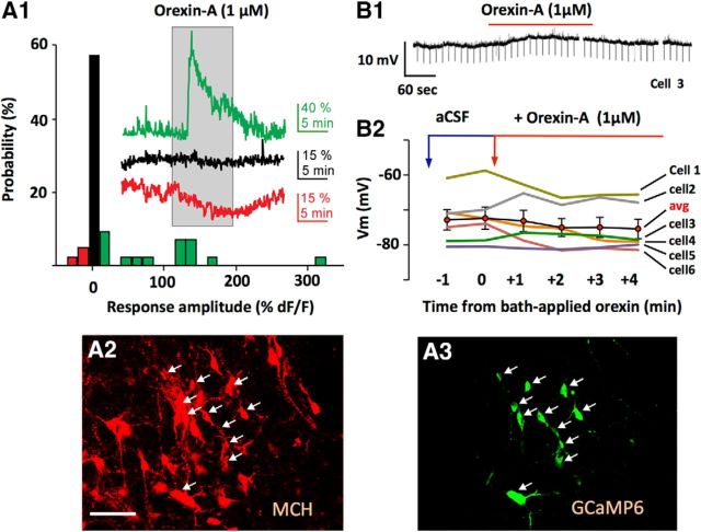
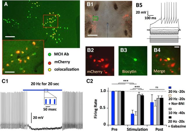
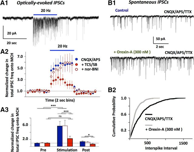
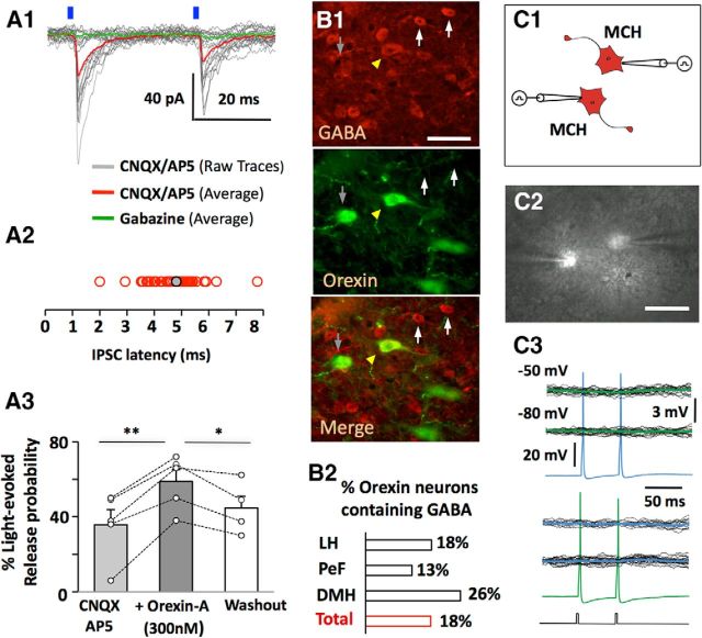
The lateral hypothalamus (LH) is a key regulator of multiple vital behaviors. The firing of brain-wide-projecting LH neurons releases neuropeptides promoting wakefulness (orexin/hypocretin; OH), or sleep (melanin-concentrating hormone; MCH). OH neurons, which coexpress glutamate and dynorphin, have been proposed to excite their neighbors, including MCH neurons, suggesting that LH may sometimes coengage its antagonistic outputs. However, it remains unclear if, when, and how OH actions promote temporal separation of the sleep and wake signals, a process that fails in narcolepsy caused by OH loss. To explore this directly, we paired optogenetic stimulation of OH cells (at rates that promoted awakening in vivo) with electrical monitoring of MCH cells in mouse brain slices. Membrane potential recordings showed that OH cell firing inhibited action potential firing in most MCH neurons, an effect that required GABAA but not dynorphin receptors. Membrane current analysis showed that OH cell firing increased the frequency of fast GABAergic currents in MCH cells, an effect blocked by antagonists of OH but not dynorphin or glutamate receptors, and mimicked by bath-applied OH peptide. In turn, neural network imaging with a calcium indicator genetically targeted to MCH neurons showed that excitation by bath-applied OH peptides occurs in a minority of MCH cells. Collectively, our data provide functional microcircuit evidence that intra-LH feedforward loops may facilitate appropriate switching between sleep and wake signals, potentially preventing sleep disorders.

Keywords: GABA; hypocretin; hypothalamus; melanin-concentrating hormone; optogenetics; orexin.


Figures

Figure 1.
Figure 1.
Responses of MCH neurons to bath-applied orexin peptide. A, Summary of intracellular calcium responses of individual MCH neurons to 1 μm bath-applied orexin-A (A1; n = 42 cells). Traces show examples of excitation (green), inhibition (red), and no response (black). A2, A3, Confirmation of localization of GCaMP6s calcium indicator to MCH neurons (see Materials and Methods). Scale bar, 80 μm. B, Effect of bath applied orexin-A on MCH cell membrane potential; typical example (B1) and group data (B2).
Figure 2.
Figure 2.
Membrane potential effects in MCH neurons of activation of intrinsic OH neurons. A, Targeted expression of mCherry in MCH neurons (n = 3 animals, 630/638 mCherry cells colocalised with MCH). Scale bars: top, 200 μm; inset, 40 μm. B, Whole-cell recordings in mCherry-expressing neurons (B2) were confirmed by filling neurons with biocytin (B3, B4). Membrane potential responses to current injections (B5); n = 50 cells. C, Effect of optogenetic stimulation of OH neurons. Typical example (C1) and summary of effects of different stimulation frequencies and drug conditions on firing rate, normalized to prestimulation firing (C2); n = 7 cells, two-way ANOVA, F(2,42) = 21.37, Bonferroni post-test; ***p < 0.001; ns, nonsignificant (p > 0.05).
Figure 3.
Figure 3.
Membrane current effects in MCH neurons of activating intrinsic OH neurons, and of bath-applied OH peptide. A1, Typical example of MCH cell IPSCs caused by optical stimulation of OH cells (n = 26 cells). IPSCs were recorded in the presence of CNQX/AP5, and confirmed as GABAergic by blockade with gabazine. A2, Summary of effects of OH cell stimulation on MCH cell IPSC tone, under different drug conditions (n = 26 cells). A3, Comparison of IPSC tone at different times relative to the optical stimulation (6 s before, last 6 s of stimulation, and 6 s after the stimulation). Color codes and data are the same as in A2. Two-way ANOVA, F(2,42) = 21.37, Bonferroni post-test: ***p < 0.001; *p < 0.05; ns, nonsignificant (p > 0.05). B1, Typical example of effect of bath applied orexin-A on miniature IPSCs (n = 5 cells). B2, Comparison of miniature IPSC frequency with and without bath orexin; n = 5 cells, Kolmogorov–Smirnov test, p < 0.001.
Figure 4.
Figure 4.
Inhibitory actions and GABA content of OH neurons. A1, An example of a short-latency (<10 ms) IPSC evoked by a blue laser flashes (n = 7 cells). A2, distribution of short latencies (A2; n = 48 cells). A3, effect on bath-applied orexin-A on short-latency IPSC recorded as in A1 (n = 5 cells). Paired two-tailed Student's t test, **p < 0.005, *p < 0.05; ns, nonsignificant (p > 0.05). B, Orexin-containing neurons immunopositive for GABA (B1) are distributed throughout hypothalamic subregions. The number of OH-immunopositive cells colocalized with GABA were as follows: LH, 107/590; perifornical area (PeF), 23/88; dorsomedial hypothalamus (DMH), 29/226; total 159/908 cells (n = 3 brains; B2). Scale bar, 40 μm. C, Schematic (C1) and live brain-slice image (C2) of paired recordings from MCH-mCherry neurons. Scale bar, 50 μm. C3, representative recordings from an MCH-MCH cell pair, showing that evoked action potentials in one cell did not produce postsynaptic current in the other. Black lines are individual traces and green/blue are averaged traces. Current-clamp protocol used to evoke action potentials is shown below the traces. Similar results were obtained from 43 cell pairs.

References

    1. Adamantidis AR, Zhang F, Aravanis AM, Deisseroth K, de Lecea L. Neural substrates of awakening probed with optogenetic control of hypocretin neurons. Nature. 2007;450:420–424. doi: 10.1038/nature06310. - DOI - PMC - PubMed
    1. Balcita-Pedicino JJ, Sesack SR. Orexin axons in the rat ventral tegmental area synapse infrequently onto dopamine and gamma-aminobutyric acid neurons. J Comp Neurol. 2007;503:668–684. doi: 10.1002/cne.21420. - DOI - PubMed
    1. Baratta R, Solomonow M, Zhou BH, Letson D, Chuinard R, D'Ambrosia R. Muscular coactivation: the role of the antagonist musculature in maintaining knee stability. Am J Sports Med. 1988;16:113–122. doi: 10.1177/036354658801600205. - DOI - PubMed
    1. Bittencourt JC, Presse F, Arias C, Peto C, Vaughan J, Nahon JL, Vale W, Sawchenko PE. The melanin-concentrating hormone system of the rat brain: an immuno- and hybridization histochemical characterization. J Comp Neurol. 1992;319:218–245. doi: 10.1002/cne.903190204. - DOI - PubMed
    1. Burdakov D, Gerasimenko O, Verkhratsky A. Physiological changes in glucose differentially modulate the excitability of hypothalamic melanin-concentrating hormone and orexin neurons in situ. J Neurosci. 2005;25:2429–2433. doi: 10.1523/JNEUROSCI.4925-04.2005. - DOI - PMC - PubMed

Publication types

MeSH terms

LinkOut - more resources