. 2015 Apr 8;35(14):5693-706.
doi: 10.1523/JNEUROSCI.3397-14.2015.

Axonal amphoterin mRNA is regulated by translational control and enhances axon outgrowth

Affiliations

Axonal amphoterin mRNA is regulated by translational control and enhances axon outgrowth

Tanuja T Merianda et al. J Neurosci. .

Abstract

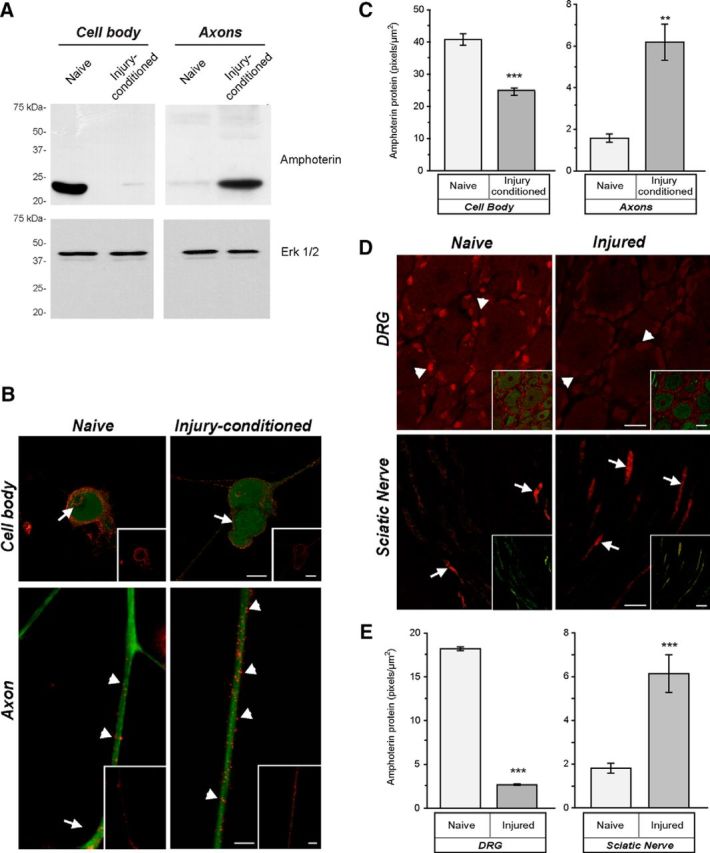
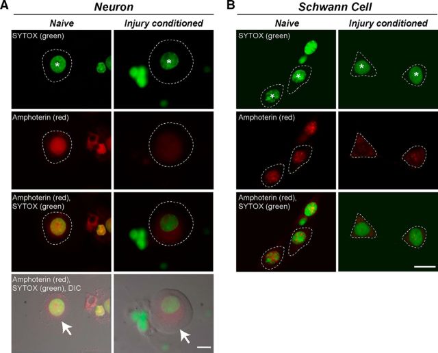
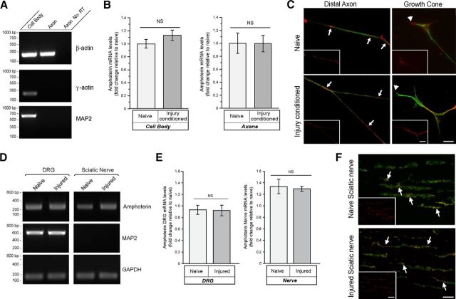
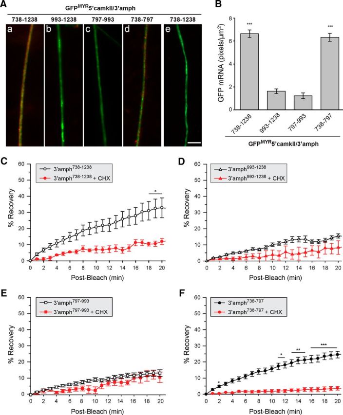
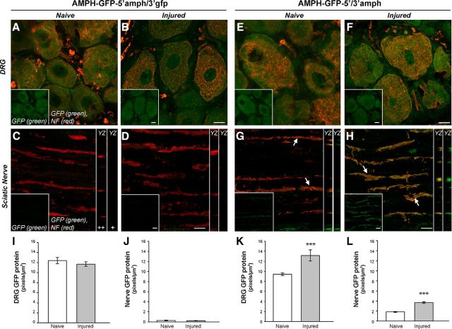
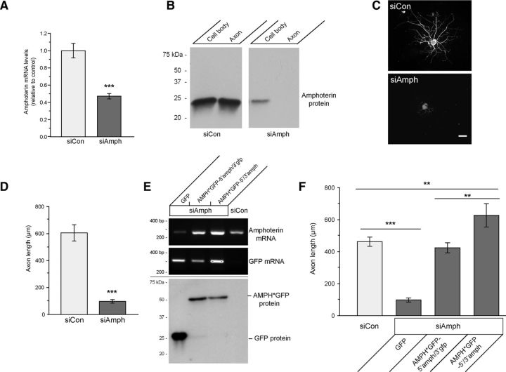
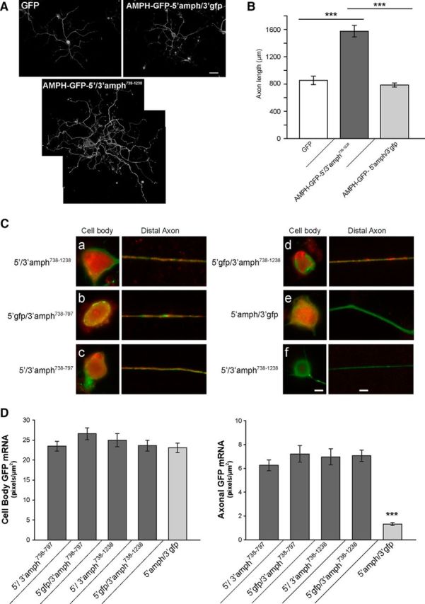
High mobility group (HMG) proteins concentrate in the nucleus, interacting with chromatin. Amphoterin is an HMG protein (HMGB1) that has been shown to have extranuclear functions and can be secreted from some cell types. Exogenous amphoterin can increase neurite growth, suggesting that the secreted protein may have growth promoting activities in neurons. Consistent with this, we show that depletion of amphoterin mRNA from cultured adult rat DRG neurons attenuates neurite outgrowth, pointing to autocrine or paracrine mechanisms for its growth-promoting effects. The mRNA encoding amphoterin localizes to axonal processes and we showed recently that its 3'-UTR is sufficient for axonal localization of heterologous transcripts (Donnelly et al., 2013). Here, we show that amphoterin mRNA is transported constitutively into axons of adult DRG neurons. A preconditioning nerve injury increases the levels of amphoterin protein in axons without a corresponding increase in amphoterin mRNA in the axons. A 60 nucleotide region of the amphoterin mRNA 3'-UTR is necessary and sufficient for its localization into axons of cultured sensory neurons. Amphoterin mRNA 3'-UTR is also sufficient for axonal localization in distal axons of DRG neurons in vivo. Overexpression of axonally targeted amphoterin mRNA increases axon outgrowth in cultured sensory neurons, but axon growth is not affected when the overexpressed mRNA is restricted to the cell body.

Keywords: HMGB1; axon regeneration; high mobility group protein; mRNA translation; mRNA transport.


Figures

Figure 1.
Figure 1.
Amphoterin protein is enriched in axons of injury-conditioned neurons. A, Axonal versus cell body compartments were used for isolation of protein from DRG cultures. By immunoblotting, amphoterin protein is shown to be cell body predominant in naive DRG cultures and axon predominant in injury-conditioned DRG cultures. Replicate blots probed with anti-Erk 1/2 shows approximately equal loading between the naive and injury-conditioned lysates. B, C, Representative, exposure-matched epifluorescent images of naive and injury-conditioned DRG cultures stained for amphoterin (red) and NF (green) protein are shown in B. Consistent with the immunoblotting in A, amphoterin protein is higher in cell bodies of naive compared with injury-conditioned neurons. There is a clear increase in amphoterin protein in the axons of the injury-conditioned neurons and the signal appears concentrated along the periphery of the axon (arrowheads). Cell body signals are noted along the cell periphery (arrows) and in the nucleus of the naive neurons, whereas the injury-conditioned neurons show predominantly cell periphery signals (arrows). Quantification immunoreactivity from exposure-matched image sets in C shows increased levels of amphoterin protein in axons of injury-conditioned versus naive DRG cultures, whereas cell bodies show a decrease in amphoterin protein in injury-conditioned versus naive DRG cultures (n ≥ 25 for cell body and n ≥ 30 for axons from at least 3 separate experiments; **p ≤ 0.01, ***p ≤ 0.001 by Student's t test) Scale bars, 10 μm. D, Representative, exposure-matched confocal images of L4 DRGs and sciatic nerves of naive and 7 d postsciatic nerve crush animals with the amphoterin protein in red and the NF protein in green. Similar to the cultured neurons in B, there is a clear increase in axonal amphoterin protein (arrows) in the injured compared with naive nerve. Amphoterin protein signals in the neuronal perikaryon also decrease in the injured compared with naive DRGs. Satellite cells in the DRG show a strong, apparently nuclear signal for amphoterin that similarly declines with injury (arrowheads). E, Quantification of amphoterin protein signals from the tissue sections comparing axonal levels in naive versus injured nerve and neuronal cell body levels in naive versus injured DRGs (n ≥ 25 for cell body and n ≥ 30 for axons from at least 3 separate experiments; **p ≤ 0.01, ***p ≤ 0.001 by Student's t test). Scale bars: DRG panels, 25 μm; sciatic nerve panels, 10 μm.
Figure 2.
Figure 2.
Nuclear amphoterin protein shifts to cytoplasm in injury-conditioned DRGs. Confocal microscopy was used to gain a better assessment of amphoterin protein localization in DRG neurons and glial cells. A, Representative single optical planes through center of neuronal perikaryon and nucleus of naive (left column) and injury-conditioned (right column) DRG cultures that were immunostained for amphoterin protein and counterstained with SYTOX to highlight the nucleus. The optically sectioned nucleus is marked with an asterisk in the first row. The cytoplasm of the neurons is outlined with a dashed line in the fluorescent images and marked with arrows in the images merged with DIC (fourth row). Note that amphoterin signal is strongly nuclear in the naive DRG neuron but shifts to more cytoplasmic in the injury-conditioned DRG neuron. B, Representative single optical planes taken through the center of the Schwann cells in these DRG cultures (left, naïve; right, injury-conditioned). The optically sectioned Schwann cell nuclei are marked with asterisks in the first row. The cytoplasm of the Schwann cell is outlined with a dashed line in the images. The Schwann cell amphoterin signal is strongly nuclear in the naive DRG neuron, but also shifts to more cytoplasmic in the Schwann cells after injury-conditioning. Scale bars, 10 μm.
Figure 3.
Figure 3.
Axonal levels of amphoterin mRNA do not change with injury. A, B, Neuronal cultures prepared from 7 d injury-conditioned versus naive DRGs were used for fractionation of cell body versus axonal RNA. RT-qPCR shows β-actin in both cell body and axonal preparations, but γ-actin and MAP2 RT-qPCR products were only detected in the cell body preparations (A). By RT-qPCR, amphoterin mRNA levels show no significant differences comparing cell body preparations or axonal preparations from the naive versus injury-conditioned neurons (B). C, Representative exposure-matched images of FISH/IF for amphoterin mRNA (red) and NF protein (green). Large panels show merged images and insets show only the RNA signal. There are overall comparable signals for amphoterin mRNA in axon shaft and growth cones of the naive versus injury-conditioned DRG neurons. Axonal amphoterin mRNA appears granular in the axon shaft (arrows) and growth cone (arrowheads) Scale bars, 10 μm. D, E, Representative RT-qPCR for naive versus 7 d postsciatic nerve crush injury for L4–L5 DRGs and sciatic nerve proximal to crush site. GAPDH was used as a loading control and the absence of MAP2 mRNA amplification from nerve samples confirms axonal nature of these nerve RNA preparations (D). RT-qPCR showed no significant alterations in amphoterin mRNA in the L4–L5 DRGs or sciatic nerve in vivo comparing the naive versus 7 d axotomized samples (E). F, Representative confocal XYZ images of FISH/IF signals for naive and 7 d post-crush-injury sciatic nerve are shown (matched for laser power, PMT gain/offset). Amphoterin mRNA is displayed in red and NF protein is shown in green, with merged mRNA and NF in main images and mRNA only for insets. Intraaxonal amphoterin mRNA (arrows) is seen in both the naive and injured nerve, with no apparent differences in abundance between the two. Scale bars, 10 μm.
Figure 4.
Figure 4.
A 60 nt segment of amphoterin 3′-UTR is sufficient for axonal localization in DRG neurons. A, Representative exposure-matched FISH/IF images for axons of neurons transfected with indicated GFPMYR-5′camkII/3′-amph constructs. GFP mRNA is shown in red and NF protein in green (antisense probes are shown in AaAd and sense cRNA probe is shown in Ae). Quantifications of GFP RNA FISH signals in DRG axons across multiple experiments are shown in B as average ± SEM (n ≥ 30 over three independent experiments; ***p ≤ 0.001 by Student's t test). Scale bar, 10 μm. CF, Postbleach signals from FRAP studies for distal axons of DRG neurons transfected with the same GFPMYR constructs as in A are shown. In each case, the ROI was at least 400 μm from the cell body. Average signal intensity normalized to prebleach and postbleach signal for each experiment is shown; error bars indicate the SEM of normalized data (n ≥ 7 over at least 3 independent experiments). CHX, Cultures pretreated with 150 μg/ml cycloheximide for 20 min before photobleaching (n ≥ 4 over at least 3 independent experiments; *p ≤ 0.05, **p ≤ 0.01, and ***p ≤ 0.001 for indicated time points compared with t = 0 min postbleach by repeated-measures ANOVA with Bonferroni post hoc comparisons).
Figure 5.
Figure 5.
Amphoterin 3′-UTR is needed for axonal localization in vivo. AH, Representative, exposure-matched confocal images of L4–L5 DRGs and distal sciatic nerve for animals transduced with LV encoding amphoterin-GFP fusion protein plus 5′-UTR of amphoterin and 3′-UTRs of GFP or amphoterin are shown [AMPH-GFP-5′amph/3′gfp (AD) and AMPH-GFP-5′/3′-amph (EH), respectively), with NF protein in red and GFP in green. All samples are 14 d post-LV transduction of L4–L5 nerve roots; naive (A, C, E, G) and 7 d after sciatic nerve crush (B, D, F, H) samples are shown. The top shows confocal XYZ images of L4–L5 DRGs, with merged channels in main panel and GFP signal only in the insets; bottom shows XYZ projections of sciatic nerve as merge of GFP and NF signals in the main panels and GFP only in the inset (lower left of each panel). YZ orthogonal projections of GFP + NF (++) and GFP only (+) are shown as strip images adjacent to each sciatic nerve image in C. GFP signals are seen in the DRGs cell bodies expressing either AMPH-GFP-5′amph/3′gfp (A, B) or AMPH-GFP-5′/3′-amph (C, D). However, the sciatic nerve only shows GFP signals in AMPH-GFP-5′/3′-amph-expressing animals (arrows), with a clear increase after nerve crush injury (G, H) Scale bars: DRG panels, 25 μm; sciatic nerve panels, 10 μm; orthogonal projections are at same scale as main panels. IL, Quantification of GFP signal intensity is shown for DRG (I, K) and sciatic nerves (J, L) for AMPH-GFP-5′amph/3′gfp-expressing animals (I, J) and AMPH-GFP-5′/3′-amph-expressing animals (K, L). Values represent mean intensity ± SEM from images matched for exposure, gain, offset, and after processing (n ≥ 30 from at least three separate experiments; **p ≤ 0.01; ***p ≤ 0.001 by Student's t test).
Figure 6.
Figure 6.
Amphoterin depletion decreases axonal outgrowth. A, B, siAmph mRNA-transfected DRG neuron cultures show significantly decreased amphoterin mRNA by RT-qPCR compared with nontargeting control (siCon) (A); there is also a clear decrease in amphoterin protein with siAmph compared with siCon in cell body and axonal preparations by immunoblotting (B). C, D, Representative images for NF immunofluorescence for DRG neurons transfected with siCon and siAmph (C); quantitation of the longest axon per neuron over multiple transfection experiments is also shown (D) for siAmph versus siCon; there is a marked reduction in axon length with depletion of amphoterin (***p ≤ 0.001 by Student's t test for n ≥ 30 over 3 independent experiments). Scale bar, 100 μm. E, F, siRNA-resistant Amphoterin-GFP constructs (AMPH*GFP) was used to test for potential off target effects of the siAmph. Cell-body-restricted (AMPH*GFP-5′amph/3′gfp) or axonally targeted (AMPH*GFP-5′/3′-amph) constructs were used to distinguish rescue effects of axonally synthesized AMPH*GFP. RT-qPCR shows rescue of amphoterin mRNA levels upon expression of AMPH*GFP-5′amph/3′gfp and AMPH*GFP-5′/3′-amph (E, top and middle, respectively). AMPH*GFP protein expression is comparable between the siAmph + AMPH*GFP-5′amph/3′gfp and siAmph + AMPH*GFP-5′/3′-amph transfected cultures (E, bottom). Length of longest axon in siAmph + GFP, AMPH*GFP-5′amph/3gfp, and AMPH*GFP-5′/3′-amph cotransfected cultures is shown in F. Both AMPH*GFP-5′amph/3′gfp and the AMPH*GFP-5′/3′-amph transfection rescue the axon growth deficit of siAmph transfection; however, axon lengths in the cultures expressing the axonally targeted AMPH*GFP-5′/3′-amph exceed those in control cultures (n ≥ 30 over 3 independent experiments; **p ≤ 0.01 and ***p ≤ 0.001 by Student's t test).
Figure 7.
Figure 7.
Overexpression of axonally targeted amphoterin mRNA increases axon growth. A, B, Representative montage images of NF-H-immunostained DRG neurons are shown in A for transfections with GFP, AMPH-GFP-5′/3′-amph738–1238, AMPH-GFP-5′amph/3′gfp. Average length of the longest axon ± SEM for the neurons expressing GFP, AMPH-GFP-5′/3′-amph738–1238, AMPH-GFP-5′amph/3′gfp is shown in B (n ≥ 30 neurons in 3 separate experiments; ***p ≤ 0.001 by Student's t test). Scale bar, 100 μm. C, D, Exposure-matched FISH/IF images for cell body (left) and distal axon (right) of DRG neurons transfected with AMPH-GFP plus the indicated 5′ and 3′-UTRs are shown in C. GFP mRNA is shown red and NF protein is shown in green (ae show antisense GFP probes and f shows sense GFP probe). Axonal GFP mRNA is only seen for AMPH-GFP that includes nt 738–797 from amphoterin mRNA 3′-UTR. Quantitation of GFP mRNA intensity in DRG axons and cell body is shown as average ± SEM for cell bodies and axons as indicated (D). There are no significant differences in cell body GFP RNA levels between the different UTR-containing constructs. All constructs containing amphoterin nt 738–797 and 738–1238 show significantly higher GFP mRNA in axons than the AMPH-GFP-5′amph/3′gfp. The axonal levels for AMPH-GFP-5′amph/3′gfp are comparable to those for GFPMYR3′-amph797–993 and GFPMYR3′-amph993–1238 shown in Figure 3F (n ≥ 30 processes in three experiments; ***p ≤ 0.001 by Student's t test). Scale bars: cell body, 20 μm; distal axon, 10 μm.

References

    1. Aakalu G, Smith WB, Nguyen N, Jiang C, Schuman EM. Dynamic visualization of local protein synthesis in hippocampal neurons. Neuron. 2001;30:489–502. doi: 10.1016/S0896-6273(01)00295-1. - DOI - PubMed
    1. Andreassi C, Zimmermann C, Mitter R, Fusco S, De Vita S, Saiardi A, Riccio A. An NGF-responsive element targets myo-inositol monophosphatase-1 mRNA to sympathetic neuron axons. Nat Neurosci. 2010;13:291–301. doi: 10.1038/nn.2486. - DOI - PubMed
    1. Bell CW, Jiang W, Reich CF, 3rd, Pisetsky DS. The extracellular release of HMGB1 during apoptotic cell death. Am J Physiol Cell Physiol. 2006;291:C1318–C1325. doi: 10.1152/ajpcell.00616.2005. - DOI - PubMed
    1. Ben-Yaakov K, Dagan SY, Segal-Ruder Y, Shalem O, Vuppalanchi D, Willis DE, Yudin D, Rishal I, Rother F, Bader M, Blesch A, Pilpel Y, Twiss JL, Fainzilber M. Axonal transcription factors signal retrogradely in lesioned peripheral nerve. EMBO J. 2012;31:1350–1363. doi: 10.1038/emboj.2011.494. - DOI - PMC - PubMed
    1. Blesch A. Lentiviral and MLV based retroviral vectors for ex vivo and in vivo gene transfer. Methods. 2004;33:164–172. doi: 10.1016/j.ymeth.2003.11.005. - DOI - PubMed

Publication types

MeSH terms

LinkOut - more resources