. 2015 Jun 15;308(12):R1021-33.
doi: 10.1152/ajpregu.00445.2014. Epub 2015 Apr 8.

The effect of spinal cord injury on the neurochemical properties of vagal sensory neurons

Affiliations

The effect of spinal cord injury on the neurochemical properties of vagal sensory neurons

April N Herrity et al. Am J Physiol Regul Integr Comp Physiol. .

Abstract

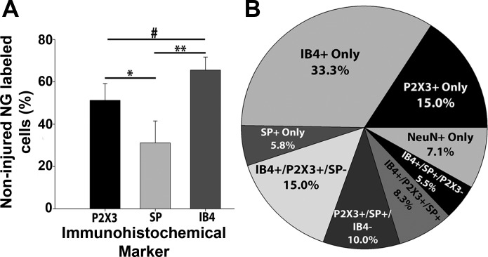
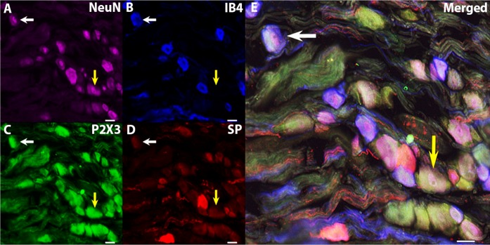
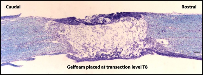
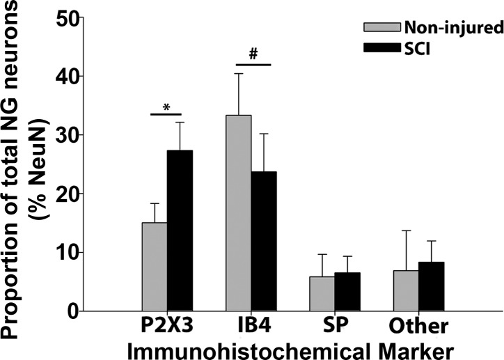
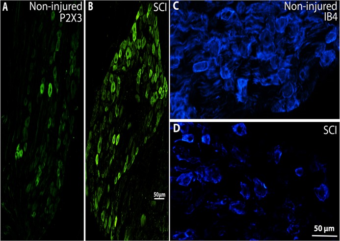
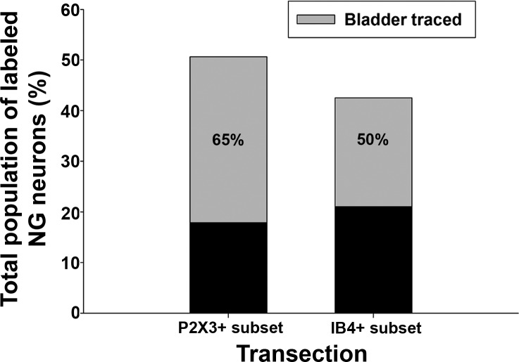
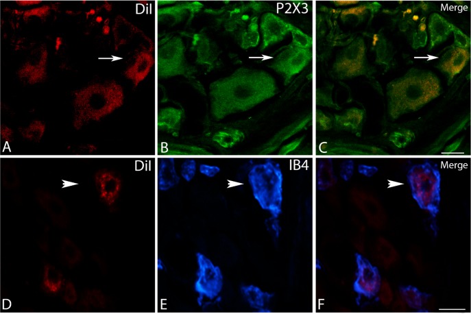
The vagus nerve is composed primarily of nonmyelinated sensory neurons whose cell bodies are located in the nodose ganglion (NG). The vagus has widespread projections that supply most visceral organs, including the bladder. Because of its nonspinal route, the vagus nerve itself is not directly damaged from spinal cord injury (SCI). Because most viscera, including bladder, are dually innervated by spinal and vagal sensory neurons, an impact of SCI on the sensory component of vagal circuitry may contribute to post-SCI visceral pathologies. To determine whether SCI, in male Wistar rats, might impact neurochemical characteristics of NG neurons, immunohistochemical assessments were performed for P2X3 receptor expression, isolectin B4 (IB4) binding, and substance P expression, three known injury-responsive markers in sensory neuronal subpopulations. In addition to examining the overall population of NG neurons, those innervating the urinary bladder also were assessed separately. All three of the molecular markers were represented in the NG from noninjured animals, with the majority of the neurons binding IB4. In the chronically injured rats, there was a significant increase in the number of NG neurons expressing P2X3 and a significant decrease in the number binding IB4 compared with noninjured animals, a finding that held true also for the bladder-innervating population. Overall, these results indicate that vagal afferents, including those innervating the bladder, display neurochemical plasticity post-SCI that may have implications for visceral homeostatic mechanisms and nociceptive signaling.

Keywords: bladder; immunohistochemical phenotype; nodose ganglion; spinal cord injury; vagus nerve.


Figures

Fig. 1.
Fig. 1.
Immunohistochemical representation of P2X3, isolectin B4 (IB4), and substance P (SP) in nodose ganglion (NG) neurons. A: following staining of the immunohistochemical markers P2X3, SP, and IB4, the bar graph demonstrates that all three were well represented in the NG, with the majority being IB4+ (IB4 vs. P2X3, #P < 0.05; IB4 vs. SP, **P < 0.001; P2X3 vs. SP, *P < 0.01). Values are means ± SD; n = 3 rats and 6 ganglia (one-way ANOVA with Tukey post hoc t-tests). B: pie graph depicting all possible combinations of the molecular targets in the NG. Note that neurons that were IB4+ only were the most prevalent, and most NG neurons were labeled with at least one of the three cellular targets examined.
Fig. 2.
Fig. 2.
Quadruple immunohistochemical staining in the NG. A: NeuN. B: IB4. C: P2X3. D: SP. E: merged. A confocal image displays the typical staining within the NG. NeuN was used to label all neurons. Different histochemical combinations include neurons that were IB4+, P2X3+, but SP− (white arrows), and neurons that were IB4−, P2X3+, and SP− (yellow arrows). Scale bar indicates 25 μm (×20 objective).
Fig. 3.
Fig. 3.
Spinal transection injury at T8. An 18-μm-thick sagittal section stained with the Kluver-Barrera method illustrates a complete spinal transection at T8. Gelfoam is placed in the lesion cavity to prevent contact from between rostral and caudal spinal cord tissue. The scale bar indicates 500 μm (×4 objective).
Fig. 4.
Fig. 4.
The effect of spinal cord injury (SCI) on number of NG neurons expressing the individual markers. Following SCI, there was an increase in P2X3-immunoreactivity (ir) in the transected group relative to intact/normal (SCI, 27.3 ± 4.8% vs. noninjured, 15.0 ± 3.3%, *P < 0.001) and a decrease in IB4 binding in the transected group relative to intact/normal (SCI, 23.7 ± 6.5% vs. noninjured, 33.3 ± 7.1%, #P < 0.05). No changes were apparent for SP. The “Other” category represents neurons that were NeuN+, but did not express or bind any of the markers. Values are means ± SD; n = 6 rats and 12 ganglia.
Fig. 5.
Fig. 5.
The effect of SCI on P2X3 and IB4 in the NG. An example displaying P2X3-ir (A) and IB4 binding (C) in the NG and following chronic spinal cord transection injury at T8 (B and D, respectively) is shown. Note the presence of increased P2X3-ir and decreased IB4 binding post-SCI. Images of sections from both SCI and noninjured animals were stained and captured with the same protocols and at the same time (×10 objective).
Fig. 6.
Fig. 6.
Superior cervical ganglion. There was no evidence of 1,1′-dilinoleyl-3,3,3′,3′-tetramethylindo-carbocyanine perchlorate (DiI) punctate labeling present in the superior cervical ganglion. The scale bar represents 20 μm (×20 objective).
Fig. 7.
Fig. 7.
Effect of SCI on bladder-traced NG neurons. The graph demonstrates that, out of the total percentage of either P2X3 or IB4 subsets after injury, more than one-half of the neurons were traced from bladder. Bladder innervating neurons in the P2X3+ subset represent 32.8 ± 1.1%, while, in the IB4+ subset, they represent 42.6 ± 5.1%. n = 3 rats and 6 ganglia.
Fig. 8.
Fig. 8.
P2X3-ir and IB4 binding in bladder-traced NG neurons after transection. A confocal image illustrates a DiI+ neuron in A that is also immunoreactive for P2X3 in B (white arrows). C: demonstration of the overlay. An image from the inverted Nikon microscope illustrates a DiI+ neuron in D that also binds IB4 in E (white arrowhead). F: demonstration of the overlay. One example of each is displayed. In both images, the scale bar indicates 25 μm (×20 objective).

References

    1. Altschuler SM, Bao XM, Bieger D, Hopkins DA, Miselis RR. Viscerotopic representation of the upper alimentary tract in the rat: sensory ganglia and nuclei of the solitary and spinal trigeminal tracts. J Comp Neurol 283: 248–268, 1989. - PubMed
    1. Altschuler SM, Escardo J, Lynn RB, Miselis RR. The central organization of the vagus nerve innervating the colon of the rat. Gastroenterology 104: 502–509, 1993. - PubMed
    1. Amir R, Devor M. Functional cross-excitation between afferent A- and C-neurons in dorsal root ganglia. Neuroscience 95: 189–195, 2000. - PubMed
    1. Anderson KD. Targeting recovery: priorities of the spinal cord-injured population. J Neurotrauma 21: 1371–1383, 2004. - PubMed
    1. Apodaca G, Kiss S, Ruiz W, Meyers S, Zeidel M, Birder L. Disruption of bladder epithelium barrier function after spinal cord injury. Am J Physiol Renal Physiol 284: F966–F976, 2003. - PubMed

Publication types

LinkOut - more resources