. 2015 Apr 28;112(17):5395-400.
doi: 10.1073/pnas.1420508112. Epub 2015 Apr 8.

Cadherin flexibility provides a key difference between desmosomes and adherens junctions

Affiliations

Cadherin flexibility provides a key difference between desmosomes and adherens junctions

Humera Tariq et al. Proc Natl Acad Sci U S A. .

Abstract

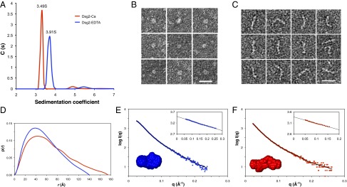
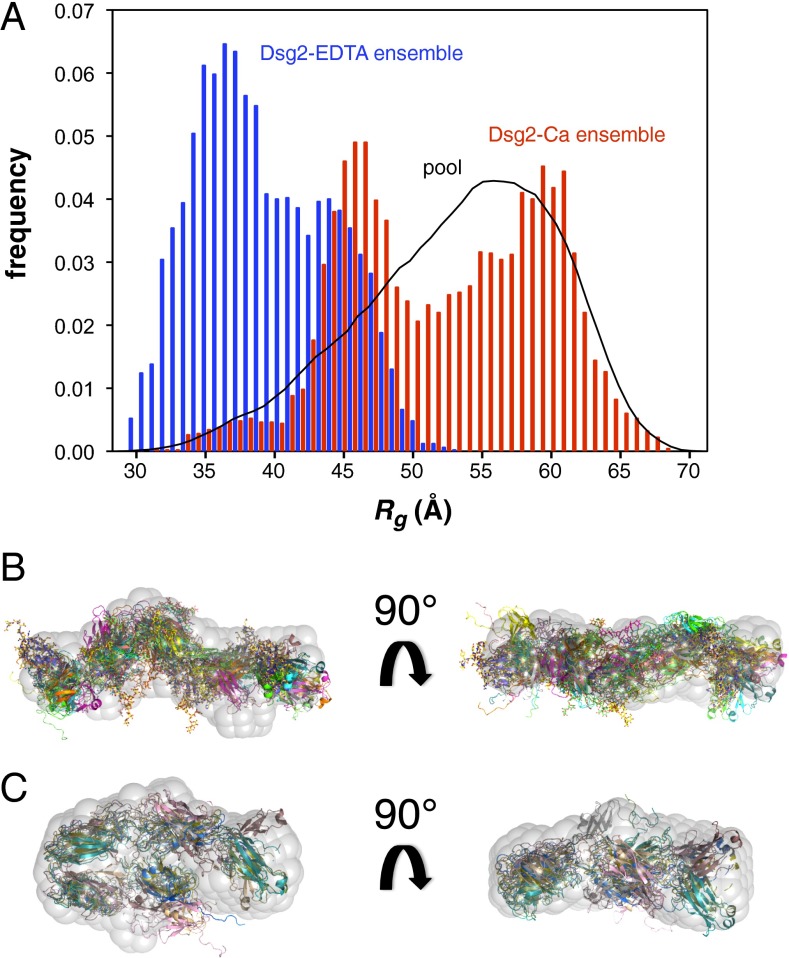
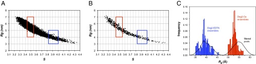
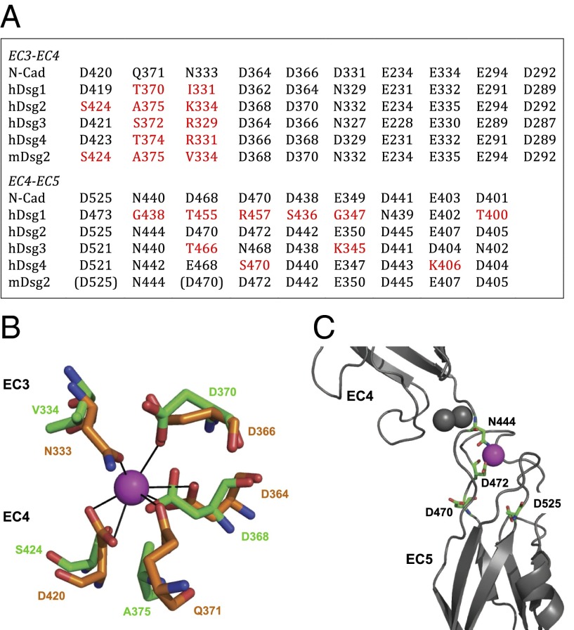
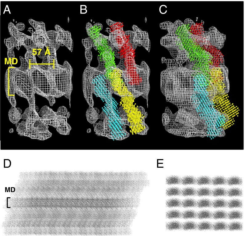
Desmosomes and adherens junctions are intercellular adhesive structures essential for the development and integrity of vertebrate tissue, including the epidermis and heart. Their cell adhesion molecules are cadherins: type 1 cadherins in adherens junctions and desmosomal cadherins in desmosomes. A fundamental difference is that desmosomes have a highly ordered structure in their extracellular region and exhibit calcium-independent hyperadhesion, whereas adherens junctions appear to lack such ordered arrays, and their adhesion is always calcium-dependent. We present here the structure of the entire ectodomain of desmosomal cadherin desmoglein 2 (Dsg2), using a combination of small-angle X-ray scattering, electron microscopy, and solution-based biophysical techniques. This structure reveals that the ectodomain of Dsg2 is flexible even in the calcium-bound state and, on average, is shorter than the type 1 cadherin crystal structures. The Dsg2 structure has an excellent fit with the electron tomography reconstructions of human desmosomes. This fit suggests an arrangement in which desmosomal cadherins form trans interactions but are too far apart to interact in cis, in agreement with previously reported observations. Cadherin flexibility may be key to explaining the plasticity of desmosomes that maintain tissue integrity in their hyperadhesive form, but can adopt a weaker, calcium-dependent adhesion during wound healing and early development.

Keywords: adhesion; cadherins; desmosomes; small-angle X-ray scattering; structure.


Conflict of interest statement

The authors declare no conflict of interest.

Figures

Fig. 1.
Fig. 1.
Structural and biophysical analyses of mDsg2. (A) Fitted distribution of the sedimentation coefficients for Dsg2-EDTA (blue) and Dsg2-Ca (red). The higher sedimentation coefficient in the absence of Ca2+ indicates a more compact particle, consistent with the lower Rh of 4.1 nm and a f/f0 of 1.54 compared with the sample with Ca2+ (Rh, 4.6 nm; f/f0, 1.72). Representative EM images of Dsg2-EDTA (B) and Dsg2-Ca (C) samples after negative staining. The Dsg2-EDTA particles appear as compact globules, whereas the Dsg2-Ca particles display a range of morphologies, some of them bent or curled at the ends. (Scale bar, 20 nm.) (D) The p(r) distribution plot for Dsg2-EDTA (blue) and Dsg2-Ca (red) shows Dmax of 140 and 175 Å, respectively. SAXS profiles showing the log of X-ray scattering intensity (logI) as a function of the scattering vector q for the experimental scattering data Dsg2-EDTA (blue) (E) and Dsg2-Ca (red) (F). The normalized fit to the experimental data (from the program GNOM) is superimposed as a black line). (Inset, top right) Guinier plot (logI vs. q2) of the low q region of the X-ray scattering data where the radius of gyration (Rg) can be measured from the gradient of the slope (Rg2/3). (Inset, bottom left) Average DAMMIN models for Dsg2-Ca (red) and Dsg2-EDTA (blue).
Fig. 2.
Fig. 2.
EOM ensemble analysis. (A) Rg distribution of the random pool and selected ensembles for Dsg2-EDTA (blue) and Dsg2-Ca (red) generated with EOM. Ab initio shape restoration of Dsg2-Ca (B) and Dsg2-EDTA (C); superimposed are the models of the final ensemble.
Fig. 3.
Fig. 3.
Pool filtering analysis. Plots of Rg vs. sedimentation coefficients (s) as calculated with SoMo for the Ranch model pool (A) and the TAMD model pool (B). Dots represent the pairwise values for each individual model generated. The range of experimentally determined values for s (±1σ) is shown as red boxes for Dsg2-Ca and blue boxes for Dsg2-EDTA. (C) Rg distribution of EOM ensembles for the filtered subsets generated by Ranch (solid bars) and by TAMD with CNS (open bars).
Fig. 4.
Fig. 4.
Calcium coordination in Dsg2 is not conserved. (A) Table listing the residues involved in EC3–EC4 and EC4–EC5 interdomain calcium coordination. Nonconservative substitutions in mDsg2 and hDsgs are in red; residues located too far for proper coordination are in brackets. (B) Side chain coordination of one of the three Ca2+ ions at the EC3–EC4 region; residues coordinating the same Ca2+ ion (purple) in N-cadherin (PDB ID code 3Q2W, orange chain) and in the mDsg2 homology model in green. (C) Coordination of one of the three Ca2+ ions at the EC4–EC5 region; the two Ca2+ binding residues D470 and D525 located in loop regions are too distant from the calcium ion, leaving only residues N444 and D472 available for coordination. Gray spheres are the other two Ca2+ ions.
Fig. 5.
Fig. 5.
Fitting of the ab initio model into the desmosome tomography maps. (A) The electron density maps derived from the ET of the human native epidermal desmosome (14) were used to fit the DAMMIN model for Dsg2-Ca. Excellent fit was obtained, as shown in B, where four DAMMIN models illustrate the trans dimer interactions across the midline (MD). (C) Side view of the four DAMMIN models shown in B. Side (D) and top (E) view projections of an idealized array generated with the DAMMIN models from the fitting into the ET maps. A midline (MD) is clearly visible on the side view projection.

References

    1. Ivanov AI, Naydenov NG. Dynamics and regulation of epithelial adherens junctions: Recent discoveries and controversies. Int Rev Cell Mol Biol. 2013;303:27–99. - PubMed
    1. Kowalczyk AP, Green KJ. Structure, function, and regulation of desmosomes. Prog Mol Biol Transl Sci. 2013;116:95–118. - PMC - PubMed
    1. Boggon TJ, et al. C-cadherin ectodomain structure and implications for cell adhesion mechanisms. Science. 2002;296(5571):1308–1313. - PubMed
    1. Taveau JC, et al. Structure of artificial and natural VE-cadherin-based adherens junctions. Biochem Soc Trans. 2008;36(Pt 2):189–193. - PMC - PubMed
    1. Rayns DG, Simpson FO, Ledingham JM. Ultrastructure of desmosomes in mammalian intercalated disc; appearances after lanthanum treatment. J Cell Biol. 1969;42(1):322–326. - PMC - PubMed

Publication types

LinkOut - more resources