. 2015 Jun 1;113(10):3943-53.
doi: 10.1152/jn.01055.2014. Epub 2015 Apr 8.

Closed-loop, ultraprecise, automated craniotomies

Affiliations

Closed-loop, ultraprecise, automated craniotomies

Nikita Pak et al. J Neurophysiol. .

Abstract

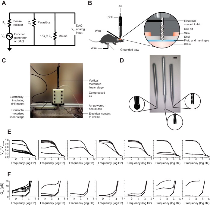
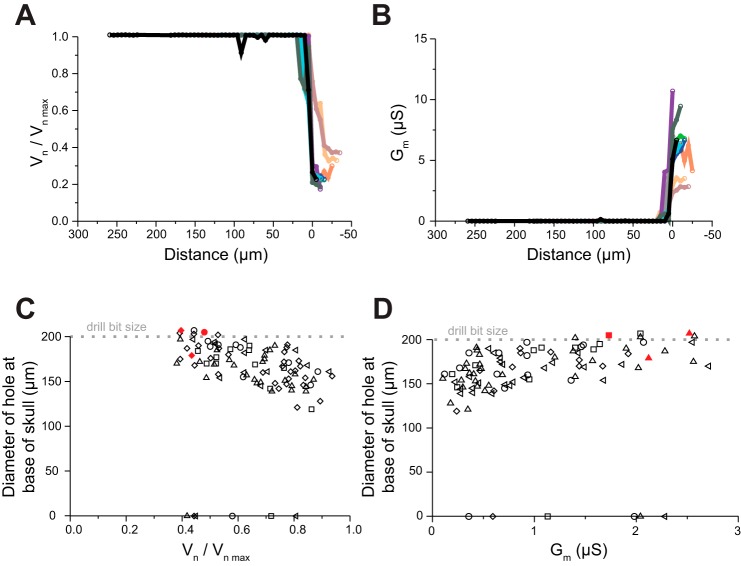
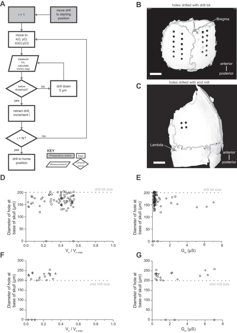
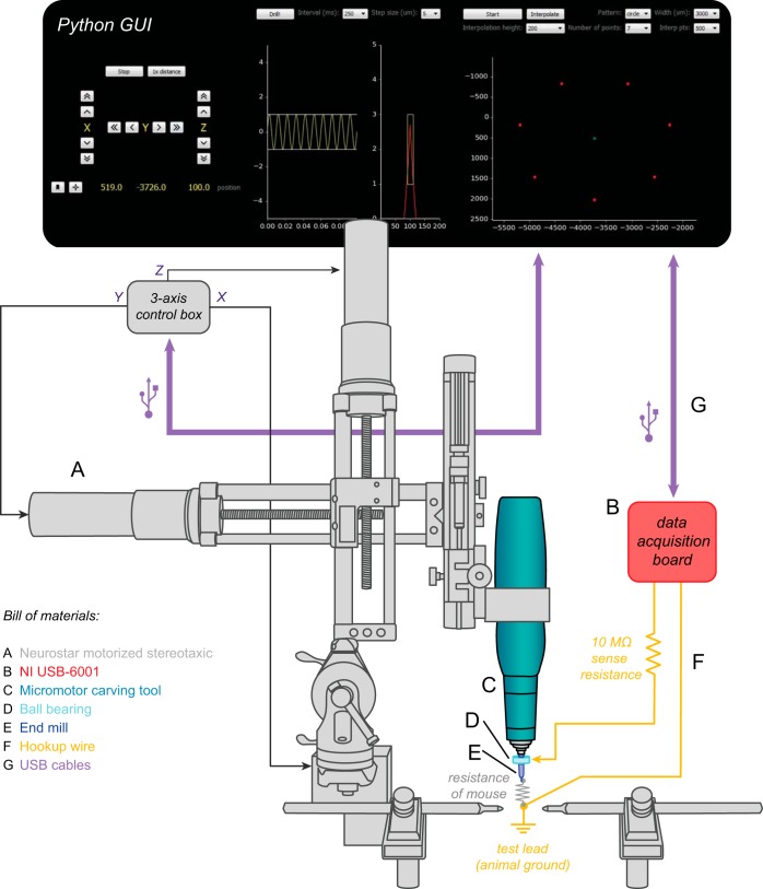
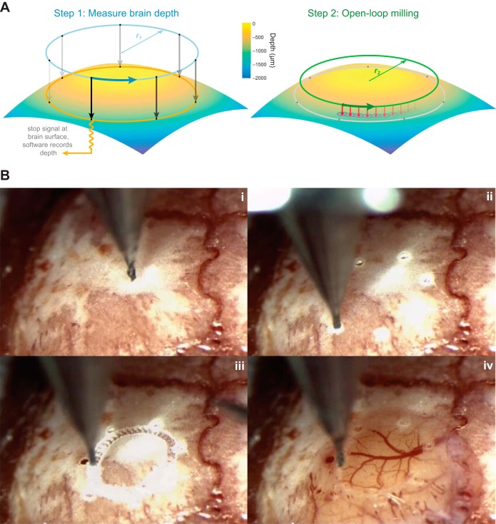
A large array of neuroscientific techniques, including in vivo electrophysiology, two-photon imaging, optogenetics, lesions, and microdialysis, require access to the brain through the skull. Ideally, the necessary craniotomies could be performed in a repeatable and automated fashion, without damaging the underlying brain tissue. Here we report that when drilling through the skull a stereotypical increase in conductance can be observed when the drill bit passes through the skull base. We present an architecture for a robotic device that can perform this algorithm, along with two implementations--one based on homebuilt hardware and one based on commercially available hardware--that can automatically detect such changes and create large numbers of precise craniotomies, even in a single skull. We also show that this technique can be adapted to automatically drill cranial windows several millimeters in diameter. Such robots will not only be useful for helping neuroscientists perform both small and large craniotomies more reliably but can also be used to create precisely aligned arrays of craniotomies with stereotaxic registration to standard brain atlases that would be difficult to drill by hand.

Keywords: automation; cranial windows; craniotomy; robotics.


Figures

Fig. 1.
Fig. 1.
Automated craniotomy robot design and implementation. A: diagram of electrical impedance measurement circuit. B: illustration of the experimental setup. C: photograph of the homebuilt automated craniotomy robot. D: drill bits used in this report, including a commercially available dental burr, 500 μm in diameter (left), a custom drill bit fusing a 200-μm tip with a custom aluminum dental drill adapter (center), and a custom 200-μm end mill created by using a lathe to turn down a commercially available bit (right). Scale bar, 1/16 in. E: normalized electric potential across the drill (acquired as schematized in B but with the electrical contact on the drill itself rather than the bit) and mouse, as a function of frequency, as a 500-μm dental burr is lowered into the skull for 7 different mice (step size: 10 μm for 6 mice, 50 μm for the 7th). Lower lines indicate lower drilling depth. F: electrical conductance vs. frequency (as in E) for all 7 mice. Higher lines indicate lower drilling depth.
Fig. 2.
Fig. 2.
The automated craniotomy threshold. A: normalized electric potential vs. distance traveled for 10 holes in 1 mouse skull, each represented by a different color (step size 5 μm, frequency 100 Hz). Traces were aligned (at x-axis = 0) at the point in the curve of maximum slope. B: electrical conductance vs. distance traveled (as in A) for all 10 craniotomies. C: hole size, measured at the base of the skull, measured with X-ray micro-computed tomography (CT), as a function of final normalized electric potential, with the drill stopping when various normalized electrical potentials were reached. n = 98 craniotomies in 5 mice; 200-μm drill bit (width indicated by dotted line). Each mouse is represented by a different shape, with red fill indicating visible blood related to the use of the standard pointed drill bit. For 6 of the 98 craniotomies the drill bit did not pass the bottom of the skull, and thus they are on the y = 0 line. D: hole size vs. electrical conductance for the data in C.
Fig. 3.
Fig. 3.
Implementation and validation of automated craniotomy algorithm. A: automated craniotomy algorithm flowchart. B: representative CT scan of a skull from D. Scale bar, 1 mm. C: representative CT scan of a skull from F. Scale bar, 1 mm. D: hole size as a function of final stopping normalized electric potential for 72 craniotomies in 3 mice with a 200-μm drill bit, step size of 5 μm, and normalized electrical potential threshold of 0.65. Each mouse is represented by a different shape. For 2 of the 72 craniotomies the drill bit did not pass the bottom of the skull, and thus they are at the y = 0 line. E: hole size vs. electrical conductance for the data in D. F: hole size as a function of final stopping normalized electric potential for 20 craniotomies in 5 mice with a 200-μm flat-end end mill, step size of 5 μm, and normalized electrical potential threshold of 0.45. Each mouse is represented by a different shape. For 3 of the 20 craniotomies the drill bit did not pass the bottom of the skull, and thus they are at the y = 0 line. G: electrical conductance vs. hole size for the data in F.
Fig. 4.
Fig. 4.
Implementation of automated craniotomy algorithm on a commercial motorized stereotaxic. A graphical user interface (GUI, top) controls the movement of the stereotaxic frame (A) via a 3-axis control box. A micromotor carving drill (C) with adjustable rotation speeds up to 45,000 rpm is attached to the stereotaxic via a custom adapter. The drill turns an end mill (E) with a tip diameter of 200 μm. When the end mill breaks through the skull, it completes the circuit formed between a wire carrying the 100-Hz test signal from a data acquisition board (B) and a test lead connected to the animal (F). The signal wire is attached to the drill bit via a ball bearing (D), allowing continuous impedance testing without the need to stop the drill.
Fig. 5.
Fig. 5.
Automated formation of large cranial windows. A: windows are created after drilling a series of test holes to account for the curvature of the skull (left). In this example, holes are drilled at 7 points along the circumference of a 3-mm circle (dots). Impedance-based feedback is used to measure the location of the brain along the z-axis at each point. Then, cubic spline-based interpolation is used to compute the optimal path for the drill to create a circular pattern in the bone without contacting the underlying tissue (right). The best results were achieved when the drill made a series of shallow, overlapping holes (red) rather than milling horizontally through the bone. B: the steps involved in creating a cranial window in an actual mouse skull (see Supplemental Movie S1 for a video of the windowing process). i: The skull is exposed and cleaned. ii: The center point and diameter of the desired cranial window are manually chosen by the surgeon, and several holes are automatically drilled along its circumference. iii: The drill automatically interpolates between the hole locations at the appropriate depth. iv: The skull is manually removed under saline.

References

    1. Brunoni AR, Nitsche MA, Bolognini N, Bikson M, Wagner T, Merabet L, Edwards DJ, Valero-Cabre A, Rotenberg A, Pascual-Leone A, Ferrucci R, Priori A, Boggio PS, Fregni F. Clinical research with transcranial direct current stimulation (tDCS): challenges and future directions. Brain Stimul 5: 175–195, 2012. - PMC - PubMed
    1. Chaieb L, Antal A, Paulus W. Transcranial alternating current stimulation in the low kHz range increases motor cortex excitability. Restor Neurol Neurosci 29: 167–175, 2011. - PubMed
    1. Chan SY, Bernstein JG, Boyden ES. Scalable fluidic injector arrays for viral targeting of intact 3-D brain circuits. J Vis Exp 35: 1489, 2010. - PMC - PubMed
    1. Cunha-Cruz V, Follmann A, Popovic A, Bast P, Wu T, Heger S, Engelhardt M, Schmieder K, Radermacher K. Robot- and computer-assisted craniotomy (CRANIO): from active systems to synergistic man-machine interaction. Proc Inst Mech Eng H 224: 441–452, 2010. - PubMed
    1. Holtmaat A, Bonhoeffer T, Chow DK, Chuckowree J, De Paola V, Hofer SB, Hübener M, Keck T, Knott G, Lee WC, Mostany R, Mrsic-Flogel TD, Nedivi E, Portera-Cailliau C, Svoboda K, Trachtenberg JT, Wilbrecht L. Long-term, high-resolution imaging in the mouse neocortex through a chronic cranial window. Nat Protoc 4: 1128–1144, 2009. - PMC - PubMed

Publication types