. 2015 May 22;290(21):13144-56.
doi: 10.1074/jbc.M115.647446. Epub 2015 Apr 8.

Hyaluronidase Hyal1 Increases Tumor Cell Proliferation and Motility through Accelerated Vesicle Trafficking

Affiliations

Hyaluronidase Hyal1 Increases Tumor Cell Proliferation and Motility through Accelerated Vesicle Trafficking

Caitlin O McAtee et al. J Biol Chem. .

Abstract

Hyaluronan (HA) turnover accelerates metastatic progression of prostate cancer in part by increasing rates of tumor cell proliferation and motility. To determine the mechanism, we overexpressed hyaluronidase 1 (Hyal1) as a fluorescent fusion protein and examined its impact on endocytosis and vesicular trafficking. Overexpression of Hyal1 led to increased rates of internalization of HA and the endocytic recycling marker transferrin. Live imaging of Hyal1, sucrose gradient centrifugation, and specific colocalization of Rab GTPases defined the subcellular distribution of Hyal1 as early and late endosomes, lysosomes, and recycling vesicles. Manipulation of vesicular trafficking by chemical inhibitors or with constitutively active and dominant negative Rab expression constructs caused atypical localization of Hyal1. Using the catalytically inactive point mutant Hyal1-E131Q, we found that enzymatic activity of Hyal1 was necessary for normal localization within the cell as Hyal1-E131Q was mainly detected within the endoplasmic reticulum. Expression of a HA-binding point mutant, Hyal1-Y202F, revealed that secretion of Hyal1 and concurrent reuptake from the extracellular space are critical for rapid HA internalization and cell proliferation. Overall, excess Hyal1 secretion accelerates endocytic vesicle trafficking in a substrate-dependent manner, promoting aggressive tumor cell behavior.

Keywords: cell motility; endocytosis; hyaluronan; hyaluronidase; lysosome; prostate cancer.


Figures

FIGURE 1.
FIGURE 1.
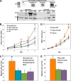
Expression of catalytically active Hyal1 increases proliferation and motility of prostate tumor cells. 22Rv1 prostate carcinoma cells were selected for stable overexpression of the empty vector encoding the reporter tdT or the wild-type Hyal1 (WT and Hyal1WT-tdT), Hyal1Y202F, or Hyal1E131Q, each as a fusion with tdT. A, soluble cell lysates, membrane-enriched fractions, and concentrated conditioned media were analyzed by Western blotting with equal amounts of total protein probed for Hyal1 (fusion protein molecular mass of ≈100 kDa) and normalized to tubulin. The specific band for Hyal1-tdT fusion protein in conditioned media is indicated by an arrowhead. B, proliferation was assayed by daily manual counts. Equal numbers of cells were seeded in quadruplicate wells of 96-well plates. Cell numbers are plotted as mean ± S.D. (error bars). C, cell motility was compared using a modified Boyden chamber assay. Lower wells contained type IV collagen as a chemotactic agent. Single cell suspensions were placed in the upper wells separated from the lower wells by a polycarbonate membrane with 8-μm pore size. The mean ± S.D. (error bars) is plotted for quadruplicate manual counts per cell line. D and E, proliferation and motility, respectively, were quantified for 22Rv1 cells stably expressing empty vector (pIRES2-EGFP), Hyal1WT, or Hyal1E131Q unfused proteins as indicated. *, p < 0.01 for Hyal1WT-tdT transfectant values relative to those of the tdT control line. MWM, molecular weight markers; IB, immunoblot.
FIGURE 2.
FIGURE 2.
Subcellular distribution of Hyal1 is affected by its catalytic competence. Stable 22Rv1 cell lines expressing an empty vector encoding the tdTomato fluorescent reporter or C-terminal fusion constructs of tdTomato with wild-type or mutant Hyal1 were imaged by live cell confocal fluorescence microscopy. Red fluorescence (left panel of each set) indicates localization of Hyal1WT-tdT fusion protein (A), Hyal1Y202F-tdT (B), Hyal1E131Q-tdT (C), or tdTomato protein (empty vector; D). Inset panels are zoomed images of the individual cells designated by a corresponding white square. In the right panels of each set, red channel fluorescence is superimposed on the corresponding transmitted light image. Scale bars, 10 μm.
FIGURE 3.
FIGURE 3.
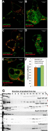
Characterization of intracellular Hyal1 distribution by confocal microscopy and biochemical fractionation. A–F, still images of live 22Rv1 cells expressing Hyal1WT-tdT (A and B), Hyal1Y202F-tdT (C and D), or Hyal1E131Q-tdT (E) were captured by confocal microscopy following treatment with either LysoTracker Green DND-26 (A and C) or ER-Tracker Green BODIPY FL glibenclamide (B, D, and E). Yellow color represents colocalization of signals. Strong colocalization of Hyal1WT-tdT and Hyal1Y202F-tdT was observed with LysoTracker Green (Lyso), whereas the Hyal1-E131Q-tdT merged with ER-Tracker Green (ER) as indicated by Pearson's correlation coefficient (F). Error bars indicate S.E. G, stable 22Rv1 transfectants expressing Hyal1WT-tdT or Hyal1E131Q-tdT were lysed and fractionated on a continuous sucrose gradient. Fractions were collected and analyzed by Western blotting with anti-Hyal1 polyclonal antibody. Blots were stripped and reprobed with antibodies to cathepsin D (lysosomes), calnexin (ER), and EEA1 (early endosomes). In each panel, MWM is the molecular weight marker, 800×g is the insoluble fraction of the cell lysate, and PNS indicates a sample of the postnuclear supernatant (soluble lysate) that was loaded on the gradient. Numbers correspond to the density fraction following centrifugation. Increasing numbers indicate increasing density. Scale bars, 10 μm.
FIGURE 4.
FIGURE 4.
Examination of Hyal1 trafficking itineraries by chemical inhibition of vesicular transport or vesicle acidification. A–D, stable 22Rv1 cell lines expressing Hyal1-tdTomato fusions were incubated in the absence (row A) or presence of brefeldin A (BFA) (row B), chloroquine (Chl) (row C), or bafilomycin A1 (BafA1) (row D). Representative images of cells are shown from cultures of Hyal1WT-tdT (left column), Hyal1Y202F-tdT (center column), and Hyal1E131Q-tdT (right column). Inset panels show magnified images of individual cells designated in white squares. Images are presented with red channel fluorescence isolated in black and white to increase sensitivity of visualization and facilitate analysis. Average vesicle sizes for Hyal1WT-tdT (E) and Hyal1Y202F-tdT (F) were quantified and compared in each condition. Error bars indicate S.E. Statistical significance is indicated on the graphs. Hyal1WT-tdT (G) and Hyal1E131Q-tdT (H) cells were cultured in standard media or the indicated inhibitor, then lysed, and fractionated by centrifugation on a continuous sucrose gradient. In each panel, MWM is the molecular weight marker, 800×g is the insoluble fraction of the cell lysate, and PNS indicates a sample of the postnuclear supernatant (soluble lysate) that was loaded on the gradient. Numbers correspond to fractions of increasing density following centrifugation. Fractions were analyzed by Western blotting with the polyclonal anti-Hyal1 antibody. Arrows indicate bands corresponding to the full-length (black) and processed (red) forms of Hyal1.
FIGURE 5.
FIGURE 5.
Colocalization of wild-type Hyal1 with Rab GTPases demonstrates its presence in endocytic and slow recycling vesicles as well as late endosomes and lysosomes. Stable Hyal1WT-tdT cells were transfected with constructs encoding GFP fusions to Rab5, Rab7, or Rab11. Colocalization of each Rab protein is shown by confocal imaging of Rab-GFP fusions (green) and Hyal1-tdT fusions (red) for Rab5 (A), Rab7 (B), and Rab11 (C). Yellow color indicates colocalization. Each set of images shows colocalization of wild-type Rab (left column), constitutively active (CA) Rab (center column), or dominant negative (DN) Rab (right column). Inset panels are magnified images of individual cells indicated by white squares. D, colocalization of Hyal1WT-tdT with each Rab-GFP construct was quantified by Pearson's coefficient and plotted as mean ± S.E. (error bars). Scale bars, 10 μm.
FIGURE 6.
FIGURE 6.
Hyaluronan internalization requires catalytically active Hyal1, and internalized HA vesicles colocalize with Hyal1. Confocal images are shown for uptake of fHA (green) by prostate tumor cells stably expressing Hyal1WT-tdT (A), Hyal1Y202F-tdT (B), Hyal1E131Q-tdT (C), or empty vector (D; tdTomato). Left panels show fHA uptake alone in green, and right panels are the corresponding merge. Inset panels are magnified images of individual cells designated by white squares in the merged panels. Scale bars, 10 μm.
FIGURE 7.
FIGURE 7.
Secreted Hyal1 is essential for efficient HA internalization. Stable 22Rv1 lines were incubated with fHA without prior treatment (A), immediately following treatment with brefeldin A (B), or after treatment with brefeldin A followed by a 30-min chase with media to allow recovery (C). In each row, representative Hyal1WT-tdT cell images are in the left column, Hyal1Y202F-tdT images are in the center column, and Hyal1E131Q-tdT images are on the right. Inset panels are magnified images of individual cells designated by white squares in the merged panels. The left inset shows the isolated green channel fluorescence in black and white indicating fHA uptake, and the right inset is the corresponding red channel signal indicating tdT localization. D, number of green vesicles per cell was determined using ImageJ for at least 10 cells per condition, each repeated three times and compared for statistical significance by one-way analysis of variance. Error bars indicate S.E. Scale bars, 10 μm. NT, no treatment; BFA, brefeldin A; rec, recovery.
FIGURE 8.
FIGURE 8.
Stable Hyal1 expression increases rates of HA uptake and transferrin internalization by prostate tumor cells. A, single cell suspensions of 22Rv1 prostate tumor cells stably expressing empty vector (tdT), Hyal1WT-tdT, Hyal1Y202F-tdT, or Hyal1E131Q-tdT were incubated in serum-free media (negative) or with media containing 100 nm fHA for the indicated times. HA uptake was monitored via internalized fluorescence intensity using flow cytometry. B, prostate tumor cells in A were incubated in serum-free media (negative) or with media containing 2.5 μg/ml transferrin-Alexa Fluor 633 for the incubation times indicated, and transferrin uptake was monitored via internalized fluorescence intensity using flow cytometry. In both panels, mean fluorescence intensity as plotted is 1000× higher than the value on the axis for presentation. Data are representative of experiments repeated at least four times each.
FIGURE 9.
FIGURE 9.
Proposed model for Hyal1 impact on vesicle trafficking and tumor cell motility. Hyal1 activity is virtually undetectable (basal) in parental 22Rv1 cells, which retain low levels of cell surface high molecular weight (HMW) HA and are poorly motile. When Hyal1 activity is elevated by overexpression, the increased secretion of Hyal1 leads to absence of surface high molecular weight HA; increased motility, proliferation, and receptor recycling; and accelerated vesicular trafficking that depends on Hyal1 binding to HA and/or chondroitin sulfate proteoglycans (CSPG) and may result in increased nutrient availability for biosynthesis as well as the release of partially digested, biologically potent low molecular weight (LMW) HA signals. EE, early endosome; LE, late endosome; RE, recycling endosome; PM, plasma membrane.

References

    1. Siegel R., Ma J., Zou Z., Jemal A. (2014) Cancer statistics, 2014. CA Cancer J. Clin. 64, 9–29 - PubMed
    1. McAtee C. O., Barycki J. J., Simpson M. A. (2014) Emerging roles for hyaluronidase in cancer metastasis and therapy. Adv. Cancer Res. 123, 1–34 - PMC - PubMed
    1. Aaltomaa S., Lipponen P., Tammi R., Tammi M., Viitanen J., Kankkunen J. P., Kosma V. M. (2002) Strong stromal hyaluronan expression is associated with PSA recurrence in local prostate cancer. Urol. Int. 69, 266–272 - PubMed
    1. Anttila M. A., Tammi R. H., Tammi M. I., Syrjänen K. J., Saarikoski S. V., Kosma V. M. (2000) High levels of stromal hyaluronan predict poor disease outcome in epithelial ovarian cancer. Cancer Res. 60, 150–155 - PubMed
    1. Auvinen P., Tammi R., Parkkinen J., Tammi M., Agren U., Johansson R., Hirvikoski P., Eskelinen M., Kosma V. M. (2000) Hyaluronan in peritumoral stroma and malignant cells associates with breast cancer spreading and predicts survival. Am. J. Pathol. 156, 529–536 - PMC - PubMed

Publication types

MeSH terms