. 2015 Apr 9;10(4):e0122723.
doi: 10.1371/journal.pone.0122723. eCollection 2015.

T lymphocyte density and distribution in human colorectal mucosa, and inefficiency of current cell isolation protocols

Affiliations

T lymphocyte density and distribution in human colorectal mucosa, and inefficiency of current cell isolation protocols

Gloria Cuevas Preza et al. PLoS One. .

Abstract

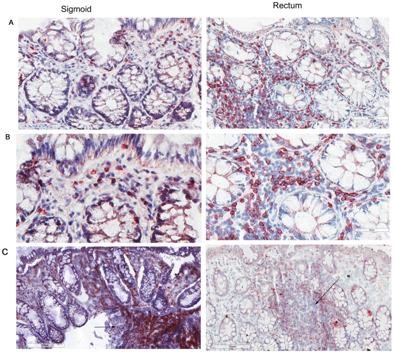
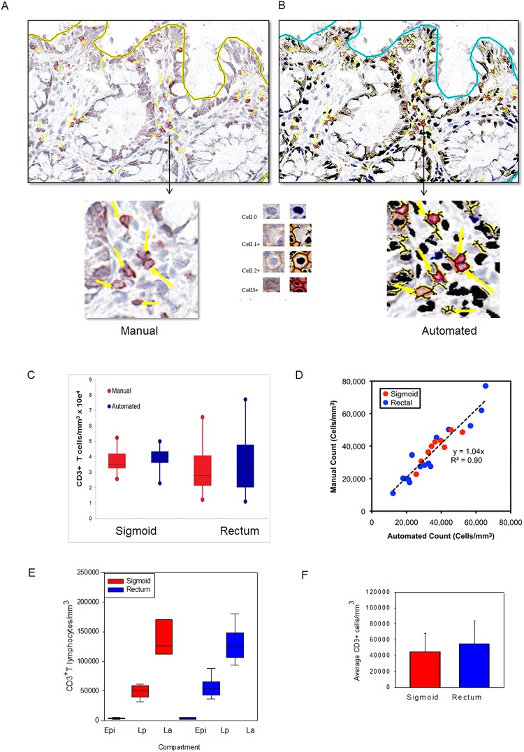
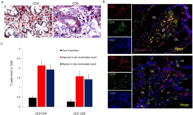
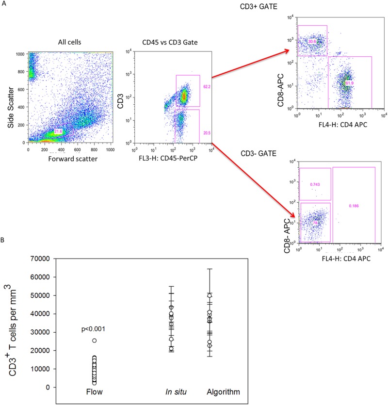
Mucosal tissues are critical immune effector sites containing complex populations of leukocytes in a tissue microenvironment that remains incompletely understood. We identify and quantify in human distal colorectal tissue absolute mucosal CD3+ lymphocytes, including CD4+ and CD8+ subsets, by direct visualization using immunohistochemistry (IHC), immunofluorescence (IF), and an automated counting protocol (r2=0.90). Sigmoid and rectal mucosal tissues are both densely packed with T lymphocytes in the mucosal compartment. Both compartments had similar densities of CD3+ T lymphocytes with 37,400 ± 2,801 cells/mm3 and 33,700 ± 4,324 cell/mm3, respectively. Sigmoid mucosa contained 57% CD3+CD4+ and 40% CD3+CD8+ T lymphocytes which calculates to 21,300 ± 1,476/mm3 and 15,000 ± 275/mm3 T lymphocytes, respectively. Rectal mucosa had 57% CD3+CD4+ and 42% CD3+CD8+ or 21,577 ± 332, and 17,090 ± 1,206 cells/mm3, respectively. By comparison, sigmoid mucosal biopsies subjected to conventional collagenase digestion, mononuclear cell (MMC) isolation and staining for flow cytometry yielded 4,549 ± 381/mm3 and 2,708 ± 245/mm3 CD4+ and CD8+ T lymphocytes. These data suggest only ~20.7% recovery compared to IHC results for these markers. Further studies will determine if this reflects a selective bias in only CD3+, CD4+ and CD8+ T cells or can be generalized to all flow-analyzed cells from mucosal tissues for phenotyping and functional testing.


Conflict of interest statement

Competing Interests: The authors have declared that no competing interests exist.

Figures

Fig 1
Fig 1. CD3+ lymphocytes are similar in normal human sigmoid colonic and rectal mucosa.
Sigmoid colonic and rectal mucosal sections immunostained for CD3. The membrane red/brown colors denote positive staining (red/brown, AEC chromogen). Specimens were counterstained with hematoxylin. (A)Upper panel original magnification 200x (B) lower panel 400x (C) lower magnification showing distribution of lymphoid aggregates in both sigmoid and rectum (100x). Arrows represent lymphoid aggregates.
Fig 2
Fig 2. Application of an automated algorithm to quantitate T lymphocytes in immunostained sigmoid colonic and rectal mucosa.
A membrane detection algorithm was applied to CD3-immunostained sigmoid colonic and rectal mucosal biopsies from 5 healthy donors (5 sigmoid colon, 5 rectum). (A) Digital images with AEC chromogen staining for CD3 and hematoxylin counterstaining. (B) Mark-up images with red, orange, and yellow pixels depicting immunopositive cells (strong, moderate, and weak intensity, respectively), and black pixels depicting nuclei for immunonegative cells. Bar = 100 μm. Arrowheads denote manual enumeration. (C) Tissue concentrations of T lymphocytes obtained using both methods and analyzed by paired t test. (D) Correlation between counts obtained by manual and automated enumeration of the same regions. R2 value = 0.90 (E) Tissue concentrations of T lymphocytes in epidermis (Epi), lamina propria (Lp), and lymphoid aggregates (La; when present) of sigmoid colon and rectum. (F) Average T lymphocyte concentrations obtained from averaging the various compartments from the sigmoid colon and rectum.
Fig 3
Fig 3. Distribution and number of CD4+ and CD8+ T lymphocytes in sigmoid mucosal and rectal mucosa.
(A) Immunoperoxidase for CD4 and CD8 with hematoxylin counterstaining. (B) Distribution of CD4+ and CD8+ cells in lymphoid aggregates. (C) Confocal microscopy of CD3+CD4+ and CD3+CD8+ subsets of T lymphocytes. Bar = 50μm. (D) Densities of CD4+ and CD8+ T lymphocytes compared to densities obtained from flow analysis. GL = gland; Lu = lumen
Fig 4
Fig 4. Comparison of automated counts of in situ sigmoid colonic and rectal mucosal T lymphocytes versus isolation and flow cytometric counting.
Mucosal CD3+ T lymphocytes were isolated by mechanical tissue disruption and collagenase digestion. (A) Gating strategy for flow cytometric analysis of isolated lymphocytes. (B) Comparison of the mean numbers of sigmoid colonic mucosal CD3+ lymphocytes isolated from 30 sigmoid biopsies from similar persons versus automated (n = 48) and manual counting of 5 sigmoid biopsies and 5 rectal biopsies from 5 subjects. Dots represent the means of 3–5 regions analyzed with bars representing standard error of the means. (Mann-Whitney Rank Sum test p<0.001).

References

    1. Geho DH, Bandle RW, Clair T, Liotta LA. Physiological mechanisms of tumor-cell invasion and migration. Physiol. Bethesda Md, 2005. (20):194–200. - PubMed
    1. Espina V, Wulfkuhle JD, Calvert VS, VanMeter A, Zhou W, Coukos G, et al. Laser-capture microdissection. Nat. Protoc. 2006. 1(2):586–603. - PubMed
    1. Gerner MY, Kastenmuller W, Ifrim I, Kabat J, Germain RN. Histo-cytometry: a method for highly multiplex quantitative tissue imaging analysis applied to dendritic cell subset microanatomy in lymph nodes. Immunity. 2012. 37(2):364–376. 10.1016/j.immuni.2012.07.011 - DOI - PMC - PubMed
    1. Trepel F. Number and distribution of lymphocytes in man. A critical analysis. Klin Wochenschr. 1974. June 1;52(11):511–5. - PubMed
    1. Westermann J, Pabst R. Distribution of lymphocyte subsets and natural killer cells in the human body. Clin Investig. 1992. July;70(7):539–44. - PubMed

Publication types