Review
. 2015 Apr 8;86(1):92-105.
doi: 10.1016/j.neuron.2015.01.028.

Tools for probing local circuits: high-density silicon probes combined with optogenetics

Affiliations
Review

Tools for probing local circuits: high-density silicon probes combined with optogenetics

György Buzsáki et al. Neuron. .

Abstract

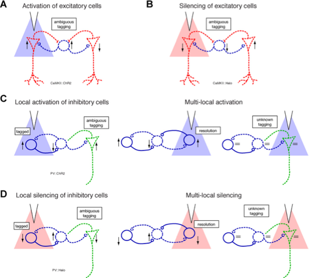
To understand how function arises from the interactions between neurons, it is necessary to use methods that allow the monitoring of brain activity at the single-neuron, single-spike level and the targeted manipulation of the diverse neuron types selectively in a closed-loop manner. Large-scale recordings of neuronal spiking combined with optogenetic perturbation of identified individual neurons has emerged as a suitable method for such tasks in behaving animals. To fully exploit the potential power of these methods, multiple steps of technical innovation are needed. We highlight the current state of the art in electrophysiological recording methods, combined with optogenetics, and discuss directions for progress. In addition, we point to areas where rapid development is in progress and discuss topics where near-term improvements are possible and needed.


Conflict of interest statement

Competing financial interests

Antal Berenyi is the founder and owner of Amplipex Ltd., Szeged, Hungary, which manufactures signal-multiplexed head stages and demultiplexing systems. Daryl Kipke is the founder and current Executive Director of NeuroNexus Technologies, Inc., a subsidiary of Greatbatch, Inc. The other authors declare the absence of any commercial or financial relationships that could be construed as a potential conflict of interest.

Figures

Figure 1
Figure 1. Flow chart of large-scale silicon probe recordings of unit and LFP activity combined with optogenetic manipulation of circuits
The components of the chart are discussed in this Primer. While large-scale recordings and optogenetic perturbations can be implemented separately, their combination provides a powerful tool for circuit analysis. FPGA, field-programmable gate array.
Figure 2
Figure 2. Electric signals in the extracellular space
(A) Simultaneous intracellular and extracellular recordings of action potential in a hippocampal CA1 neuron in vivo. Note that the trough and peak of the extracellular unit spike (0.1 Hz-5 kHz) correspond to the maximum rate of rise and maximum rate of fall of the intracellular action potential. (B) Extracellular contribution of an action potential (‘spike’) to the LFP in the vicinity of the spiking pyramidal cell. The magnitude of the spike is normalized. The peak-to-peak voltage range is indicated by the color of the traces. Note that the spike amplitude decreases rapidly with distance from the soma. The distance-dependence of the spike amplitude within the pyramidal layer is shown (bottom left panel) with voltages drawn to scale, using the same color identity as the traces in the boxed area. The same traces are shown normalized to the negative peak (bottom right panel). (C) Multisite electrodes can estimate the position of the recorded neurons by triangulation of extracellular voltage measurements. Distance of the electrode tips from a single pyramidal cell is indicated by arrows. The spike amplitude of neurons (>60 µV) within the gray cylinder (50 µ m radius), containing ∼100 neurons, can be used for effective separation by current clustering methods. (D) Coherence maps of gamma activity (30–90 Hz) in the hippocampus of a freely behaving rat. The 10 example sites (black dots) served as reference sites, and coherence was calculated between the reference site and the remaining 255 locations of an 8-shank (32-site vertical linear spacing at 50 µm) probe. (A) Reproduced from (Buzsáki et al., 1996). (B) Reproduced from (Buzsáki et al., 2012). (C) Reproduced from (Buzsáki, 2004). (D) Reproduced from (Berenyi et al., 2014).
Figure 3
Figure 3. Silicon probes designed for unit sampling and high spatial density monitoring of LFP
A. Six-shank ‘decatrodes’ (Buzsáki64sp probe from NeuroNexus) for recording and effective separation of single neurons spikes. Vertical separation of the recording sites (160 µm2): 20 µm. Shanks are 15 µm thick and 52 µm wide. B. Eight-shank probe for large spatial coverage (2.1 mm × 1.6 mm; Buzsáki256 probe from NeuroNexus). Recording sites (160 µm2) are separated by 50 µm. Shanks taper from 96 µm to 13 µm at the tip.
Figure 4
Figure 4
High-quality unit recordings from the superficial layers of the prefrontal cortex (traces 1–64) and the hippocampal CA1 pyramidal layer (traces 65–96). 400 msec epoch during sleep. * indicates sharp wave ripple. Data reproduced from (Fujisawa et al., 2008).
Figure 5
Figure 5. Ambiguity and disambiguation of neuron identity by optical tagging
(A) Identification of ChR2-expressing excitatory cells (red) by light pulses is often ambiguous because they can drive nearby interneurons (blue) at a short-latency Since opsin expression level and spiking threshold may vary among principal cells, and since synaptic transmission from principal cells to interneurons is be rapid and strong, some principal cells may show decreased firing rates. (B) Similarly, silencing of several excitatory cells can bring about a short-latency rate decrease of interneurons and circuit-mediated rate increase of excitatory cells. (C) Strong optogenetic activation of inhibitory cells may also be ambiguous, since disynaptic disinhibition of opsin-free cells (dashed green; recorded on another shank) may also occur at a short latency. The ambiguity may be resolved or reduced if the neuron responds at a shorter latency upon direct illumination of the neighboring shank (central panel) but not if no change in firing rate is detected (right panel). (D) Silencing of inhibitory cells results in decreased spiking of illuminated opsin-expressing cells (direct effect), increased spiking of opsin-free cells (synaptic effect), and perhaps interneuron-mediated spiking decrease of other cells (disynaptic effect; green). The direct and disynaptic effects are particularly hard to differentiate upon prolonged global illumination, but may be disambiguated using sequential multi-site illumination.
Figure 6
Figure 6. Diode probes for focal in/activation of neurons
(A) Schematic of a single LED-fiber assembly. The LED is coupled to a 50-µm multimode fiber, etched to a point at the distal (brain) end. (B) Schematic of a drive equipped with a 6-shank diode probe with LED-fibers mounted on each shank. Etched optical fibers are attached ∼40 µm above the recordings sites on the silicon probe shanks. (C) Recording silicon probe integrated with a waveguide. Light transmission through the optical splitter waveguides integrated on the fabricated neural probe: (C, E) bright field microscope images; (D, F) dark field images. Light can be delivered to multiple shanks from a single fiber source via an optical splitter or different wave-length lights can be delivered to the same shank through an optical mixer. A-B, reproduced after (Stark et al., 2012), C-F, after (Wu et al., 2013).
Figure 7
Figure 7. Optogenetic activation of a single neuron in the behaving mouse
Autocorrelograms and optogenetically evoked responses (light blue rectangles) of pyramidal cells (red) and putative interneurons (blue) in a freely-moving CaMKII::ChR2 mouse during weak 50 ms light pulses (0.01 mW/mm2 at the center of the CA1 pyramidal layer). All neurons were recorded from the middle shank of the diode-probe (inset). Note robust response of a single pyramidal cell (boxed). Ten repetitions. No neurons were activated by light on the adjacent shanks. At higher intensities (>0.05 mW/mm2) multiple other neurons also increased their firing.

References

    1. Abidian MR, Martin DC. Experimental and theoretical characterization of implantable neural microelectrodes modified with conducting polymer nanotubes. Biomaterials. 2008;29:1273–1283. - PMC - PubMed
    1. Adrian ED, Moruzzi G. Impulses in the pyramidal tract. The Journal of physiology. 1939;97:153–199. - PMC - PubMed
    1. Agarwal G, Stevenson IH, Berenyi A, Mizuseki K, Buzsáki G, Sommer FT. Spatially distributed local fields in the hippocampus encode rat position. Science. 2014;344:626–630. - PMC - PubMed
    1. Al-Ashmouny KM, Chang SI, Yoon E. A 4 muW/Ch analog front-end module with moderate inversion and power-scalable sampling operation for 3-D neural microsystems. IEEE transactions on biomedical circuits and systems. 2012;6:403–413. - PubMed
    1. Alivisatos AP, Chun M, Church GM, Deisseroth K, Donoghue JP, Greenspan RJ, McEuen PL, Roukes ML, Sejnowski TJ, Weiss PS, Yuste R. Neuroscience. The brain activity map. Science. 2013;339:1284–1285. - PMC - PubMed

Publication types