. 2015 Jul;172(14):3579-95.
doi: 10.1111/bph.13159. Epub 2015 May 20.

A Sativex(®) -like combination of phytocannabinoids as a disease-modifying therapy in a viral model of multiple sclerosis

Affiliations

A Sativex(®) -like combination of phytocannabinoids as a disease-modifying therapy in a viral model of multiple sclerosis

A Feliú et al. Br J Pharmacol. 2015 Jul.

Abstract

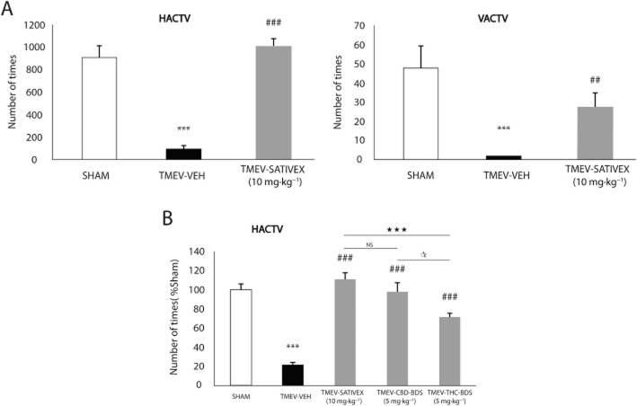
Background and purpose: Sativex(®) is an oromucosal spray, containing equivalent amounts of Δ(9) -tetrahydrocannabinol (Δ(9) -THC) and cannabidiol (CBD)-botanical drug substance (BDS), which has been approved for the treatment of spasticity and pain associated to multiple sclerosis (MS). In this study, we investigated whether Sativex may also serve as a disease-modifying agent in the Theiler's murine encephalomyelitis virus-induced demyelinating disease model of MS.

Experimental approach: A Sativex-like combination of phytocannabinoids and each phytocannabinoid alone were administered to mice once they had established MS-like symptoms. Motor activity and the putative targets of these cannabinoids were assessed to evaluate therapeutic efficacy. The accumulation of chondroitin sulfate proteoglycans (CSPGs) and astrogliosis were assessed in the spinal cord and the effect of Sativex on CSPGs production was evaluated in astrocyte cultures.

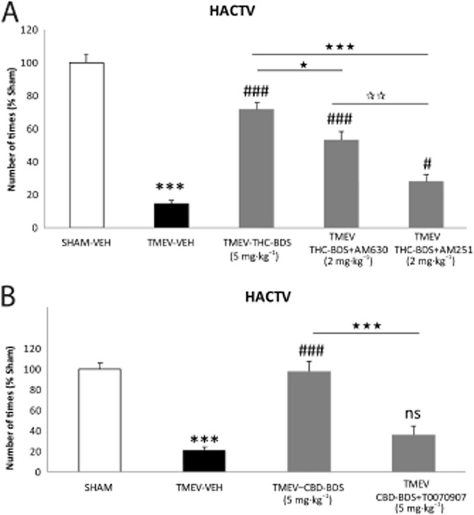
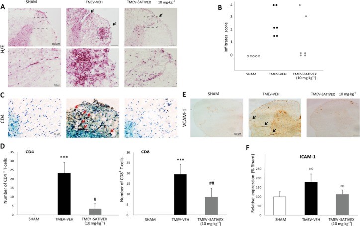
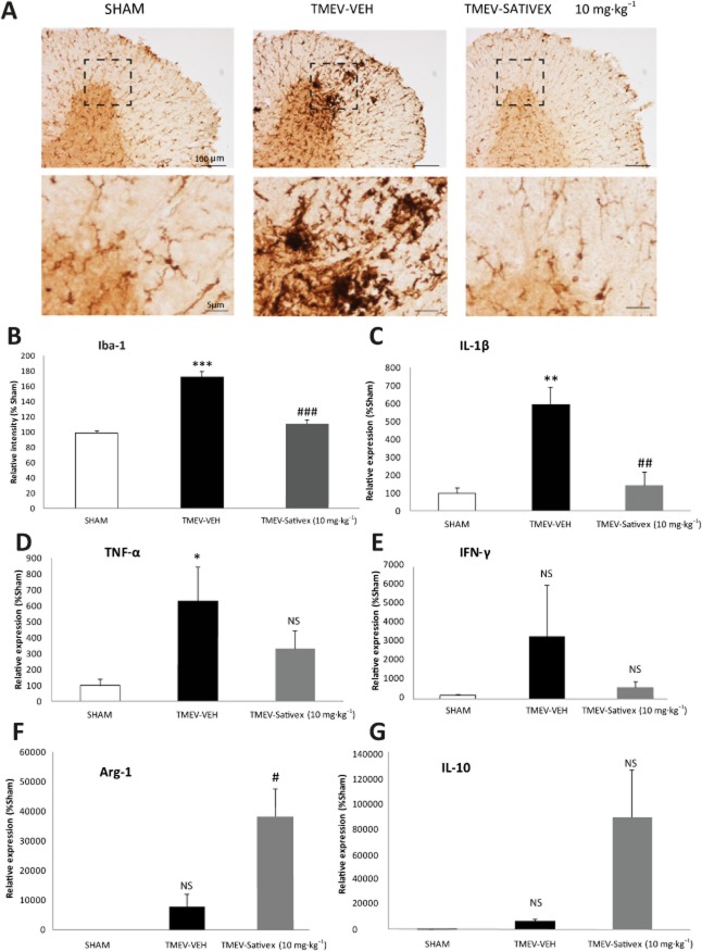
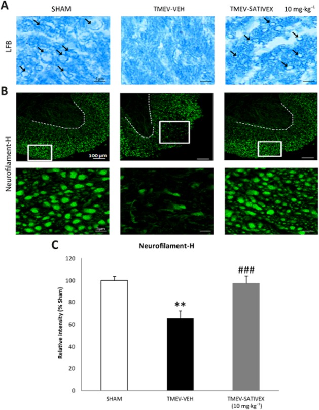
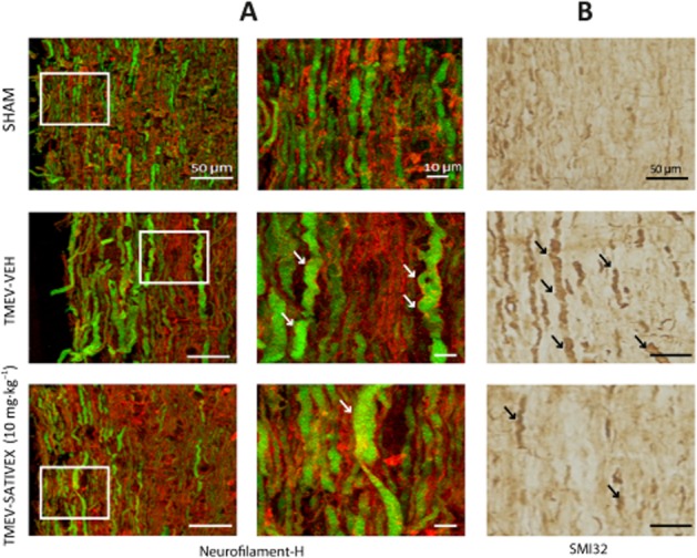
Key results: Sativex improved motor activity - reduced CNS infiltrates, microglial activity, axonal damage - and restored myelin morphology. Similarly, we found weaker vascular cell adhesion molecule-1 staining and IL-1β gene expression but an up-regulation of arginase-1. The astrogliosis and accumulation of CSPGs in the spinal cord in vehicle-infected animals were decreased by Sativex, as was the synthesis and release of CSPGs by astrocytes in culture. We found that CBD-BDS alone alleviated motor deterioration to a similar extent as Sativex, acting through PPARγ receptors whereas Δ(9) -THC-BDS produced weaker effects, acting through CB2 and primarily CB1 receptors.

Conclusions and implications: The data support the therapeutic potential of Sativex to slow MS progression and its relevance in CNS repair.


Figures

Figure 1
Figure 1
A Sativex®-like combination of phytocannabinoids, Δ9-THC-BDS or CBD-BDS treatments alone, significantly improved motor deficits in the chronic phase of TMEV infection. SJL/J mice were intracranially inoculated with TMEV or the vehicle alone and after 70 days, once the symptomatology was established, the mice were treated i.p. for 10 consecutive days with a Sativex-like combination of phytocannabinoids (10 mg·kg−1), CBD-BDS (5 mg·kg−1), Δ9-THC-BDS (5 mg·kg−1) or the vehicle alone. On the last day of treatment, motor function was evaluated by measuring horizontal (HACTV) and vertical activity (VACTV) in the activity cage test (A) which revealed a significant attenuation of motor deficits in the TMEV-IDD model following Sativex administration. In a comparative analysis, Sativex and CBD-BDS alone were the most effective treatments as they abolished the decreased motor activity and restored the normal levels of HACTV (B). Activity parameters were recorded for 10 min and the results are represented as the mean ± SEM of nine mice per group: ***P ≤ 0.001 versus Sham; ##P ≤ 0.01; ###P ≤ 0.001 versus TMEV-VEH animals; ⋆⋆⋆P ≤ 0.001 versus TMEV-Sativex; *P ≤ 0.05 TMEV-CBD-BDS versus TMEV-THC-BDS (non parametric Kruskal–Wallis test). NS, not significant.
Figure 2
Figure 2
The involvement of the CB1, CB2 receptors and PPARγ in the effect of the Sativex®-like phytocannabinoid combination. To determine the receptors involved in the beneficial effects of Sativex, we administered Δ9-THC-BDS (5 mg·kg−1) together with a selective antagonist for the CB2 receptor (AM630, 2 mg·kg−1) and an antagonist of the CB1 receptor (AM251, 2 mg·kg−1). CBD-BDS (5 mg·kg−1) was administered in combination with a PPARγ receptor antagonist (T0070907, 5 mg·kg−1) for 10 days once the symptomatology had been established and motor function was evaluated by measuring horizontal activity (HACTV) on day 80 post-infection. The positive effect of Δ9-THC-BDS was significantly blocked by the administration of the CB1 receptor antagonist and less so by the CB2 receptor antagonist (A). Likewise, the beneficial effect of CBD-BDS was significantly attenuated by the antagonist of PPARγ (B). Activity parameters were recorded for 10 min and the results represent the mean ± SEM of six mice per group: ***P ≤ 0.001 versus Sham; #P ≤ 0.05 ###P ≤ 0.001 versus TMEV-VEH animals; ⋆P ≤ 0.05 versus TMEV-THC-BDS; ⋆⋆⋆P ≤ 0.001 versus TMEV-Δ9-THC-BDS; versus TMEV-CBD-BDS; **P ≤ 0.01 TMEV-THC-BDS-AM630 versus TMEV-THC-BDS-AM251 (one-way anova followed by Tukey's and Bonferroni's test: CBD-BDS and antagonist analysis; non-parametric Kruskal–Wallis test: Δ9-THC-BDS and antagonist analysis). ns, not significant.
Figure 3
Figure 3
A Sativex®-like phytocannabinoid combination treatment decreased leukocyte infiltration and down-regulated the adhesion molecules in the spinal cord of TMEV-infected animals. Transverse cervical spinal cord sections (30 μm thick) obtained at day 80 post-infection were stained with H&E (A). TMEV infection increased leukocyte infiltration, an effect that was attenuated by Sativex (10 mg·kg−1), as indicated by the infiltrate score (B) and by the analysis of CD4 and CD8 lymphocyte staining (representative microphotographs of CD4 staining, C; quantification of CD4 and CD8 number of positive cells, D). Sativex also showed a tendency to decrease mRNA expression of the ICAM-1 adhesion molecule as determined by RT-PCR on spinal cord of TMEV-infected animals (n = 6 per group) normalizing mRNA expression to that of the 18S gene, (F). Sativex significantly reduced the expression of VCAM-1 adhesion molecule (representative microphotographs of VCAM-1 staining, E). The data represent the mean ± SEM: ***P ≤ 0.001 versus Sham; #P ≤ 0.05, ##P ≤ 0.01 versus TMEV-VEH animals (non-parametric Kruskal–Wallis test: CD4 and CD8 analysis). For histology analysis, five to six spinal cord slices per animal were analysed (n = 5–6 animals per group): Scale bar = 100 μm; 30 μm. NS, not significant.
Figure 4
Figure 4
A Sativex®-like phytocannabinoid combination attenuated the microglial response, down-regulated proinflammatory cytokines and up-regulated Arg-1 and IL-10 in the chronic phases of TMEV-IDD. Transverse cervical spinal cord sections (30 μm thick) obtained at day 80 post-infection were stained for Iba-1. (A) Representative microphotographs of Iba-1 immunostaining showing morphological changes in the microglial cells of infected animals that were reversed by Sativex treatment (10 mg·kg−1). (B) Quantification of the percentage area occupied by microglia in the spinal cord white matter per field (n = 5–6 animals per group). Sativex treatment also decreased IL-1β (C) TNF-α (D) and IFN-γ (E) mRNA induction and increased Arg-1 (F) and IL-10 (G) as determined by RT-PCR in the spinal cord of TMEV-infected animals, normalizing mRNA expression to that of the 18S gene (n = 6 per group). The data represent the mean ± SEM: *P ≤ 0.05, **P ≤ 0.01, ***P ≤ 0.001 versus Sham; #P ≤ 0.05, ##P ≤ 0.01, ###P ≤ 0.001 versus TMEV-VEH animals (one-way anova followed by Tukey's and Bonferroni's test: IL-1β analysis; non-parametric Kruskal–Wallis test: Iba-1 analysis; and unpaired two-tailed Student's t-test: TNF-α and Arg-1 analysis). For histology analysis, five to six spinal cord slices were examined per animal (n = 6 animals per group): Scale bar = 100 μm; 5 μm. NS, not significant.
Figure 5
Figure 5
Sativex®-like phytocannabinoid combination treatment restored myelin morphology and prevented axonal damage in TMEV-infected mice. Transverse cervical spinal cord sections were obtained at day 80 post-infection and stained with LFB (15 μm thick) and Neurofilament-H (30 μm thick). The myelin was clearly disrupted in cervical spinal cord sections from infected animals treated with the vehicle while Sativex (10 mg·kg−1) treatment contributed to maintain the myelin structure (A, arrows indicate myelin sheaths. (B) Representative images of Neurofilament-H staining showing that there is prominent axonal damage in the spinal cord white matter of vehicle-treated infected animals that diminished significantly following Sativex treatment. The area occupied by Neurofilament-H was quantified in the spinal cord white matter of each field (5–6 slices, n = 5–6 animals per group) (C). The data represent the mean ± SEM: **P ≤ 0.01 versus Sham; ###P ≤ 0.001 versus TMEV-VEH animals (non-parametric Kruskal–Wallis test): Scale bar = 5 μm; 100 μm.
Figure 6
Figure 6
A Sativex®-like phytocannabinoid combination prevented the swelling and ovoid formations in axons from TMEV-infected animals. Longitudinal cervical spinal cord sections (30 μm thick) stained for Neurofilament-H (green) and RIP (red) (A) revealed the axons to be swollen, deformed and with ovoid formation in TMEV vehicle-treated mice. Lesions in the axons of the Sativex-treated group (10 mg·kg−1) were less frequent and axons displayed markedly reduced damage as confirmed in longitudinal spinal cord sections labelled with SMI32 (B): Scale bar = 10 μm; 50 μm.
Figure 7
Figure 7
A Sativex®-like phytocannabinoid combination reduced astrocyte reactivity and the accumulation of CSPGs. Transverse cervical spinal cord sections (30 μm) were obtained on day 80 post-infection and stained for GFAP, vimentin and CS56. There was a prominent astrogliosis in the spinal cord of vehicle-treated infected animals evident in the representative microphotographs of GFAP staining (A). This is also reflected in the area occupied by GFAP+ astrocytes in the spinal cord white matter in each field (5–6 slices, n = 5–6 animals per group) (B), as well as in the vimentin staining (C). This astrogliosis is associated with an accumulation of CSPGs (CS56 staining, D; brevican gene expression, E) which is impeded by Sativex treatment (10 mg·kg−1). The data represent the mean ± SEM: **P ≤ 0.01, ***P ≤ 0.001 versus Sham; #P ≤ 0.05, ###P ≤ 0.001 versus TMEV-VEH animals (one-way anova followed by Tukey's and Bonferroni test: GFAP analysis; Non parametric Kruskal–Wallis test: brevican). WM, white matter; GM, gray matter. Scale bar = 100 μm; 20 μm.
Figure 8
Figure 8
A Sativex®-like phytocannabinoid combination treatment reduced the synthesis and release of CSPGs by astrocytes in vitro. CSPG, brevican and XT-I enzyme syntheses are up-regulated in primary rat astrocytes stimulated with TGFβ1 (10 ng·mL−1) plus bFGF (10 ng·mL−1) and significantly down-regulated by Sativex at a dose of 100 nM and 0.5 μM or 1 μM in the case of XT-I after 24 h of treatment. Brevican and XT-I mRNA induction was determined by RT-PCR, normalizing mRNA expression (n = 10 per group) to that of the 18S gene (A). Representative microphotographs of primary astrocytes after 48 h in the presence of Sativex (0.5 μM) show that neurocan immunocytochemistry is less intense than in control astrocytes, both at the intracellular level and in the culture substrate (B). In the presence of Sativex (0.5 μM) for 72 h, activated astrocytes release less neurocan to the supernatant, as evident in Western blots. Representative Western blots (C) and quantification of the OD (D). Every assay was performed in duplicate, in three independent experiments. The data represent the mean ± SEM: *P ≤ 0.05, **P ≤ 0.01, ***P ≤ 0.001 versus CTL (control); #P ≤ 0.05, ##P ≤ 0.01, ###P ≤ 0.001 versus TGFβ1+ bFGF (non-parametric Kruskal–Wallis test). ns, not significant.

Similar articles

Cited by

References

    1. Alexander SPH, Benson HE, Faccenda E, Pawson AJ, Sharman JL, Spedding M, et al. The Concise Guide to PHARMACOLOGY 2013/14: G Protein-Coupled Receptors. Br J Pharmacol. 2013a;170:1459–1581. - PMC - PubMed
    1. Alexander SPH, Benson HE, Faccenda E, Pawson AJ, Sharman JL, Spedding M, et al. The Concise Guide to PHARMACOLOGY 2013/14: Nuclear Hormone Receptors. Br J Pharmacol. 2013b;170:1652–1675. - PMC - PubMed
    1. Alexander SPH, Benson HE, Faccenda E, Pawson AJ, Sharman JL, Spedding M, et al. The Concise Guide to PHARMACOLOGY 2013/14: Enzymes. Br J Pharmacol. 2013c;170:1797–1867. - PMC - PubMed
    1. Arévalo-Martín A, Vela JM, Molina-Holgado E, Borrell J, Guaza C. Therapeutic action of cannabinoids in a murine model of multiple sclerosis. J Neurosci. 2003;23:2511–2516. - PMC - PubMed
    1. Arévalo-Martín A, García-Ovejero D, Gómez O, Rubio-Araiz A, Navarro-Galve B, Guaza C, et al. CB2 cannabinoid receptors as an emerging target for demyelinating diseases: from neuroimmune interactions to cell replacement strategies. Br J Pharmacol. 2008;153:216–225. - PMC - PubMed

Publication types