Review
. 2015 Jul:130:29-70.
doi: 10.1016/j.pneurobio.2015.03.005. Epub 2015 Apr 6.

The ventral pallidum: Subregion-specific functional anatomy and roles in motivated behaviors

Affiliations
Review

The ventral pallidum: Subregion-specific functional anatomy and roles in motivated behaviors

David H Root et al. Prog Neurobiol. 2015 Jul.

Abstract

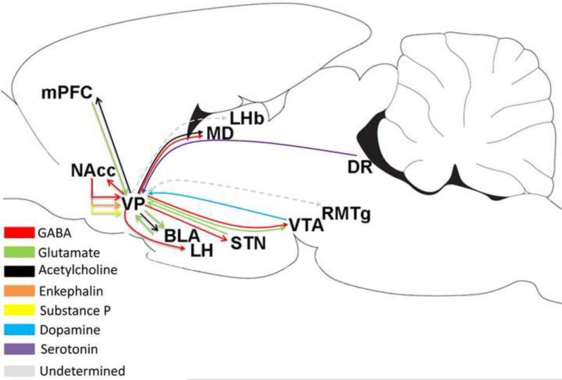
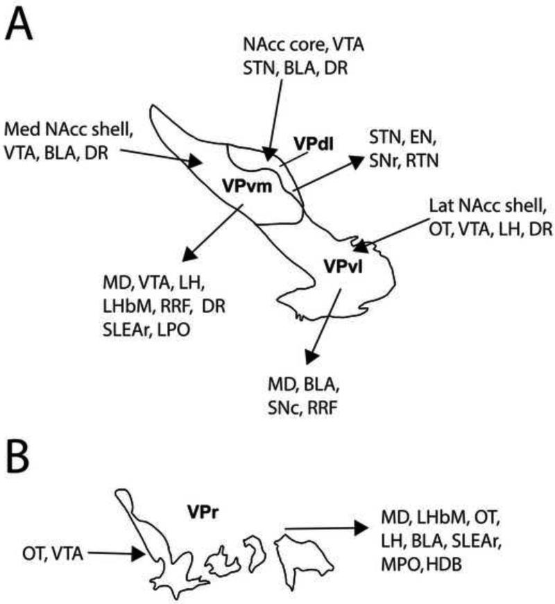
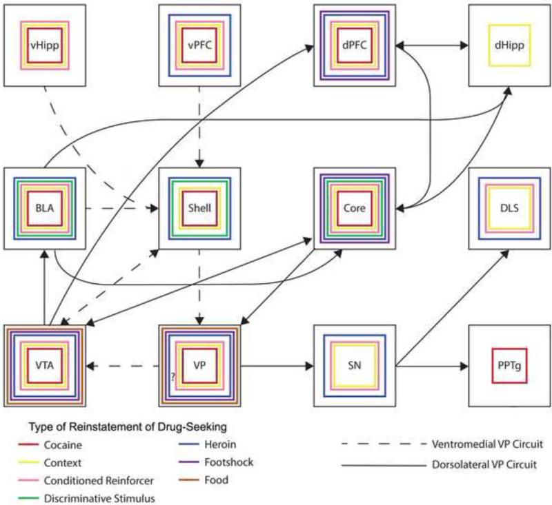
The ventral pallidum (VP) plays a critical role in the processing and execution of motivated behaviors. Yet this brain region is often overlooked in published discussions of the neurobiology of mental health (e.g., addiction, depression). This contributes to a gap in understanding the neurobiological mechanisms of psychiatric disorders. This review is presented to help bridge the gap by providing a resource for current knowledge of VP anatomy, projection patterns and subregional circuits, and how this organization relates to the function of VP neurons and ultimately behavior. For example, ventromedial (VPvm) and dorsolateral (VPdl) VP subregions receive projections from nucleus accumbens shell and core, respectively. Inhibitory GABAergic neurons of the VPvm project to mediodorsal thalamus, lateral hypothalamus, and ventral tegmental area, and this VP subregion helps discriminate the appropriate conditions to acquire natural rewards or drugs of abuse, consume preferred foods, and perform working memory tasks. GABAergic neurons of the VPdl project to subthalamic nucleus and substantia nigra pars reticulata, and this VP subregion is modulated by, and is necessary for, drug-seeking behavior. Additional circuits arise from nonGABAergic neuronal phenotypes that are likely to excite rather than inhibit their targets. These subregional and neuronal phenotypic circuits place the VP in a unique position to process motivationally relevant stimuli and coherent adaptive behaviors.

Keywords: Addiction; Dopamine; GABA; Glutamate; Nucleus accumbens; Ventral tegmental area.


Figures

Figure 1
Figure 1. Delineation of the ventral pallidum and topographic input from nucleus accumbens core to the dorsolateral ventral pallidum subregion
A-D. Four anteroposterior planes of the VP, defined by the presence of substance P-IR (black outline). A. Plane of the “finger-like” rostral VP subregion. B-D. Planes of the VP that contain the ventromedial, dorsolateral, and ventrolateral VP subregions (shown in Figure 2). In the caudal extreme of the VP (D), substance P-IR is also observed in the more dorsally located globus pallidus (D). E-H. Four anteroposterior planes showing labeling of the anterograde tracer phaseolus vulgaris leucoagglutinin (black label) at the injection site within the AcbC (E) and efferent fibers within the dorsolateral VP (F-H). Note labeled cells at the injection site are concentrated in the heavily stained area, slight enhancement of background labeling due to local edema. Labeling in panels G-H are from a slightly medially-shifted AcbC injection compared to labeling from case in panels E-F. Brown labeling indicates neurons immunolabeled for choline acetyl transferase (ChAT; a marker for cholinergic elements). Material from Dr. Zaborszky.
Figure 2
Figure 2. Subregions of the ventral pallidum
A,D. Two anteroposterior levels of the VP, defined by the presence of substance P-IR, at approximately +0.36 mm (A) and −0.12 mm (D). B-C. Sections proximal to tissue in panel D showing calbindin d28k-IR (B) and neurotensin-IR (C). E-F. Sections proximal to tissue in panel D showing calbindin d28k-IR (E) and neurotensin-IR (F). The VPdl subregion exhibits fibers with calbindin-d28k-IR but not neurotensin-IR. The VPvm subregion exhibits fibers with neurotensin-IR but not calbindin-d28k-IR, and the VPr and VPvl subregions do not express calbindin-d28k-IR or neurotensin-IR. This compartmentation of VP is observed across the anteroposterior extent of VP, except in the VPr (Zahm and Heimer, 1988, ; Zahm 1989; Zahm et al., 1996; Riedel et al., 2002; Tripathi et al., 2010, 2013). All sections are 30 μm thick. Neighboring locations to the VP are demarcated by dotted lines. Material from Dr. Root (Morales laboratory, NIDA).
Figure 3
Figure 3. Neuronal phenotypes of the ventral pallidum
A-B. GABAergic neurons. Double in situ hybridization; the purple labeled neurons display digoxigenin-labeling for GAD 65 and GAD 67 mRNAs. Box in A showing VP regions displayed in B, C, and D. B. Higher resolution photograph of GAD mRNA neurons. Note abundance of GABAergic neurons, two examples shown by red thin arrows. C-D. Glutamatergic neurons. The same section as B further processed with radioactive in situ hybridization for VGluT2 mRNA under brightfield (C) and darkfield (D) illumination. Clusters of green grains (C) or white grains (D) indicate VGluT2 mRNA neurons. Note abundance of glutamatergic neurons, uniquely localized within VPvm. Examples of VGluT2-expressing neurons indicated by green small arrows. E-F. Immunohistochemistry for tyrosine hydroxylase (TH; a marker for noradrenergic/dopaminergic elements) and choline acetyl transferase (ChAT; a marker for cholinergic elements). Boxed in region in the low power photomicrograph (E) is the region shown in the high power photomicrograph (F). The VP is delineated by fewer TH-fibers than neighboring structures (i.e., bed nucleus of the stria terminalis, interstitial nucleus of the anterior commissure, striatum, and tubercle). ChAT-labeled soma (brown diaminobenzadine reaction); two examples of cholinergic neurons are indicated by black arrow heads. Material from Dr. Root (Morales laboratory, NIDA).
Figure 4
Figure 4. Accumbens neurons establish inhibitory synapses onto VP GABA neurons
A-C. Accumbal to ventral pallidal projections determined by lesion studies. A. Electrolytic lesion locations in the AcbC (horizontal hatching) and AcbSh (vertical hatching). B. Degenerating terminals in VP after AcbC lesion using a silver-impregnation method (Gallyas et al., 1980). C. Loss of GAD-IR (arrow) in VP after lesion of the AcbSh. D-F. Electron microscopy evidence of a GABAergic AcbSh and AcbC projection to GABAergic VP neurons. D. A large degenerating bouton establishing a symmetric synapse with a GAD-positive dendrite in the VP after lesion of the AcbC. E. A GABAergic cell and dendrite ensheathed by GAD-expressing terminals in the VP. F. Small degenerating bouton contacting a GAD-expressing dendrite after lesion of the AcbSh. Arrows in D and F point to the postsynaptic membrane. Scale bars in B,C: 1 mm, F: 1 μm (also refers to D). Material from Dr. Zaborszky.
Figure 5
Figure 5. General overview of afferents and efferents of the ventral pallidum
Nonsubregional illustration of the major transmitter phenotype and associated brain structures of the projections. Supporting literature includes: VP/mPFC (medial prefrontal cortex) - Carlsen et al. 1985; Hur and Zaborszky, 2005. VP/Acb Haber and Nauta, 1983; Zahm et al. 1985; Churchill et al. 1990; Kalivas et al. 1993; Groenewegen and Russchen 1984; Chrobak and Napier, 1993; Napier et al. 1995. VP/BLA - Fuller et al. 1987; Carlsen et al. 1985; Poulin et al. 2006; Maslowski-Cobuzzi and Napier, 1994; Mitrovic and Napier, 1998. VP/STN - Bevan et al. 1997; Ricardo et al. 1980; Turner et al., 2001, . VP/LH - Bevan et al. 1997. VP/DR - Semba et al. 1988. VP/VTA – Maslowski-Cobuzzi and Napier, 1994; Mitrovic and Napier, 2002; Klitenick et al. 1992; Geisler et al. 2005, ; Kalivas et al. 1993. VP/RMTg - Jhou et al. 2009; Taylor et al. 2014. VP/LHb - Groenwegen et al. 1993. VP/RTN - Young et al. 1984; O'Donnell et al. 1997. VP/MD - Young et al. 1984; Mariotti et al. 2001; MD/mPFC Pirot et al. 1994. The VP projection to LHb and RMTg projection to VP have not explicitly tested a GABAergic phenotype. Sagital outline modified from Paxinos and Watson (2007).
Figure 6
Figure 6. Subregional afferent and efferent connections of the ventral pallidum
A. Known afferent and efferent projections of the VPvm, VPdl, and VPvl subregions. Subregions are illustrated to represent projections from any anteroposterior location within these VP subregions. Anatomical studies have also demonstrated that the BLA projects to, and receives projections from, cholinergic neurons that reside in all VP subregions (Gritti et al., 1993; Poulin et al., 2006; Mascagni and McDonald, 2009; Záborszky et al., 1986, 1999, 2012), but electrophysiological studies consistent with monosynaptic afferents suggest a wider influence on VP neuronal populations (Maslowski-Cobuzzi and Napier, 1994; Mitrovic and Napier, 1998). Glutamatergic neuron distribution for VP subregions has yet to be validated, but appear to be located largely within the VPvm, and thus far have been shown to project to the VTA (Geisler et al. 2007). VTA and DR appear to project to all VP subregions (Maslowski-Cobuzzi and Napier, 1994; Mitrovic and Napier, 2002; Klitenick et al. 1992; Semba et al. 1988). B. Known afferent and efferent projections of the VPr subregion.
Figure 7
Figure 7. Proposed changes in firing rate during responses for drugs of abuse involving the VP subregions
Neurons of the AcbC exhibit increased firing rates during responses for cocaine (Ghitza et al. 2004), which are likely the result of dPFC and BLA activation during the same behavior (Chang et al., 1997, 2000; Carelli et al., 2003). Activation of AcbC leads to decreased firing rates of VPdl neurons during the response (Root et al., 2013) and likely disinhibits its target structures SNr and STN (Lardeux et al., 2013; Fan et al., 2012). While AcbSh neurons are less sensitive to the response compared with AcbC neurons, AcbSh neurons exhibiting changes in firing rate during the response are heterogeneous (Ghitza et al., 2004). Such heterogeneity is found within the VPvm subregion (Root et al., 2013) and is likely to continue within its target regions VTA and MD. The involvement of VPvl and its circuits (SNc and DLS; dotted lines with arrows) is unclear (white color). Dashed lines with arrows represent circuits involving VPvm. Solid lines with arrows represent circuits involving VPdl. Purple shaded structure indicates heterogeneous (increased or decreased) change in firing rate during cocaine self-administering responses. Red shaded structure and green shaded structure indicates decreased and increases change in firing rate during cocaine self-administering responses. The illustrated subregions are proposed to represent subregions throughout the anteroposterior extent of the VP.
Figure 8
Figure 8. VP subregions are differentially associated with drug-seeking behaviors during cocaine self-administration
A. Top shows illustration of behaviors used to analyze changes in firing rate recorded from single VP neurons during discrete behaviors involved in self-administering cocaine. Approach occurred when the animal initially moved towards a set of photocells used as a response device (left), response occurred when the animal emitted a learned long distance vertical head movement (middle), and retreat occurred when the animal moved away from the photocells; right). Bottom shows an example approach-related firing pattern of a VPvm neuron. For A, B, F, and G, green dot is the onset of the approach and time zero is the offset of approach (left), blue dot is the onset of response and time zero is offset of response (middle), and red dot is offset of retreat while time zero is onset of retreat (right). B. An example response-related firing pattern of a VPvm neuron. C. VPdl neurons exhibit a significantly greater correlation between directional changes in firing rate during approach and response compared to VPvm neurons. Directional change in firing rate refers to change from baseline according to a B/(A+B) formula, where ‘B’ is firing rate during movement (e.g., approach or response), and ‘A’ is firing rare during baseline. D. Neurons classified as approach-response and approach-response-retreat were observed significantly more often within VPdl compared to VPvm, χ2(1) = 4.03, p = 0.04. E. Neurons classified as retreat-only and response-retreat were observed significantly more often within VPvm compared to VPdl, χ2(1) = 5.96, p = 0.01. F. Example approach-response firing pattern of VPdl neuron. G. Example approach-response-retreat firing pattern of VPdl neuron. * p < 0.05; *** p < 0.001. Data from Dr. Root while in the West laboratory (Rutgers), published in Root et al., 2013.
Figure 9
Figure 9. Proposed reinstatement circuits involving the VP subregions
Theoretical schema of brain regions and circuits that involve VP subregions and their participation in different types of reinstatement of drug-seeking behavior. Presence of colored squares indicates region is necessary for the type of reinstatement. Question mark in VP indicates that a test has not yet been performed. Absence of colored squares in all other regions indicates either test not yet performed or region not involved. Dashed lines represent circuits involving VPvm. Solid lines represent circuits involving VPdl. Supporting literature includes the following: Ventral hippocampus (vHipp) - Lasseter et al. 2010; Sun and Rebec 2003; vPFC – Peters et al., 2008; Rodgers et al. 2008; Bossert et al. 2011; LaLumiere et al., 2012; Dorsal prefrontal cortex (dPFC) - McFarland and Kalivas 2001, 2004; Rodgers et al. 2008; Fuchs et al. 2005; Capriles et al. 2003; Dorsal hippocampus (dHipp) - Fuchs et al. 2005; dPFC/dHipp - Fuchs et al. 2007; BLA - Kantak et al. 2002; Feltenstein and See 2007; Alleweireldt et al. 2006; Fuchs et al. 2005; You and Fields, 2003; Ledford et al. 2003; McLaughlin and See, 2003; Hayes et al. 2003; Fuchs and See 2002; Kruzich and See 2001; Grimm and See, 2000; Stefanik and Kalivas, 2013. BLA/dHipp - Fuchs et al. 2007; AcbSh – Peters et al., 2008; Rodgers et al. 2008; Bossert et al. 2007; BLA/AcbC – Stefanik and Kalivas, 2013; dPFC – Stefanik and Kalivas, 2013; BLA/AcbC – Stefanik and Kalivas, 2013; dPFC/AcbC – based on Kalivas and McFarland, 2001; vPFC/AcbSh – Peters et al., 2008; Bossert et al. 2012; LaLumiere et al., 2012; AcbC - Rodgers et al. 2008; McFarland and Kalivas 2001, 2004; Bossert et al. 2007; Xie et al., 2012; Fuchs et al., 2008; AcbC/VPdl – Stefanik et al., 2013; DLS - Fuchs et al. 2006; See et al. 2007; Rodgers et al. 2008; Bossert et al. 2009; SN/DLS – based on Bossert et al., 2009; VTA - McFarland and Kalivas 2001, 2004; Sun et al. 2005; Di Ciano and Everitt 2004; Wang et al. 2009; You et al. 2007; Schmidt et al. 2009; VP/VTA - Mahler et al. 2014; VP - Tang et al. 2005; Rodgers et al. 2008; McFarland and Kalivas 2001, 2004; Li et al. 2009; Torregrossa and Kalivas 2008; Lu et al. 2012; Mahler et al. 2012; Perry and McNally, 2013; VTA/dPFC – based on McFarland and Kalivas, 2001; McFarland et al., 2004; Capriles et al., 2003; SN - Sun et al. 2007; Rodgers et al. 2008; PPTg - Schmidt et al. 2009; VTA/AcbSh - LaLumiere et al., 2012; Bossert et al., 2007; VTA/BLA - LaLumiere et al., 2012; VTA/AcbC – based on Bossert et al., 2007.

References

    1. Alesdatter JE, Kalivas PW. Inhibition of mu opioid-induced motor activity in the ventral pallidum by D1 receptor blockade. Behav Pharmacol. 1993;4(6):645–651. - PubMed
    1. Alexander GE, DeLong MR, Strick PL. Parallel organization of functionally segregated circuits linking basal ganglia and cortex. Annu Rev Neurosci. 1986;9:357–381. - PubMed
    1. Alheid GF. Extended amygdala and basal forebrain. Ann NY Acad Sci. 2003;985:185–205. - PubMed
    1. Alheid GF, Heimer L. New perspectives in basal forebrain organization of special relevance for neuropsychiatric disorders: the striatopallidal, amygdaloid, and corticopetal components of substantia innominata. Neuroscience. 1988;27(1):1–39. - PubMed
    1. Alleweireldt AT, Hobbs RJ, Taylor AR, Neisewander JL. Effects of SCH-23390 infused into the amygdala or adjacent cortex and basal ganglia on cocaine seeking and self-administration in rats. Neuropsychopharmacology. 2006;31(2):363–74. - PubMed

Publication types

MeSH terms