. 2015 Aug;23(8):1368-1379.
doi: 10.1038/mt.2015.58. Epub 2015 Apr 10.

High Local Concentrations of Intradermal MSCs Restore Skin Integrity and Facilitate Wound Healing in Dystrophic Epidermolysis Bullosa

Affiliations

High Local Concentrations of Intradermal MSCs Restore Skin Integrity and Facilitate Wound Healing in Dystrophic Epidermolysis Bullosa

Tobias Kühl et al. Mol Ther. 2015 Aug.

Abstract

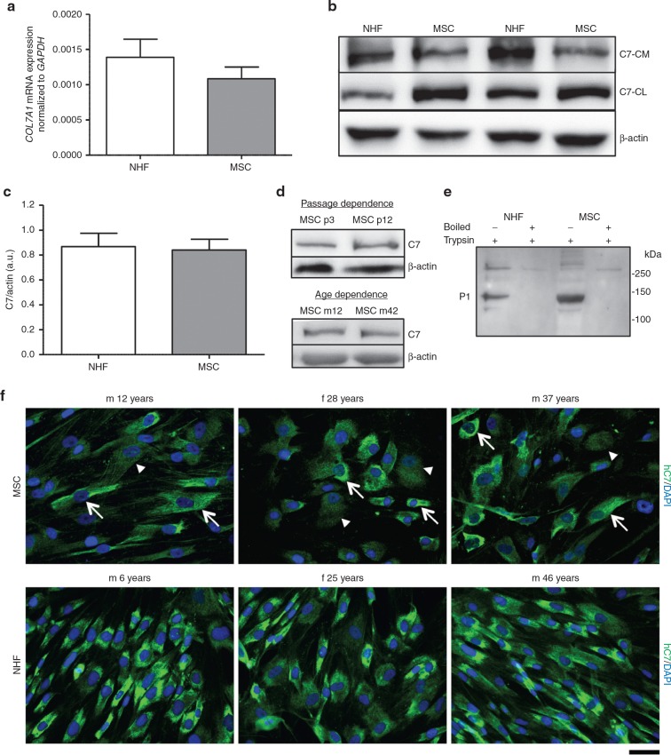
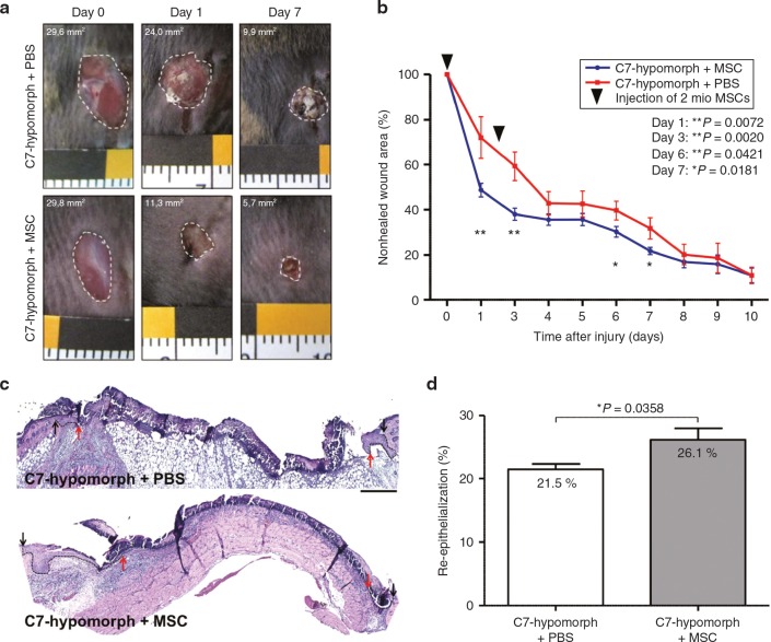
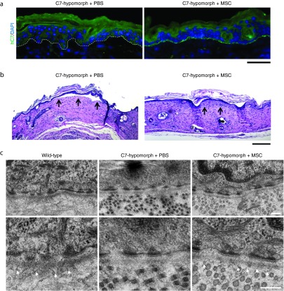
Dystrophic epidermolysis bullosa (DEB) is an incurable skin fragility disorder caused by mutations in the COL7A1 gene, coding for the anchoring fibril protein collagen VII (C7). Life-long mechanosensitivity of skin and mucosal surfaces is associated with large body surface erosions, chronic wounds, and secondary fibrosis that severely impede functionality. Here, we present the first systematic long-term evaluation of the therapeutic potential of a mesenchymal stromal cell (MSC)-based therapy for DEB. Intradermal administration of MSCs in a DEB mouse model resulted in production and deposition of C7 at the dermal-epidermal junction, the physiological site of function. The effect was dose-dependent with MSCs being up to 10-fold more potent than dermal fibroblasts. MSCs promoted regeneration of DEB wounds via normalization of dermal and epidermal healing and improved skin integrity through de novo formation of functional immature anchoring fibrils. Additional benefits were gained by MSCs' anti-inflammatory effects, which led to decreased immune cell infiltration into injured DEB skin. In our setting, the clinical benefit of MSC injections lasted for more than 3 months. We conclude that MSCs are viable options for localized DEB therapy. Importantly, however, the cell number needed to achieve therapeutic efficacy excludes the use of systemic administration.


Figures

Figure 1
Figure 1
Mesenchymal stromal cells (MSCs) stably express and secrete C7. (a) Quantitative real time polymerase chain recation of COL7A1 expression in normal human fibroblasts (NHFs) and MSCs with GAPDH as reference gene. n ≥ 9 different donors per group in three independent experiments. Values are mean ± SEM. (b) Western blots of representative experiments. NHFs and MSCs were seeded in six-well plates and grown to 80% confluence before adding serum-free culture medium + 50 µg/ml ascorbic acid for 48 hours. Conditioned medium was precipitated with ammonium sulfate and analyzed by western blotting, cells and matrix were extracted and processed for western blot analysis, too. Western blots were run under reducing conditions. Blots were probed with antibody against C7 and β-actin as loading control. (c) Densitometric quantification of three independent western blots of cell and matrix lysates. n ≥ 9 different donors per group in three independent experiments. Values represent mean ± SEM of C7 expression in NHFs and MSCs normalized to β-actin expression. (d) C7 expression in MSCs in correlation to passage number and donor age. A representative western blot of cell and matrix lysates of the same donor in a low (passage 3) and a high (passage 12) passage number and between a young (12 years) and an older (42 years) donor are shown. Western blots were run under reducing conditions. Similar C7 expression was detectable in all presented settings. (e) Limited trypsin digestion assay of secreted C7. Conditioned medium was pelleted as described above (b); the pellets were resuspended in TBS and digested with 0.05% trypsin at 30 °C for 10 minutes. Positive controls were boiled prior to trypsin digestion. Western blotting was performed as described above and probed with antibody recognizing the P1 fragment of C7. MSC-derived C7 was resistant to trypsin proteolysis suggesting stable triple helical configuration. (f) Representative images of NHFs and MSCs from different donors seeded on collagen I coated glass slides for 48 hours in the presence of 50 µg/ml ascorbic acid and stained for human C7 (green). In MSCs, a mix of C7 high (white arrows) and low (white arrowheads) expressing cells was detected. Nuclei (blue), bar = 50 µm.
Figure 2
Figure 2
C7 is deposited at the dermal-epidermal junction zone after injection of mesenchymal stromal cells (MSCs). (a) Different cell concentrations and phosphate-buffered saline (PBS) alone were injected intradermally in the back skin of C7-hypomorphic mice twice with a 7-day interval. One week after, the last injection skin was analyzed for presence of C7 with an antibody detecting both human and murine C7 (green). MSC-injected mice showed dose-dependent C7 deposition at the dermal-epidermal junction zone with a maximum at 2 × 106 injected MSCs. Arrows depict C7-positive areas. (b) Photographs of skin sections from (a) were taken with identical image settings and C7 deposition was quantified with Image J. Quantification revealed the highest C7 deposition after injection of 2 × 106 MSCs. n = 3 repeats of each cell concentration in three independent experiments. Values represent mean ± SEM of C7 expression normalized to PBS-injected hypomorphic skin. Asterisk above the data points indicate P values of cell concentrations compared with PBS. n.s., not significant, ***P = <0.001. P values in the box represent differences between the most efficient cell number (2 × 106 MSCs) and other treatment regimens as indicated. (c) Single dose of 2 × 106 MSCs expressing the red fluorophore mCherry (RFP) were injected intradermally in the back skin of C7-hypomorphic mice. Three to 28 days after injection, skin samples were analyzed for RFP+ cells (red) and human C7 using human-specific antibody (green). RFP+ cells, which simultaneously express human C7 (green), decrease over time but a few are still visible after 28 days. (d) C7 stability was assessed by immunofluorescence analysis 2–12 weeks after a repeated intradermal injection of 2 × 106 MSCs in the back skin of C7-hypomorphic mice. Human C7 deposition detected as in (c) (green) is clearly visible 2 and 8 weeks after MSC injections and in a reduced amount after 12 weeks. In contrast, in PBS-injected mice, a weak barely detectable C7 signal was seen at all time points. Arrows indicate C7-positive areas at the dermal-epidermal junction zone. Skin sections as in (d) stained with an antibody detecting both sources of C7. Pictures were taken with identical image settings and C7 deposition was quantified with Image J. Quantification revealed significant increase in C7 deposition 2, 8, and 12 weeks after injections of MSCs, as compared to PBS-injected mice. n ≥ 3 per group. Values represent mean ± SEM of C7 expression normalized to uninjected wild-type skin. ***P < 0.001. Bar = 50 µm.
Figure 3
Figure 3
Mesenchymal stromal cells (MSCs) normalize gross wound closure and re-epithelialization. (a) Closure of 6-mm full thickness punch biopsies on the back of C7-hypomorphic mice over time. Mice were injected either with PBS or MSCs. (b) Quantification of the wound area revealed an accelerated wound closure in mice injected with MSCs compared to phosphate-buffered saline (PBS)-injected wounds. n ≥ 12 wounds per group; values represent mean ± SEM. P values are stated in the figure. (c) H&E staining of 3-day-old wounds from C7-hypomorphic mice injected either with PBS or MSCs. Arrows indicating wound width (initial wound boarder; black arrows, epithelial front; red arrows, re-epithelialized area is highlighted with dotted black line). Starting with the same wound size, MSC-injected animals show an accelerated re-epithelialization of wounded tissue after 3 days. Bar = 250 µm. (d) Quantification of re-epithelialized area 3 days after wounding in percent of the maximal wound size. n = 5 wounds per group. Values represent mean ± SEM,*P = 0.0358.
Figure 4
Figure 4
Mesenchymal stromal cells (MSCs) ameliorate inflammation and granulation tissue maturation in C7-hypomorphic mouse wounds. (a) CD11b staining (red) in 7-day-old wounds revealed prolonged inflammation in C7-hypomorphic mice compared to wild-type animals as previously described in ref. . Treatment with MSCs alleviated inflammation in C7-hypomorphic wounds. Bar 200 µm. Cell nuclei are stained with 4',6-diamidino-2-phenylindole (DAPI) (blue). (b) α-SMA staining (red) indicated presence of myofibroblasts in the wound center in wild-type mice compared to peripheral distribution in phosphate-buffered saline (PBS)-injected C7-hypomorphic mice. In MSC-injected wounds, myfibroblasts were more abundant and centrally localized indicating a more matured granulation tissue. Bar = 100 µm. (c) Quantification of total amount of CD11b+ cells in the wound area (left, *P = 0.0364) and α-SMA-positive area (right, *P = 0.044). n ≥ 3 wounds per group; values represent mean ± SEM.
Figure 5
Figure 5
Mesenchymal stromal cells (MSCs) increase skin stability of healed wounds. (a) Human C7 deposition at re-epithelialized 7-day-old wounds. In phosphate-buffered saline (PBS)-injected wounds, there is no sign of C7 deposition, whereas healed wounds treated with MSCs show a clear deposition of human C7 at the dermal-epidermal junction zone of healed wounds. Bar = 50 µm. (b) Tape strip assay of 4-week-old healed skin wounds revealed increased skin stability in MSC-injected wounds compared to PBS-injected skin. Healed full thickness skin wounds were mechanically stressed through repeated tape stripping and skin sections were analyzed for epidermal-dermal detachment (black arrows). Representative pictures of four wounds per group are shown. Bar = 200 µm. (c) Transmission electron microscopy images of skin from wild-type mouse and 12-week-old healed wounds from C7-hypomorphic mice treated either with PBS or MSCs. Analysis revealed absence or formation of scarce amounts of mainly unstructured anchoring fibrils in PBS-injected mice, whereas in MSC-injected mice, immature anchoring fibrils were visible in some areas 12 weeks after wounding (white arrowheads). Pictures were taken at 15,000× magnification. Bar = 200 nm.

Comment in

References

    1. Aymé, S, Rodwell, C, eds. (2013). 2013 Report on the State of the Art of Rare Disease Activities in Europe. Publisher: European Union, Brussels.
    1. Griggs, RC, Batshaw, M, Dunkle, M, Gopal-Srivastava, R, Kaye, E, Krischer, J et al.; Rare Diseases Clinical Research Network. (2009). Clinical research for rare disease: opportunities, challenges, and solutions. Mol Genet Metab 96: 20–26. - PMC - PubMed
    1. Fine, JD, Bruckner-Tuderman, L, Eady, RA, Bauer, EA, Bauer, JW, Has, C et al. (2014). Inherited epidermolysis bullosa: updated recommendations on diagnosis and classification. J Am Acad Dermatol 70: 1103–1126. - PubMed
    1. Has, C and Bruckner-Tuderman, L (2014). The genetics of skin fragility. Annu Rev Genomics Hum Genet 15: 245–268. - PubMed
    1. Van Agtmael, T and Bruckner-Tuderman, L (2010). Basement membranes and human disease. Cell Tissue Res 339: 167–188. - PubMed

Publication types