Review
. 2015 Apr 10;116(8):1413-30.
doi: 10.1161/CIRCRESAHA.116.303614.

Use of mesenchymal stem cells for therapy of cardiac disease

Affiliations
Review

Use of mesenchymal stem cells for therapy of cardiac disease

Vasileios Karantalis et al. Circ Res. .

Abstract

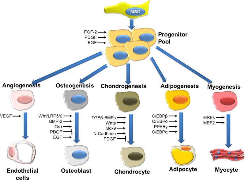
Despite substantial clinical advances over the past 65 years, cardiovascular disease remains the leading cause of death in America. The past 15 years has witnessed major basic and translational interest in the use of stem and precursor cells as a therapeutic agent for chronically injured organs. Among the cell types under investigation, adult mesenchymal stem cells are widely studied, and in early stage, clinical studies show promise for repair and regeneration of cardiac tissues. The ability of mesenchymal stem cells to differentiate into mesoderm- and nonmesoderm-derived tissues, their immunomodulatory effects, their availability, and their key role in maintaining and replenishing endogenous stem cell niches have rendered them one of the most heavily investigated and clinically tested type of stem cell. Accumulating data from preclinical and early phase clinical trials document their safety when delivered as either autologous or allogeneic forms in a range of cardiovascular diseases, but also importantly define parameters of clinical efficacy that justify further investigation in larger clinical trials. Here, we review the biology of mesenchymal stem cells, their interaction with endogenous molecular and cellular pathways, and their modulation of immune responses. Additionally, we discuss factors that enhance their proliferative and regenerative ability and factors that may hinder their effectiveness in the clinical setting.

Keywords: cell differentiation; mesenchymal stem cell; regeneration; stem cells.


Figures

Figure 1
Figure 1. Schematic overview of signaling molecules and transcription factors involved in the regulation of differentiation of MSCs
VEGF, vascular endothelial growth factor; BMP-2, bone morphogenetic protein-2; EGF, epidermal growth factor; FGF-2, Fibroblast growth factor-2; LRP5/6, low-density lipoprotein receptor-related protein-5/6; Osx, Osterix; PDGF, platelet-derived growth factor; RUNX2, runt-related transcription factor-2; TGF-b, transforming growth factor-b; MRF, myogenic regulatory factors; MEF2, myocyte enhancer factor-2. Figure modified and reporduced with permission from Augello et al. Hum Gene Ther 2010
Figure 2
Figure 2. Immunomodulatory capabilities of MSCs
Schematic overview of the interactions between MSC and the immune system. Via multiple pathways MSC suppress proliferation of both T helper (TH) and cytotoxic T cells (Tc). In addition, differentiation to TH2 and regulatory T-cells (Treg) is triggered, resulting in an anti-inflammatory environment. Maturation of dendritic cells (DC) is inhibited via IL-6, blocking upregulation of CD40, CD80, and CD86, which in turn reduce T-cell activation. Monocytes are induced by MSC to differentiate preferentially towards the M2 phenotype. IL-10 produced by the M2 macrophages can boost the formation of Treg while simultaneously reducing tissue migration of neutrophils. Neutrophils (polymorphonuclear granulocytes; PMN) are allowed longer life span but reactive oxygen species production is decreased. Natural Killer (NK) cell proliferation is suppressed as well as their cytotoxic activity. B-cell proliferation is inhibited and the production of antibodies is reduced. Modified from van den Akker et al.
Figure 3
Figure 3. Mechanisms of action of MSCs
The proposed mechanism of action of MSCs form an intertwined cycle of paracrine, autocrine and direct effects that include vascular regeneration, myocardial protection, cardiomyocyte regeneration that ultimately lead to cardiac repair.
Figure 4
Figure 4. Direct MSC stimulation of endogenous repair
(A) Graph depicting the contribution of cardiomyocyte precursors following exogenous administration of MSCs (green line) and endogenous CSCs (orange line), during cardiac repair after MI. MSC differentiation occurs rapidly after delivery. At 2-weeks, MSCs activate endogenous expansion c-kit+ CSCs (orange line). (B) Two weeks following TEI, the number of C-kit+ cells co-expressing GATA-4 is greater in MSCs vs. non-MSCs treated hearts. The cardiac precursors are preferentially located in the IZ and BZ of the MI, indicating an active process of endogenous regeneration (‡p=0.019 and †p<0.0001) (C,D) The 2-week old chimeric myocardium contains mature cardiomyocytes (open arrow), immature MSCs (arrowheads, inset) and cardiac precursors of MSCs origin (arrow), coupled to host myocardium by connexin-43 gap junctions; Interestingly, endogenous c-kit+ CSCs are found in close proximity to MSCs (D). (E) Cluster of c-kit+ CSCs in an MSCs-treated heart; numerous CSCs are committed to cardiac lineage documented by GATA-4 and MDR-1 co-expression (arrows). (F) Few, isolated c-kit+ cells were found in non-MSC treated animals. Figure reproduced with permission from Hatzistergos et al. Circulation Research 2010
Figure 5
Figure 5. Adipose Derived MSCs (PRECISE and APOLLO trials) and autologous CSC (C-CURE) in clinical trials
(A) In the PRECISE trial, Adipose Derived Regenerative Cells (ADRCs) failed to reduce scar size in patients suffering from HF, 6 months after implantation. (B) In the APOLLO trial, where similar cells were employed in the setting of an acute myocardial infarction, there was a significant scar size reduction. (C and D) In the C-CURE trial, where autologous cardiopoietic stem cells (CSCs) were employed, LVEF increased significantly compared to baseline and to control group 6-months following treatment. That was coupled with decreases in left ventricular end-diastolic (ED) and end-systolic (ES) volumes. Panel A is reproduced with permission from Perin et al. American Heart Journal, 2014, panel B from Houtgraaf et al, Journal of American College of Cardiology, 2012 and panels C and D from Bartunek et al, JACC, 2013.
Figure 6
Figure 6. Bone marrow derived MSCs in clinical trials (POSEIDON, TAC-HFT, PROMETHEUS)
(A) In the POSEIDON trial, autologous and allogeneic MSCs both decreased the scar size (as measured by MDCT) and (B) the end-systolic volume 13 months after transplantation. (C) In the TAC-HFT trial, twelve months after injection, scar mass reduced as the percentage of left ventricular mass for patients treated with mesenchymal stem cells (MSCs) and those in the placebo group. (D) In the PROMETHEUS trial, at 18 months after injection of autologous MSCs in patients undergoing CABG, scar tissue, perfusion, wall thickness and thickening,andsystolic strain improved preferentially in the MSC injected segments. There was a progressive drop-off in regional phenotypic improvement as distance from injection site increased. Panels A and B reproduced with permission from Telukuntla et al, Journal of American Heart Association, 2013. Panel C reproduced with permission from Heldman et al. Journal of American Medical Association, 2014. Panel D reproduced with permission from Karantalis et al, Circulation Research, 2014
Figure 7
Figure 7. The evolution of MSC therapy for cardiac disease
MSCs will play the central role in combination therapies supporting and orchestrating different cell types. Cytokines may also be given with or pretreat the target organ before MSC transplantation. A more challenging but promising way is the genetic modification of MSCs.

References

    1. Mozaffarian D, Benjamin EJ, Go AS, et al. Heart disease and stroke statistics-2015 update: A report from the american heart association. Circulation. 2014 - PubMed
    1. McMurray JJ, Pfeffer MA. Heart failure. Lancet. 2005;365:1877–1889. - PubMed
    1. Karantalis V, Balkan W, Schulman IH, Hatzistergos KE, Hare JM. Cell-based therapy for prevention and reversal of myocardial remodeling. Am J Physiol Heart Circ Physiol. 2012;303:H256–270. - PMC - PubMed
    1. Usunier B, Benderitter M, Tamarat R, Chapel A. Management of fibrosis: The mesenchymal stromal cells breakthrough. Stem Cells Int. 2014;2014:340257. - PMC - PubMed
    1. Hare JM, Fishman JE, Gerstenblith G, et al. Comparison of allogeneic vs autologous bone marrow-derived mesenchymal stem cells delivered by transendocardial injection in patients with ischemic cardiomyopathy: The poseidon randomized trial. JAMA. 2012;308:2369–2379. - PMC - PubMed

Publication types