

Pharmacologically blocking p53-dependent apoptosis protects intestinal stem cells and mice from radiation
- PMID: 25858503
- PMCID: PMC4392360
- DOI: 10.1038/srep08566
Pharmacologically blocking p53-dependent apoptosis protects intestinal stem cells and mice from radiation
Abstract
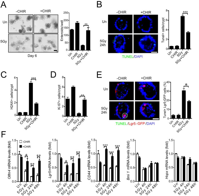
Exposure to high levels of ionizing radiation (IR) leads to debilitating and dose-limiting gastrointestinal (GI) toxicity. Using three-dimensional mouse crypt culture, we demonstrated that p53 target PUMA mediates radiation-induced apoptosis via a cell-intrinsic mechanism, and identified the GSK-3 inhibitor CHIR99021 as a potent radioprotector. CHIR99021 treatment improved Lgr5+ cell survival and crypt regeneration after radiation in culture and mice. CHIR99021 treatment specifically blocked apoptosis and PUMA induction and K120 acetylation of p53 mediated by acetyl-transferase Tip60, while it had no effect on p53 stabilization, phosphorylation or p21 induction. CHIR99021 also protected human intestinal cultures from radiation by PUMA but not p21 suppression. These results demonstrate that p53 posttranslational modifications play a key role in the pathological and apoptotic response of the intestinal stem cells to radiation and can be targeted pharmacologically.
Conflict of interest statement
The authors declare no competing financial interests.
Figures


Similar articles
-
β-Arrestin-2 modulates radiation-induced intestinal crypt progenitor/stem cell injury.Cell Death Differ. 2016 Sep 1;23(9):1529-41. doi: 10.1038/cdd.2016.38. Epub 2016 Apr 29. Cell Death Differ. 2016. PMID: 27128598 Free PMC article.
-
Inhibition of CDK4/6 protects against radiation-induced intestinal injury in mice.J Clin Invest. 2016 Nov 1;126(11):4076-4087. doi: 10.1172/JCI88410. Epub 2016 Oct 4. J Clin Invest. 2016. PMID: 27701148 Free PMC article.
-
Growth factors protect intestinal stem cells from radiation-induced apoptosis by suppressing PUMA through the PI3K/AKT/p53 axis.Oncogene. 2010 Mar 18;29(11):1622-32. doi: 10.1038/onc.2009.451. Epub 2009 Dec 7. Oncogene. 2010. PMID: 19966853 Free PMC article.
-
New Light on An Old Friend: Targeting PUMA in Radioprotection and Therapy of Cardiovascular and Neurodegenerative Diseases.Curr Drug Targets. 2018;19(16):1943-1957. doi: 10.2174/1389450119666180406110743. Curr Drug Targets. 2018. PMID: 29623837 Review.
-
The genetic basis of tissue responses to ionizing radiation.Br J Radiol. 2007 Sep;80 Spec No 1:S2-6. doi: 10.1259/bjr/60507340. Br J Radiol. 2007. PMID: 17704322 Review.
Cited by
-
4-(Nitrophenylsulfonyl)piperazines mitigate radiation damage to multiple tissues.PLoS One. 2017 Jul 21;12(7):e0181577. doi: 10.1371/journal.pone.0181577. eCollection 2017. PLoS One. 2017. PMID: 28732024 Free PMC article.
-
Interplay Between the Intestinal Microbiota and Acute Graft-Versus-Host Disease: Experimental Evidence and Clinical Significance.Front Immunol. 2021 Mar 16;12:644982. doi: 10.3389/fimmu.2021.644982. eCollection 2021. Front Immunol. 2021. PMID: 33815399 Free PMC article. Review.
-
The GS-nitroxide JP4-039 improves intestinal barrier and stem cell recovery in irradiated mice.Sci Rep. 2018 Feb 1;8(1):2072. doi: 10.1038/s41598-018-20370-9. Sci Rep. 2018. PMID: 29391546 Free PMC article.
-
The Role of Intestinal Stem Cells in Epithelial Regeneration Following Radiation-Induced Gut Injury.Curr Stem Cell Rep. 2017;3(4):320-332. doi: 10.1007/s40778-017-0103-7. Epub 2017 Oct 5. Curr Stem Cell Rep. 2017. PMID: 29497599 Free PMC article. Review.
-
Ionizing Radiation Blocks Hair Cell Regeneration in Zebrafish Lateral Line Neuromasts by Preventing Wnt Signaling.Mol Neurobiol. 2018 Feb;55(2):1639-1651. doi: 10.1007/s12035-017-0430-9. Epub 2017 Feb 13. Mol Neurobiol. 2018. PMID: 28194644
References
-
- Potten C. S. Radiation, the ideal cytotoxic agent for studying the cell biology of tissues such as the small intestine. Radiat Res 161, 123–136 (2004). - PubMed
-
- Cheng H. & Leblond C. P. Origin, differentiation and renewal of the four main epithelial cell types in the mouse small intestine. V. Unitarian Theory of the origin of the four epithelial cell types. Am J Anat 141, 537–561 (1974). - PubMed
Publication types
MeSH terms
Substances
Grants and funding
- U01 DK085535/DK/NIDDK NIH HHS/United States
- UL1 TR000005/TR/NCATS NIH HHS/United States
- U19 AI068021/AI/NIAID NIH HHS/United States
- P30 CA047904/CA/NCI NIH HHS/United States
- R01 CA121105/CA/NCI NIH HHS/United States
- P30 DK041301/DK/NIDDK NIH HHS/United States
- R01 CA172136/CA/NCI NIH HHS/United States
- T32 GM008208/GM/NIGMS NIH HHS/United States
- U01 DK085570/DK/NIDDK NIH HHS/United States
- U01 DK085507/DK/NIDDK NIH HHS/United States
- 5T32GM008208/GM/NIGMS NIH HHS/United States
- P30CA047904/CA/NCI NIH HHS/United States
- U24 DK085532/DK/NIDDK NIH HHS/United States
- CA129829/CA/NCI NIH HHS/United States
- U01DK085507/DK/NIDDK NIH HHS/United States
- U01 DK085532/DK/NIDDK NIH HHS/United States
- U01-DK085535/DK/NIDDK NIH HHS/United States
- U01-DK085570/DK/NIDDK NIH HHS/United States
- R01 CA129829/CA/NCI NIH HHS/United States
- CA106348/CA/NCI NIH HHS/United States
- R01 CA106348/CA/NCI NIH HHS/United States
LinkOut - more resources
Full Text Sources
Other Literature Sources
Medical
Molecular Biology Databases
Research Materials
Miscellaneous
