

Preoperative arterial embolization of large liver hemangiomas
- PMID: 25858526
- PMCID: PMC4463265
- DOI: 10.5152/dir.2014.14270
Preoperative arterial embolization of large liver hemangiomas
Abstract
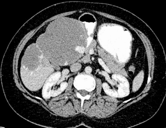
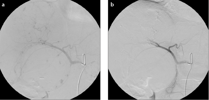
Purpose: We aimed to investigate the efficacy and safety of preoperative selective intra-arterial embolization (PSIAE) in the surgical treatment of large liver hemangiomas.
Methods: Data of 22 patients who underwent resection of large liver hemangiomas were retrospectively analyzed. PSIAE was performed in cases having a high risk of severe blood loss during surgery (n=11), while it was not applied in cases with a low risk of blood loss (n=11).
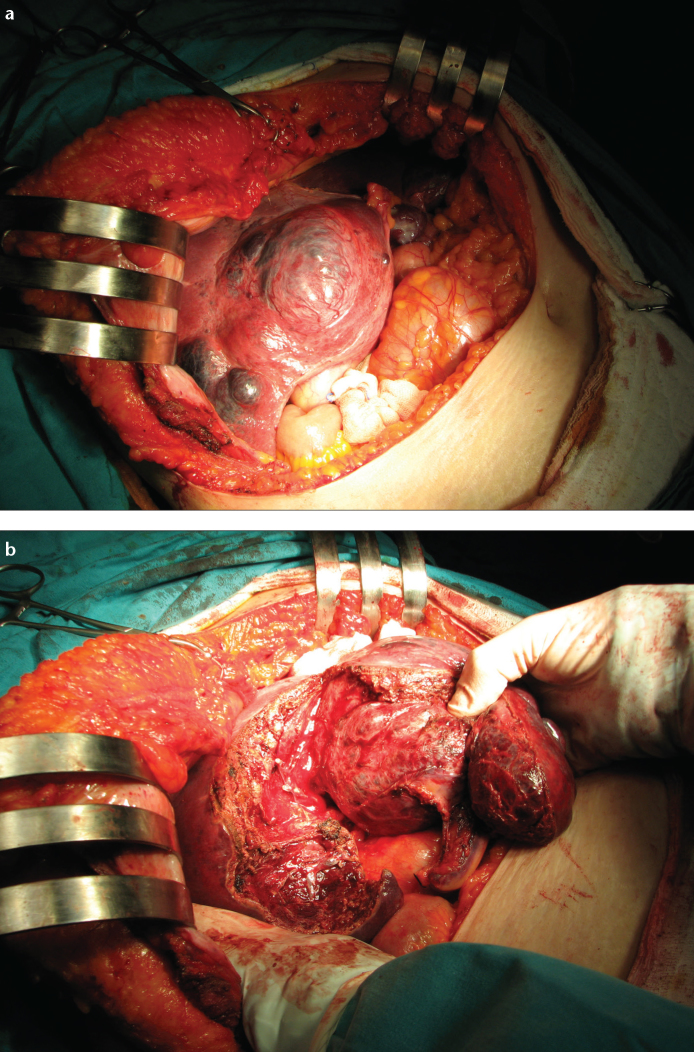
Results: A total of 19 enucleations and six anatomic resections were performed. Operative time, intraoperative bleeding amount, Pringle period, and blood transfusion were comparable between the two groups (P > 0.05, for all). The perioperative serum aspartate transaminase level was not different between groups (P = 1.000). Perioperative total bilirubin levels were significantly increased in the PSIAE group (P = 0.041). Postoperative hospital stay was longer in the PSIAE group. Surgical complications were comparable between groups (P = 0.476).
Conclusion: Patients who underwent PSIAE due to a high risk of severe blood loss during resection of large liver hemangiomas had comparable operative success as patients with a low risk of blood loss who were operated without PSIAE. Hence, PSIAE can be used for the control of intraoperative blood loss, especially in surgically difficult cases.
Figures
References
-
- Ishak KG, Rabin L. Benign tumors of the liver. Med Clin North Am. 1975;59:995–1013. http://dx.doi.org/10.1097/01.mcg.0000159226.63037.a2. - DOI - PubMed
-
- Choi BY, Nguyen MH. The diagnosis and management of benign hepatic tumors. J Clin Gastroenterol. 2005;39:401–412. http://dx.doi.org/10.1016/j.suc.2010.04.006. - DOI - PubMed
-
- Reddy KR, Kligerman S, Levi J, et al. Benign and solid tumors of the liver: relationship to sex, age, size of tumors, and outcome. Am Surg. 2001;67:173–178. - PubMed
-
- Buell JF, Tranchart H, Cannon R, Dagher I. Management of benign hepatic tumors. Surg Clin N Am. 2010;90:719–735. http://dx.doi.org/10.1016/j.suc.2010.04.006. - DOI - PubMed
-
- Strosberg JR, Choi J, Cantor AB, Kvols LK. Selective hepatic artery embolization for treatment of patients with metastatic carcinoid and pancreatic endocrine tumors. Cancer Control. 2006;13:72–78. - PubMed
MeSH terms
Substances
LinkOut - more resources
Full Text Sources
Other Literature Sources
Medical
