. 2015 Apr 10:5:9621.
doi: 10.1038/srep09621.

Galectin-1-secreting neural stem cells elicit long-term neuroprotection against ischemic brain injury

Affiliations

Galectin-1-secreting neural stem cells elicit long-term neuroprotection against ischemic brain injury

Jiayin Wang et al. Sci Rep. .

Abstract

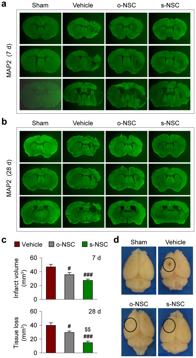
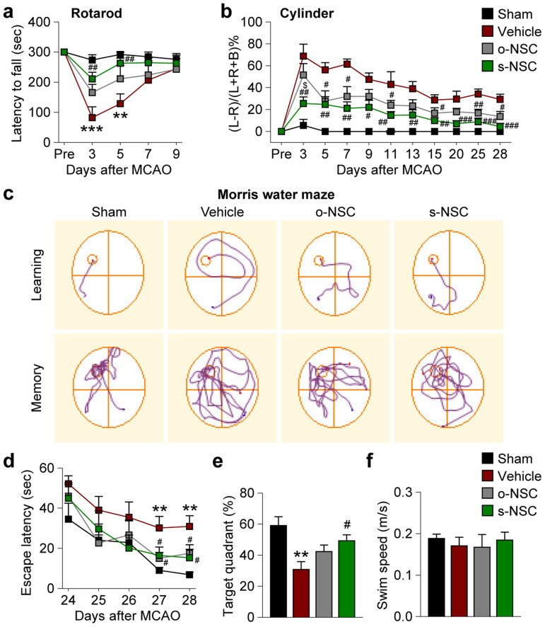
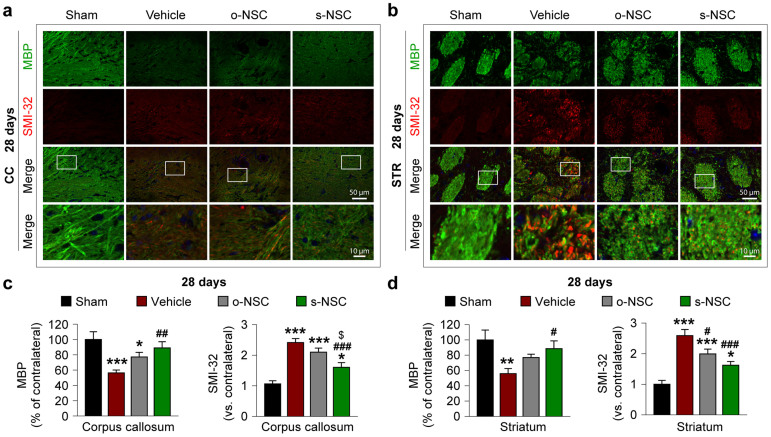
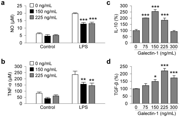
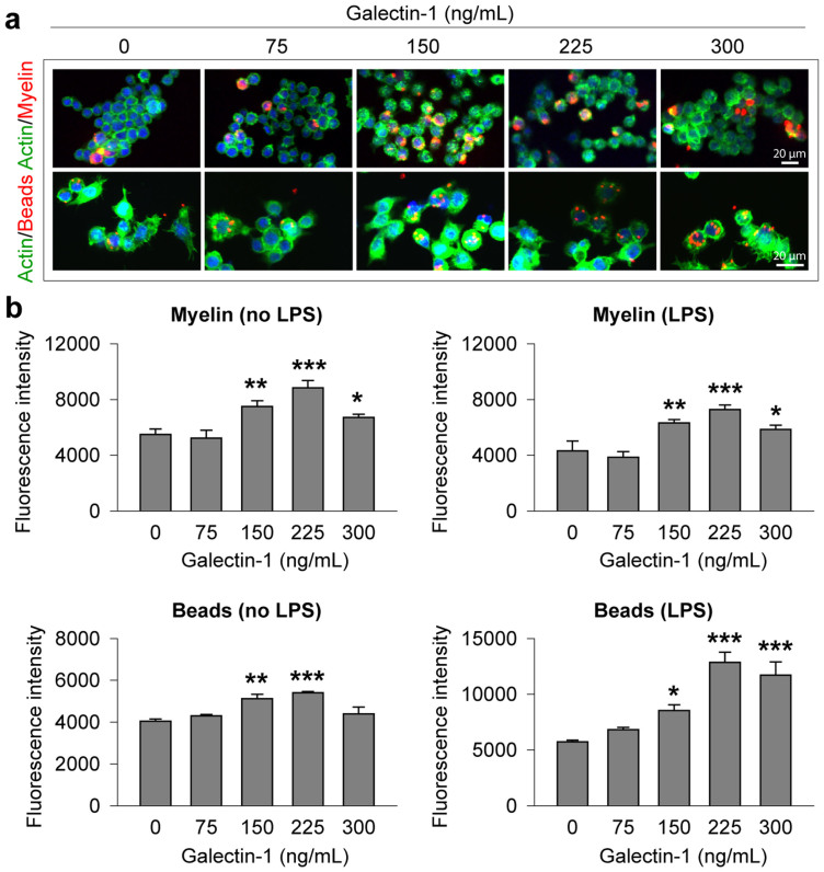
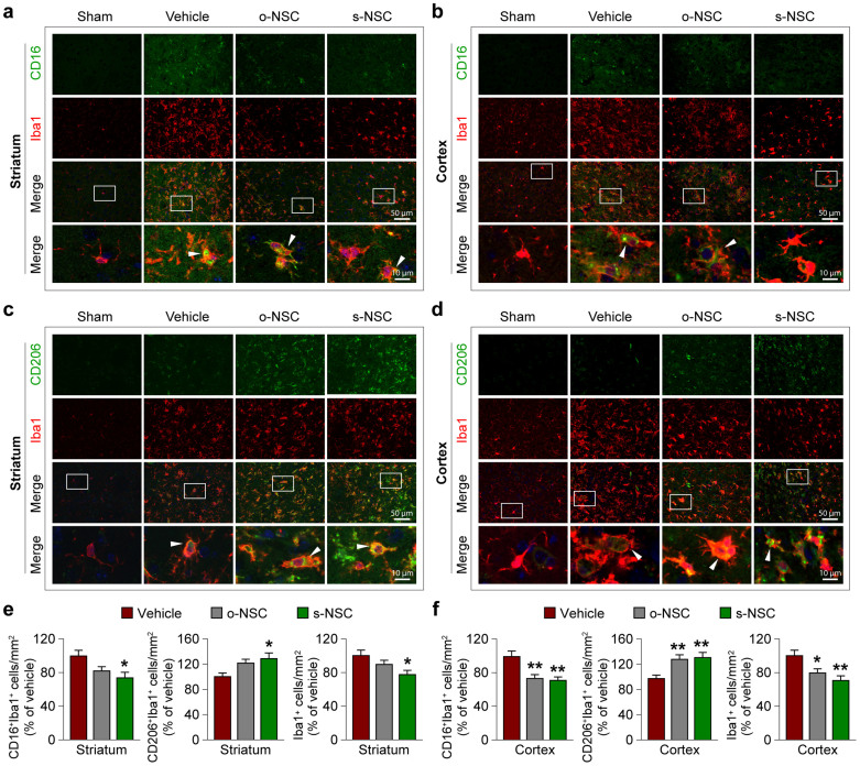
Galectin-1 (gal-1), a special lectin with high affinity to β-galactosides, is implicated in protection against ischemic brain injury. The present study investigated transplantation of gal-1-secreting neural stem cell (s-NSC) into ischemic brains and identified the mechanisms underlying protection. To accomplish this goal, secretory gal-1 was stably overexpressed in NE-4C neural stem cells. Transient cerebral ischemia was induced in mice by middle cerebral artery occlusion for 60 minutes and s-NSCs were injected into the striatum and cortex within 2 hours post-ischemia. Brain infarct volume and neurological performance were assessed up to 28 days post-ischemia. s-NSC transplantation reduced infarct volume, improved sensorimotor and cognitive functions, and provided more robust neuroprotection than non-engineered NSCs or gal-1-overexpressing (but non-secreting) NSCs. White matter injury was also ameliorated in s-NSC-treated stroke mice. Gal-1 modulated microglial function in vitro, by attenuating secretion of pro-inflammatory cytokines (TNF-α and nitric oxide) in response to LPS stimulation and enhancing production of anti-inflammatory cytokines (IL-10 and TGF-β). Gal-1 also shifted microglia/macrophage polarization toward the beneficial M2 phenotype in vivo by reducing CD16 expression and increasing CD206 expression. In sum, s-NSC transplantation confers robust neuroprotection against cerebral ischemia, probably by alleviating white matter injury and modulating microglial/macrophage function.


Figures

Figure 1
Figure 1. Transplantation of s-NSCs or o-NSCs reduces infarct volume after ischemic injury.
Mice received transplantation of o-NSCs or s-NSCs or vehicle injection 2 hours after MCAO. Brain sections were immunostained for MAP2 at 7 and 28 days after MCAO. (a–b) Representative MAP2-stained coronal sections showed smaller infarct or reduced cerebral tissue atrophy at 7 d (a) and 28 d (b) after MCAO in mice receiving transplantation of NSCs. (c) Infarct volume and tissue atrophy were significantly reduced at 7 and 28 days after MCAO, respectively, as compared to vehicle-treated mice. Data are mean ± SEM, n = 7/group, #p ≤ 0.05, ##p ≤ 0.01 versus vehicle; $$p ≤ 0.01 versus o-NSC. (d) Representative brain images illustrate less brain tissue loss in NSC-treated mice than in vehicle-treated mice.
Figure 2
Figure 2. Transplantation of s-NSCs or o-NSCs improves sensorimotor and cognitive functions after ischemic injury.
Mice received transplantation of o-NSCs or s-NSCs or vehicle injection 2 hours after MCAO. Three neurobehavioral tests were performed to assess post-stroke neurological deficits up to 28 d after MCAO. (a) The rotarod test showed significantly decreased latency to fall in vehicle-treated mice at 3 and 5 days after MCAO, whereas s-NSC-treated mice exhibited improved performance compared to vehicle-treated stroke mice. (b) In the cylinder test, the number of left, right, or both forepaw contacts were counted, and the performance asymmetry was expressed as (left-right)/(left + right + both) paw use in 5 min. Both s-NSC- and o-NSC-treated mice showed improved performance compared to vehicle-treated mice up to 28 d after ischemia. However, s-NSC-treated mice showed more consistent beneficial effects than o-NSCs-treated mice. (c) Representative images of the swim paths of mice in the Morris water maze test, when the submerged platform was present (learning phase) or after it was removed (memory phase). (d) Time to locate the submerged platform (escape latency) was measured 24–28 d after MCAO or sham surgery. Vehicle-treated mice showed significant deficits at 27 and 28 d after MCAO, whereas both o-NSC and s-NSC groups were significantly improved. (e) Spatial memory (the percentage of time spent in the goal quadrant after the platform was removed) was measured 28 d after MCAO or sham surgery. Only the s-NSC-treated group showed significant improvement compared to the vehicle-treated group after MCAO. (f) Swim speed was comparable among all groups at 27 d after MCAO or sham surgery. All data are mean ± SEM, n = 6–8 mice/group. **p ≤ 0.01 versus Sham; #p ≤ 0.05 and ##p ≤ 0.01 versus vehicle; $p ≤ 0.05 versus o-NSC.
Figure 3
Figure 3. Transplantation of s-NSCs improves white matter integrity 28 days after ischemia.
Mice received transplantation of o-NSCs or s-NSCs 2 hours after MCAO. Brain sections were dual-stained for myelin basic protein (MBP) and non-phosphorylated neurofilament H (SMI-32) on day 28 after ischemia. (a–b) Representative immunofluorescent images of MBP and SMI-32 staining in the corpus callosum (a) and striatum (b) after sham surgery or MCAO followed by transplantation of vehicle, o-NSCs, or s-NSCs. (c–d) Quantification of MBP and SMI-32 immunofluorescence in the corpus callosum (c) and striatum (d), expressed as percentages and folds of contralateral fluorescence intensities, respectively. Data are mean ± SEM, n = 6. *p ≤ 0.05, **p ≤ 0.01, ***p ≤ 0.001 versus sham; #p ≤ 0.05, ##p ≤ 0.01 versus vehicle; $p ≤ 0.05 versus o-NSC.
Figure 4
Figure 4. Galectin-1 inhibits NO and TNF-α production and enhances IL-10 and TGF-β gene expression in microglia challenged with LPS.
BV2 microglia seeded at 5 × 10⁴/well were pretreated with galectin-1 at the indicated concentrations for 1 hr and LPS (2.5 ng/mL) or its vehicle was added. (a–b) NO and TNF-α were measured in culture medium 12 h after LPS challenge or vehicle treatment. Results are mean ± SEM, from 3 independent experiments, each conducted in triplicate. **p ≤ 0.01 and ***p ≤ 0.001 versus vehicle (0 ng/mL). (c–d) Quantitative RT-PCR for IL-10 and TGF-β mRNA was performed 12 h after LPS challenge following galectine-1 pre-treatment at the indicated concentrations. Data are mean ± SEM, from 3 independent experiments, each conducted in triplicate. **p ≤ 0.01 and *p ≤ 0.05 versus LPS only.
Figure 5
Figure 5. Galectin-1 enhances the phagocytic activity of BV2 microglia.
BV2 microglia seeded at 5 × 10⁴/well were pretreated with galectin-1 at the indicated concentrations for 1 hr. LPS (2.5 ng/mL) or vehicle together with one of the phagocytic substrates, fluorescence-labeled myelin or microsphere beads, were added to cultures for 3 hours. (a) Representative fluorescent images show the phagocytosis of fluorescence-labeled myelin (upper panel) or microsphere beads (lower panel) into BV2 microglia after 3 hours of incubation. (b–c) Phagocytic activity of BV2 microglia was quantified by flow cytometry for fluorescence-labeled myelin (b) and microsphere beads (c), respectively, after 3 hours of incubation of either substrate with or without LPS (2.5 ng/mL). Data are mean ± SEM, from 4 independent experiments, each conducted in triplicate, *p ≤ 0.05, **p ≤ 0.01, ***p ≤ 0.001 versus vehicle (0 ng/mL).
Figure 6
Figure 6. Transplantation of s-NSCs enhances microglia/macrophage polarization toward M2 phenotype after ischemia.
Mice received transplantation of vehicle, o-NSCs, or s-NSCs 2 hours after MCAO or were subjected to sham surgery. Brain sections were dual-stained for CD16 and Iba-1 or CD206 and Iba-1 at 7 d after ischemia. (a–b) Representative immunofluorescence images of CD16 and Iba1 staining in the striatal (a) and cortical (b) infarct border. (c–d) Representative immunofluorescence images of CD206 and Iba1 staining in the striatal (c) and cortical (d) infarct border. (e–f) Quantification of CD16+/Iba-1+, CD206+/Iba-1+, or Iba1+ cells in the striatal (e) and cortical (f) infarct border. Data are expressed as percentages of cell numbers versus vehicle-treated stroke brains. *p ≤ 0.05 and **p ≤ 0.01 versus vehicle-treated stroke group; n = 6/group.

References

    1. DeMers G., Meurer W. J., Shih R., Rosenbaum S. & Vilke G. M. Tissue plasminogen activator and stroke: review of the literature for the clinician. J. Emerg. Med. 43, 1149–1154 (2012). - PubMed
    1. Abe K. et al. Stem cell therapy for cerebral ischemia: from basic science to clinical applications. J. Cereb. Blood. Flow. Metab. 32, 1317–1331 (2012). - PMC - PubMed
    1. Liu X. et al. Cell based therapies for ischemic stroke: from basic science to bedside. Prog. Neurobiol. 115, 92–115 (2014). - PMC - PubMed
    1. Ruggieri M. et al. Induced neural stem cells: methods of reprogramming and potential therapeutic applications. Prog. Neurobiol. 114, 15–24 (2014). - PubMed
    1. Drago D. et al. The stem cell secretome and its role in brain repair. Biochimie 95, 2271–2285 (2013). - PMC - PubMed

Publication types

MeSH terms