. 2015 Oct;30(10):1831-9.
doi: 10.1002/jbmr.2527. Epub 2015 May 6.

Experimental Myocardial Infarction Upregulates Circulating Fibroblast Growth Factor-23

Affiliations

Experimental Myocardial Infarction Upregulates Circulating Fibroblast Growth Factor-23

Olena Andrukhova et al. J Bone Miner Res. 2015 Oct.

Abstract

Myocardial infarction (MI) is a major cause of death worldwide. Epidemiological studies have linked vitamin D deficiency to MI incidence. Because fibroblast growth factor-23 (FGF23) is a master regulator of vitamin D hormone production and has been shown to be associated with cardiac hypertrophy per se, we explored the hypothesis that FGF23 may be a previously unrecognized pathophysiological factor causally linked to progression of cardiac dysfunction post-MI. Here, we show that circulating intact Fgf23 was profoundly elevated, whereas serum vitamin D hormone levels were suppressed, after induction of experimental MI in rat and mouse models, independent of changes in serum soluble Klotho or serum parathyroid hormone. Both skeletal and cardiac expression of Fgf23 was increased after MI. Although the molecular link between the cardiac lesion and circulating Fgf23 concentrations remains to be identified, our study has uncovered a novel heart-bone-kidney axis that may have important clinical implications and may inaugurate the new field of cardio-osteology.

Keywords: FGF23; MYOCARDIAL INFARCTION; VITAMIN D.


Figures

Figure 1
Figure 1
Cardiac function is impaired after MI. Fractioning shortening and left ventricular systolic internal diameter (LVIDs) were measured by echocardiography (ECG) in mouse (A) and rat (B) experimental MI models 3 weeks post‐MI or sham surgery. MI was induced in mice by permanent ligation of the left descending coronary artery and by ischemia‐reperfusion in rats. Middle panels show original ECG images. Bottom panels show blue‐stained fibrotic tissue in left ventricles of MI animals by Masson trichrome staining of paraffin sections 4 weeks post‐MI. Data represent mean ± SEM of 6 to 7 animals per group. *p < 0.05 versus Sham. Scale bar = 100 μm.
Figure 2
Figure 2
Circulating Fgf23 is increased and serum vitamin D hormone is suppressed in mouse and rat MI models. Serum Fgf23 (A), serum vitamin D hormone (B), serum PTH levels (C), serum phosphorus (D), serum calcium (E), serum sodium (H), urinary phosphate/creatinine ratio (F), urinary calcium/creatinine ratio (G), urinary sodium/creatinine ratio (I), and urinary sodium excretion per 12 hours (J) in MI‐ and sham‐operated mice (at 4 weeks postsurgery) and rats (at 2 and 4 weeks postsurgery). Data represent mean ± SEM of 6 to 7 animals in each group for A–I and of 5 to 6 animals in each group for J. *p < 0.05 versus Sham.
Figure 3
Figure 3
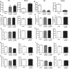
Serum Klotho abundance and biochemical markers of bone metabolism are unchanged after MI. (A) Relative serum Klotho protein abundance in mice (at 4 weeks postsurgery) and rats (at 2 and 4 weeks postsurgery). (B) Urinary deoxypyridinoline (DPD)/creatinine excretion, serum (Se) alkaline phosphatase (ALP), and serum Dickkopf‐1 (Dkk‐1) in MI‐ and sham‐operated rats and mice 4 weeks post‐MI. (C) Serum osteoprotegerin (OPG) in MI‐ and sham‐operated mice 4 weeks post‐MI. Data represent mean ± SEM of 5 to 7 animals in each group.
Figure 4
Figure 4
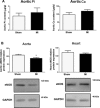
Aortic calcification is unchanged, but aortic eNOS protein expression is reduced after MI. Aortic inorganic phosphate (Pi) and calcium (Ca) content (A) and Western blotting analysis of eNOS protein expression in aorta and heart of MI and sham‐operated mice (B) 4 weeks post‐MI. Data represent mean ± SEM of 4 to 6 animals in each group. *p < 0.05 versus Sham.
Figure 5
Figure 5
Bone and cardiomyocyte Fgf23 protein expression is increased after MI. Western blotting analysis of femur and heart Fgf23 protein expression in MI‐ and sham‐operated mice (A) and rats (B) 4 weeks post‐MI. Immunohistological anti‐Fgf23 staining in femurs (C) and hearts (D) of Sham and MI mice and rats 4 weeks post‐MI. Data represent mean ± SEM of 4 to 6 animals each. *p < 0.05 versus Sham. Scale bar = 20 µm.
Figure 6
Figure 6
Role of Fgf23 in the pathophysiology of cardiac dysfunction after MI. MI increases serum intact Fgf23 possibly by inflammatory signals, ROS, or increased sympathetic tone. In addition, increased cardiac Fgf23 expression might also contribute to elevated serum Fgf23 post‐MI. Increased circulating intact Fgf23, in turn, stimulates renal NCC expression leading to salt and volume retention, suppresses renal vitamin D hormone production leading to endothelial dysfunction, and may have direct pro‐hypertrophic actions on the heart.

References

    1. Lee JH, Gadi R, Spertus JA, et al. Prevalence of vitamin D deficiency in patients with acute myocardial infarction. Am J Cardiol. 2011; 107:1636–8. - PMC - PubMed
    1. Scragg R, Sowers M, Bell C. Serum 25‐hydroxyvitamin D, ethnicity, and blood pressure in the Third National Health and Nutrition Examination Survey. Am J Hypertens. 2007; 20:713–9. - PubMed
    1. Judd SE, Nanes MS, Ziegler TR, et al. Optimal vitamin D status attenuates the age‐associated increase in systolic blood pressure in white Americans: results from the third National Health and Nutrition Examination Survey. Am J Clin Nutr. 2008; 87:136–41. - PubMed
    1. Pittas AG, Chung M, Trikalinos T, et al. Systematic review: vitamin D and cardiometabolic outcomes. Ann Intern Med. 2010; 152:307–14. - PMC - PubMed
    1. Al Mheid I, Patel R, Murrow J, et al. Vitamin D status is associated with arterial stiffness and vascular dysfunction in healthy humans. J Am Coll Cardiol. 2011; 58:186–92. - PMC - PubMed

Publication types