

Brown tumor of mandible in association with primary hyperparathyroidism: a case report
- PMID: 25859108
- PMCID: PMC4377151
Brown tumor of mandible in association with primary hyperparathyroidism: a case report
Abstract
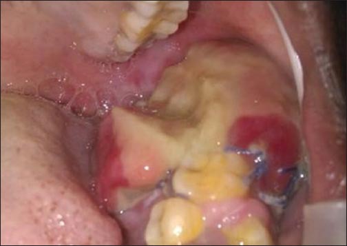
Brown tumors are giant cell focal lesion that arises as a result of abnormal bone metabolism in patients with hyperparathyroidism (HPT). The lesions localize in areas of extensive bone resorption, which is replaced by fibrovascular tissue and giant cells with abundant deposits hemorrhage and hemosiderin. A rare case of brown tumor of mandible in a 22-year-old woman is reported here. This case emphasizes the importance of a detailed systemic investigation for all lesions in the maxillofacial region and also discusses the diverse presentations associated with primary HPT.
Keywords: Brown tumor; giant cell lesion; mandible; primary hyperparathyroidism.
Conflict of interest statement
Figures


References
-
- Movahedian B, Razavi SM, Hasheminia D, Rezaei M. Simultaneous maxillary and mandibular brown tumors in secondary hyperparathyroidism: A case report. Dent Res J. 2008;5(1):41–5.
-
- Selvi F, Cakarer S, Tanakol R, Guler SD, Keskin C. Brown tumour of the maxilla and mandible: A rare complication of tertiary hyperparathyroidism. Dentomaxillofac Radiol. 2009;38(1):53–8. - PubMed
-
- Jebasingh F, Jacob JJ, Shah A, Paul TV, Seshadri MS. Bilateral maxillary brown tumours as the first presentation of primary hyperparathyroidism. Oral Maxillofac Surg. 2008;12(2):97–100. - PubMed
-
- Suarez-Cunqueiro MM, Schoen R, Kersten A, Klisch J, Schmelzeisen R. Brown tumor of the mandible as first manifestation of atypical parathyroid adenoma. J Oral Maxillofac Surg. 2004;62(8):1024–8. - PubMed
Publication types
LinkOut - more resources
Full Text Sources